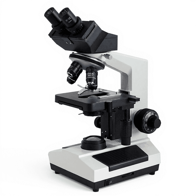
Electric Scientific Metallurgical Microscope with DIC System
Research-grade metallurgical microscope with DIC system, electric field switching, and magnification from 50x to 1000x for materials characterization and microstructural analysis.
| Automation Level | semi-automated |
| Product Type | Microscope |
| Microscope Type | Metallurgical |
| Application | Laboratory Research, Materials Science, Quality Control |
| Skill Level | Professional / Research |
| Brand | ConductScience |
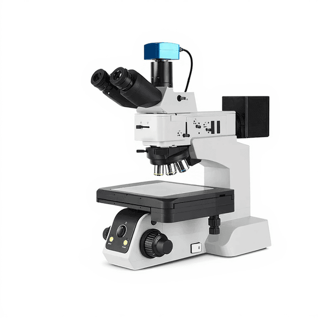
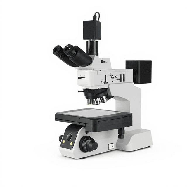
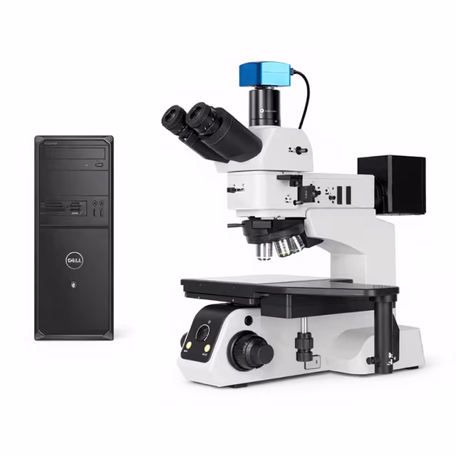
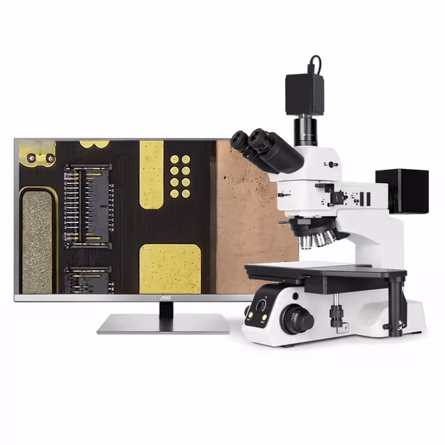
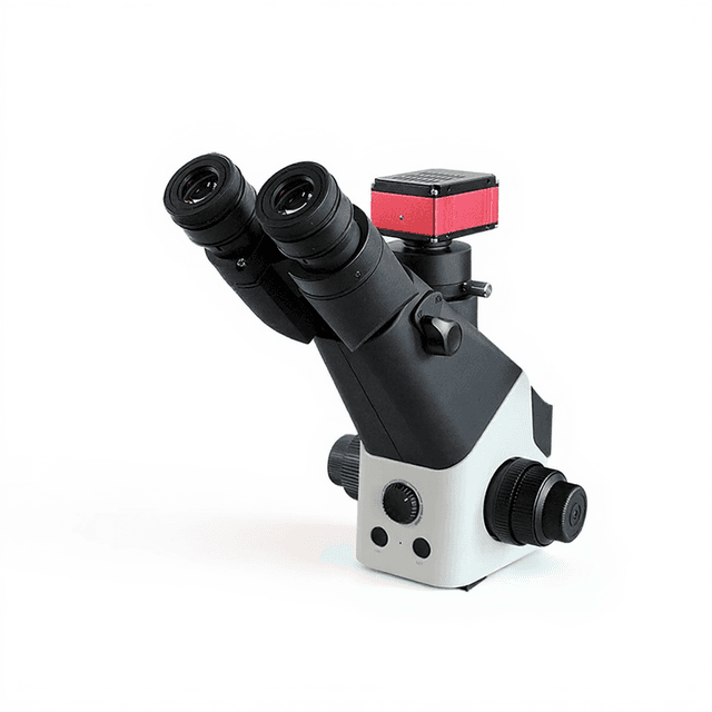
The Electric Scientific Metallurgical Microscope with DIC System is a research-grade reflected light microscope designed for materials characterization and metallurgical analysis. Built around an infinite chromatic aberration correction optical system, this instrument provides magnification from 50x to 1000x with optional high-magnification objectives. The system features electric light/dark field switching, differential interference contrast (DIC) capabilities, and both reflected and transmitted illumination for comprehensive materials examination.
The microscope incorporates a motorized 5-hole nosepiece with DIC slot, 8-inch mechanical stage with 210mm x 210mm travel range, and dual LED illumination systems (5W each) for reflected and transmitted light applications. Digital imaging capabilities extend magnification to 3000x when equipped with the optional 12MP camera system. The instrument supports polarized light microscopy through integrated polarizer assemblies and includes variable aperture controls for optimized contrast and resolution.
How It Works
The microscope operates using reflected light illumination where light from the 5W LED source passes through the objective lens and illuminates the specimen surface. Reflected light returns through the same objective, creating an image that reveals surface topography, grain boundaries, and phase contrast in opaque materials. The infinite chromatic aberration correction optical system maintains color fidelity across the magnification range while minimizing optical distortions.
Differential interference contrast (DIC) enhances surface relief and subtle structural features by introducing controlled phase shifts between polarized light beams. The system's polarizing assemblies include a 360° rotary analyzer and polarizer insert plates that enable examination of optically anisotropic materials and stress patterns. The electric field switching capability allows rapid transition between bright field and dark field illumination modes, optimizing contrast for different specimen characteristics.
The roll-out achromatic condenser (N.A. 0.9) provides controlled illumination geometry, while variable aperture and field stops optimize resolution and contrast. The coaxial X-Y mechanical stage enables precise specimen positioning across the 210mm travel range, supporting systematic examination of large samples or multiple specimen areas.
Features & Benefits
Automation Level
- semi-automated
Product Type
- Microscope
Microscope Type
- Metallurgical
Application
- Laboratory Research
- Materials Science
- Quality Control
Skill Level
- Professional / Research
Brand
- ConductScience
Research Domain
- Analytical Chemistry
- Industrial Hygiene
- Materials Science
Weight
- 1.9 kg
Dimensions
- L: 4.9 mm
- W: 6.6 mm
- H: 6.6 mm
Comparison Guide
| Feature | This Product | Typical Alternative | Advantage |
|---|---|---|---|
| Magnification Range | 50x-1000x standard, up to 3000x digital | Many entry-level models limited to 400x maximum magnification | Extended range supports both overview and high-resolution analysis without instrument changes |
| Field Switching | Electric 5-hole nosepiece with software and button control | Manual slider or rotating mechanisms common in basic models | Eliminates positioning errors and enables automated imaging protocols |
| Stage Size and Travel | 8-inch stage with 210mm x 210mm mechanical travel | Smaller stages typically offer 100-150mm travel range | Accommodates larger specimens and enables systematic examination of extended areas |
| Illumination System | Dual 5W LED systems for reflected and transmitted light | Single halogen systems with limited color stability | Provides stable, color-balanced illumination with minimal heat and maintenance requirements |
| Optical Correction | Infinite chromatic aberration correction system | Finite optical systems with more limited correction | Maintains color accuracy and reduces distortions across all magnifications for precise analysis |
| DIC Capability | Integrated DIC slot in electric nosepiece with polarizing assemblies | DIC often optional or unavailable on basic models | Reveals subtle surface features and microstructural details not visible with conventional contrast methods |
This microscope offers research-grade optical performance with automated features typically found in higher-end instruments. The combination of electric field switching, extended magnification range, and integrated DIC capabilities provides comprehensive materials characterization in a single platform.
Practical Tips
Verify magnification calibration using certified stage micrometers at multiple objective levels, particularly after any optical component changes.
Why: Ensures measurement accuracy and maintains traceability for quantitative analysis applications.
Clean objective lenses monthly using appropriate optical solvents and lens tissue, working from center outward in circular motions.
Why: Prevents contamination buildup that can degrade image quality and cause optical artifacts.
Allow LED illumination systems to stabilize for 10-15 minutes before critical observations or documentation.
Why: Ensures consistent color temperature and intensity for reproducible imaging results.
If DIC contrast appears uneven, check polarizer alignment and ensure specimen surface is properly leveled on the stage.
Why: DIC effectiveness depends on consistent optical path geometry and specimen orientation.
Document illumination settings and optical configurations used for each imaging session to ensure reproducible results.
Why: Maintains consistency across multiple operators and enables accurate comparison of results over time.
Use appropriate eye protection when operating high-intensity illumination modes and avoid direct LED exposure.
Why: Prevents eye strain and potential retinal damage from concentrated light sources.
Store DIC prisms and polarizing components in dust-free environments when not in use to maintain optical quality.
Why: Preserves critical optical surfaces that are difficult to clean and expensive to replace.
Lubricate stage mechanisms annually with recommended precision lubricants to maintain smooth operation.
Why: Prevents mechanical wear and ensures precise positioning accuracy for measurement applications.
Setup Guide
What’s in the Box
- Electric Scientific Metallurgical Microscope main unit
- SWH10X/25mm high eye point eyepieces (pair)
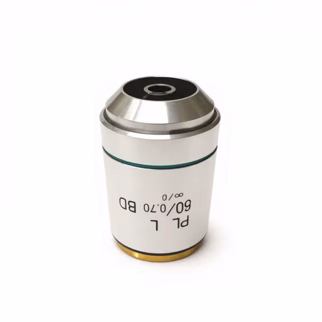
- Long Working Distance BD Semi-Apo 5X objective lens
- DIC prism set and polarizing accessories
- Stage clips and specimen holders (typical)
- Power cable and LED driver unit
- User manual and optical alignment tools (typical)
- Dust covers for optical components (typical)
Compliance
Warranty & ConductCare
ConductScience provides a comprehensive 1-year manufacturer warranty covering optical and mechanical components, with technical support for setup and operational guidance. Extended warranty options and calibration services are available for laboratory compliance requirements.
What specimen preparation is required for optimal DIC imaging?
Specimens require flat, polished surfaces with minimal relief. Standard metallographic preparation through progressive grinding and polishing to 0.25-1 micron finish is recommended. Etching may be applied to reveal grain boundaries, but excessive relief can reduce DIC effectiveness.
How does the electric nosepiece control compare to manual systems?
The electric system provides reproducible positioning and rapid field switching via software or physical buttons. This eliminates manual adjustment errors and enables automated imaging protocols, though it requires power and adds system complexity compared to manual nosepieces.
What is the maximum specimen thickness that can be accommodated?
The 8-inch stage with 210mm travel and long working distance objectives can accommodate specimens several centimeters thick. Actual thickness limits depend on objective working distance and stage clearance, with the 5X LWD objective providing 15mm working distance.
Can the system perform quantitative measurements?
Yes, when equipped with the optional 12MP camera and measurement software. The calibrated optical system supports linear measurements, area calculations, and statistical analysis of microstructural features. Consult specifications for measurement accuracy limits.
How stable are the LED illumination systems over time?
LED systems provide stable output with minimal drift after initial warm-up period. Color temperature remains consistent throughout LED lifetime, eliminating frequent recalibration needs common with tungsten-halogen systems.
What maintenance is required for the DIC components?
DIC prisms require periodic cleaning with appropriate optical solvents and lint-free materials. Polarizer assemblies should be inspected for stress-induced birefringence and replaced if optical quality degrades. Avoid mechanical stress on DIC elements during handling.
Is the microscope compatible with third-party camera systems?
The C-mount camera adapter (0.65x) is compatible with standard industrial cameras. However, magnification calibration and software integration may require adjustment. Consult ConductScience for validated camera configurations.
How does this compare to inverted metallurgical microscopes?
This upright configuration is optimal for standard prepared specimens and provides better access to sample surfaces. Inverted systems are preferred for large, heavy specimens or in-situ observations where sample orientation cannot be changed.
Have a question about this product?
Frequently Bought Together
Upgrade Options
Related Products