Paper Microfluidic Chip
Cellulose-based microfluidic chip with 800-2000 μm features for point-of-care diagnostics and colorimetric detection applications.
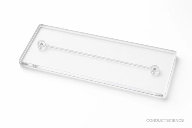
Reusable chip — designed for multiple experimental runs. Compatible with standard microfluidic tubing: steel pins (0.7 mm ID / 1.0 mm OD) and silicone tubing (0.8 mm ID / 1.9 mm OD). Available in bulk packs (5‑, 10‑, and 25‑unit) for lab-scale and consumable workflows.

Louise Corscadden, PhD
Director of Science · ConductScience
Ask Louise about Paper Microfluidic Chip fit, setup, configuration, or quote prep.
Already working with us? Sign in to connect this with My Scientist.
Key Specifications
Full details →- Model fit
- 1 selectable configurations
- SKU family
- WHM-0124
- Sizing
- 181.8 x 136.3 x 90.9 cm
- Ordering
- Quote-reviewed before fulfillment
- Category
- Microfluidic Chips
- Build notes
- Confirm accessories, station layout, and support needs before purchase
The Paper Microfluidic Chip (WHM-0124) is a cellulose-based analytical device designed for point-of-care diagnostic applications. Fabricated using wax printing, inkjet patterning, plasma treatment, or cutting techniques, this disposable chip features microchannels with minimum feature sizes ranging from 800-2000 micrometers. The paper substrate enables capillary-driven fluid transport without external pumping, making it suitable for field testing and resource-limited settings.
This microfluidic paper analytical device (μPAD) serves as a low-cost alternative to traditional lateral flow assays and glass-based microfluidic systems. The cellulose substrate provides excellent compatibility with aqueous samples and supports colorimetric detection methods commonly used in diagnostic applications. The chip's design allows for sample preparation, mixing, and detection within a single disposable platform.
How It Works
Paper microfluidic chips operate through capillary action within the cellulose fiber matrix. The hydrophilic cellulose substrate naturally wicks aqueous solutions through defined channels, eliminating the need for external pumps or pressure systems. Channel patterns are created by depositing hydrophobic barriers using wax printing, inkjet printing with hydrophobic inks, plasma treatment to create hydrophobic regions, or physical cutting of the paper substrate.
When a sample is applied to the inlet, capillary forces drive the fluid through the microchannels at a rate determined by the channel geometry, paper porosity, and fluid properties. Reagents can be pre-deposited and dried within specific zones of the chip, becoming rehydrated as the sample flows past. Detection typically occurs through colorimetric changes visible to the naked eye or quantifiable using optical readers.
The 800-2000 micrometer feature size allows for controlled fluid flow while maintaining structural integrity of the paper substrate. Multiple detection zones can be incorporated for simultaneous analysis of different analytes or for internal quality controls within a single chip.
Features & Benefits
Pack Size
- 25-Pack
Weight
- 3.3 kg
Dimensions
- L: 181.8 mm
- W: 136.3 mm
- H: 90.9 mm
| Feature | This Product | Typical Alternative | Advantage |
|---|---|---|---|
| Minimum Feature Size | 800-2000 μm | Glass/polymer chips often achieve sub-100 μm features | Larger features provide more robust fluid flow and easier visual detection for point-of-care applications |
| Fabrication Methods | Wax print, inkjet, plasma treatment, cutting | Traditional microfluidics require cleanroom photolithography | Multiple accessible fabrication options enable rapid prototyping without specialized facilities |
| Substrate Material | Cellulose paper | Glass, silicon, or polymer substrates are common | Paper substrate enables passive capillary flow without external pumping systems |
| Cost Structure | Low-cost disposable platform | Glass devices typically require higher initial investment | Enables high-volume testing and field deployment in resource-limited settings |
| Application Focus | Point-of-care diagnostics | Traditional devices often target laboratory-based analysis | Designed specifically for field testing and decentralized diagnostic applications |
The Paper Microfluidic Chip offers a cost-effective, pump-free alternative to traditional microfluidic devices. With 800-2000 μm features fabricated using accessible methods on cellulose substrate, it prioritizes ease of use and field deployment over ultra-high resolution applications.
Practical Tips
Pre-wet channels with buffer before sample application to ensure uniform flow and prevent air bubble formation.
Why: Even wetting reduces flow irregularities that can affect assay reproducibility.
Apply samples slowly to prevent flooding and allow proper capillary wicking through the paper matrix.
Why: Controlled sample introduction ensures optimal contact time with pre-deposited reagents.
Store unused chips in desiccant-containing packages to prevent humidity-induced degradation of hydrophobic barriers.
Why: Moisture exposure can compromise channel definition and affect fluid flow patterns.
Include positive and negative control zones on each chip to verify assay performance.
Why: Built-in controls help identify chip defects or reagent degradation issues.
Use consistent lighting conditions when performing visual or photometric detection to ensure reproducible results.
Why: Color development can appear different under varying lighting conditions, affecting quantitative measurements.
If flow stops prematurely, check for air bubbles or verify that sample volume is sufficient for complete channel filling.
Why: Flow interruption prevents complete mixing and can lead to false negative results.
Dispose of used chips immediately after reading to prevent cross-contamination between samples.
Why: Paper chips cannot be cleaned for reuse and may retain sample residues.
Setup Guide
What’s in the Box
- Paper microfluidic chips (quantity varies by package)
- User instruction sheet (typical)
- Storage packaging (typical)
Warranty
ConductScience provides a standard warranty covering manufacturing defects and material quality. Technical support is available for fabrication guidance and application development.
Compliance
References
Background reading relevant to this product:
What sample volumes are compatible with the 800-2000 μm channel dimensions?
Sample volume requirements depend on channel length and assay design. Typical volumes range from 1-50 μL. Consult product datasheet for specific chip configurations and recommended sample volumes.
How do I select the appropriate fabrication method for my application?
Wax printing provides sharp hydrophobic barriers for simple designs. Inkjet printing offers high-resolution patterning. Plasma treatment creates selective hydrophobic regions. Cutting provides physical channel separation. Method selection depends on resolution requirements and available equipment.
What detection methods are compatible with these paper chips?
The cellulose substrate supports colorimetric, fluorescence, and electrochemical detection methods. Colorimetric detection is most common for point-of-care applications. Signal intensity can be quantified using smartphone cameras or dedicated optical readers.
How should the chips be stored to maintain performance?
Store chips in dry conditions at room temperature, protected from light and humidity. Sealed packaging prevents contamination and maintains hydrophobic barrier integrity. Avoid extreme temperatures that may affect paper substrate properties.
Can reagents be pre-loaded onto the chip?
Yes, reagents can be spotted and dried in detection zones before use. This enables ready-to-use assays where sample addition initiates the analytical reaction. Reagent stability should be validated for the intended storage period.
What are the limitations compared to glass microfluidic devices?
Paper chips have lower resolution than glass devices and are limited to aqueous solutions. They cannot handle organic solvents or high temperatures. However, they offer significant cost advantages and do not require external pumps.
How do I optimize fluid flow for my specific assay?
Flow rate depends on channel width, paper porosity, and fluid properties. Narrower channels increase flow resistance while wider channels may reduce mixing efficiency. Test different geometries to balance flow rate with analytical performance.
Have a question about this product?
Have a question? Just ask.
Send it over and we'll email you a personalized answer — no call, no scheduling.
Prefer to talk it through?