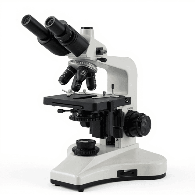
Trinocular UIS Biological Microscope
Trinocular biological microscope with UIS optics for brightfield imaging, featuring dual eyepieces and dedicated camera port for simultaneous observation and digital capture.

Louise Corscadden, PhD
Director of Science · ConductScience
Ask Louise about Trinocular UIS Biological Microscope fit, setup, configuration, or quote prep.
Already working with us? Sign in to connect this with My Scientist.
Key Specifications
Full details →- Model fit
- Configured during quote
- SKU family
- BIO-0530
- Sizing
- 43.0 x 43.0 x 33.0 cm
- Ordering
- Online checkout and quote request available
- Category
- Microscopy
- Build notes
- Confirm accessories, station layout, and support needs before purchase
The Trinocular UIS Biological Microscope is a versatile brightfield microscope designed for routine biological observation and documentation in research and clinical laboratories. The trinocular configuration provides simultaneous viewing through dual eyepieces while enabling digital capture through the dedicated camera port, facilitating both real-time observation and image acquisition workflows.
Built with UIS (Universal Infinity System) optics, this microscope delivers consistent optical performance across magnification ranges commonly used in cell biology, histopathology, and microbiological applications. The system supports standard brightfield illumination for transmitted light microscopy of stained specimens, unstained cells, and tissue sections.
How It Works
The microscope operates on the principle of transmitted light brightfield illumination, where light from the substage illuminator passes through the specimen and into the objective lens system. The UIS (Universal Infinity System) optical design employs infinity-corrected objectives that project parallel light rays to a tube lens, which focuses the image at the intermediate image plane.
Light intensity and contrast are controlled through the substage condenser system, which can be adjusted for optimal Köhler illumination. The trinocular head splits the light path, directing approximately 80% of the light to the binocular eyepieces for visual observation while sending 20% to the trinocular port for camera attachment and digital imaging.
Magnification is achieved through the combination of objective lens power and eyepiece magnification, with the UIS system maintaining optical corrections and field flatness across the magnification range.
Features & Benefits
Automation Level
- manual
Research Domain
- Cell Biology
- Clinical Diagnostics
- Developmental Biology
- Histopathology
- Materials Science
- Microbiology
Weight
- 9.0 kg
Dimensions
- L: 43.0 mm
- W: 43.0 mm
- H: 33.0 mm
| Feature | This Product | Typical Alternative | Advantage |
|---|---|---|---|
| Optical Head Configuration | Trinocular head with dedicated camera port | Entry-level models often provide binocular heads without camera ports | Enables simultaneous visual examination and digital documentation without switching between observation modes. |
| Optical System Design | UIS infinity-corrected optics | Basic models may use finite tube length optics | Provides superior image quality with reduced optical aberrations and consistent performance across magnification ranges. |
| Illumination Type | Transmitted brightfield illumination | Some models offer only basic illumination without advanced condenser systems | Supports proper Köhler illumination for optimal contrast and resolution in biological specimen examination. |
| Stage Type | Mechanical stage with precision controls | Basic models may have fixed stages with manual specimen positioning | Allows precise, reproducible positioning essential for systematic specimen examination and documentation. |
This microscope provides trinocular optics with UIS infinity correction in a compact 9.0 kg package measuring 43.0 x 43.0 x 33.0 cm. The system combines visual observation capabilities with dedicated digital imaging support, making it suitable for routine biological microscopy applications requiring both examination and documentation workflows.
Practical Tips
Verify objective parfocality by ensuring specimens remain in focus when switching between objectives.
Why: Proper parfocality reduces refocusing time and prevents mechanical damage to specimens.
Clean objective lenses weekly using appropriate lens cleaning solutions and lens tissue, working from center outward.
Why: Contaminated optics significantly degrade image quality and contrast in transmitted light microscopy.
Allow illumination system to warm up for 15 minutes before critical observations to ensure stable light output.
Why: Thermal stabilization prevents color temperature drift and intensity variations during extended observation sessions.
If images appear dim or uneven, check condenser alignment and aperture diaphragm settings for proper Köhler illumination.
Why: Misaligned illumination is the most common cause of poor contrast and uneven field brightness.
Use appropriate specimen preparation thickness to avoid over-staining or optical artifacts in transmitted light.
Why: Specimen thickness directly affects image contrast and the ability to resolve cellular details.
Always use lowest effective illumination intensity to prevent photodamage to specimens and reduce heat generation.
Why: Excessive illumination can cause specimen fading and thermal damage, particularly in living cell observations.
Setup Guide
What’s in the Box
- Trinocular biological microscope (main unit)
- Mechanical stage with specimen clips (typical)
- Substage condenser assembly (typical)
- Illumination system with power supply (typical)
- User manual and optical care instructions (typical)
- Dust cover (typical)
Warranty
ConductScience provides standard one-year manufacturer warranty covering optical and mechanical components, with technical support for setup and operational guidance.
Compliance
References
Background reading relevant to this product:
What types of specimens can be examined with this brightfield microscope?
The system supports examination of stained histological sections, cultured cells, bacterial preparations, blood smears, and other specimens suitable for transmitted light microscopy. Unstained specimens with natural contrast can also be observed.
Is the trinocular port compatible with standard digital cameras?
The trinocular port accepts standard C-mount cameras and microscopy-specific digital imaging systems. Verify mechanical compatibility and optical magnification factors for your specific camera model.
What maintenance is required for the UIS optical system?
Clean objective lenses and eyepieces with lens tissue and appropriate solvents. Keep dust covers in place when not in use and periodically clean illumination path components to maintain image quality.
Can fluorescence filters be added to this microscope?
This brightfield microscope is designed for transmitted light applications. Fluorescence capabilities would require additional filter cubes and appropriate illumination, which may not be compatible with this model.
What is the typical magnification range available?
Magnification depends on objective and eyepiece combinations used. Consult product datasheet for specific objective mounting specifications and recommended eyepiece magnifications.
How is Köhler illumination achieved with this system?
Adjust substage condenser height and center position, then use field and aperture diaphragms to achieve uniform illumination. Proper alignment ensures optimal contrast and resolution.
Is the mechanical stage suitable for slide scanning applications?
The mechanical stage provides precise positioning for manual examination. Automated scanning would require motorized stage components not included with this manual system.
What power requirements does the illumination system have?
Consult product specifications for electrical requirements. Most biological microscopes operate on standard laboratory power with halogen or LED illumination systems.
Have a question about this product?
Have a question? Just ask.
Send it over and we'll email you a personalized answer — no call, no scheduling.
Prefer to talk it through?
Accessories
Enhance your setup with compatible accessories


Replacement Parts & Consumables


Frequently Bought Together
Related Products