Wound Healing Research Microfluidic Chip
Microfluidic chip designed for wound healing research, cell migration studies, and drug delivery applications with controlled microenvironments for reproducible assays.
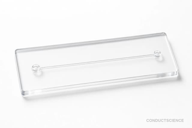
Reusable chip — designed for multiple experimental runs. Compatible with standard microfluidic tubing: steel pins (0.7 mm ID / 1.0 mm OD) and silicone tubing (0.8 mm ID / 1.9 mm OD). Available in bulk packs (5‑, 10‑, and 25‑unit) for lab-scale and consumable workflows.

Louise Corscadden, PhD
Director of Science · ConductScience
Ask Louise about Wound Healing Research Microfluidic Chip fit, setup, configuration, or quote prep.
Already working with us? Sign in to connect this with My Scientist.
Key Specifications
Full details →- Model fit
- 3 selectable configurations
- SKU family
- WHM-0080
- Sizing
- 181.8 x 136.3 x 90.9 cm
- Ordering
- Online checkout and quote request available
- Category
- Cell Culture Chips
- Build notes
- Confirm accessories, station layout, and support needs before purchase
The Wound Healing Research Microfluidic Chip provides a controlled platform for studying cell migration, wound closure dynamics, and tissue repair mechanisms at the microscale. This microfluidic device enables researchers to create reproducible scratch wound models and monitor healing processes in real-time under precisely controlled conditions.
The chip supports applications in wound healing research, cell migration studies, and drug delivery investigations by providing standardized microenvironments for cell culture and analysis. Researchers can evaluate therapeutic compounds, study cellular responses to mechanical or chemical stimuli, and quantify migration parameters with improved reproducibility compared to traditional scratch assays.
How It Works
The microfluidic chip utilizes precisely fabricated channels and chambers to create controlled cellular microenvironments that mimic physiological conditions. Cell monolayers are cultured within designated areas of the chip, and mechanical or chemical wounds can be introduced through controlled disruption or selective cell removal techniques.
Fluid flow within the microchannels enables the establishment of chemical gradients, nutrient delivery, and waste removal while maintaining sterile conditions. The transparent chip material allows for real-time microscopic observation and image-based quantification of cell migration, proliferation, and wound closure dynamics.
Multiple experimental conditions can be tested simultaneously within a single chip through parallel channel designs, enabling comparative studies of different treatments, cell types, or environmental parameters while maintaining consistent experimental conditions across all test areas.
Features & Benefits
Pack Size
- 5-Pack
- 10-Pack
- 25-Pack
Weight
- 3.3 kg
Dimensions
- L: 181.8 mm
- W: 136.3 mm
- H: 90.9 mm
| Feature | This Product | Typical Alternative | Advantage |
|---|---|---|---|
| Experimental Standardization | Provides controlled microenvironments for wound healing and cell migration studies | Traditional scratch assays often have variable wound dimensions and inconsistent conditions | Improves reproducibility and enables quantitative comparison across experiments and research groups |
| Real-time Monitoring | Transparent design allows continuous microscopic observation | Conventional assays require endpoint or periodic sampling | Enables dynamic analysis of healing kinetics and cellular responses throughout the experiment |
| Chemical Gradient Control | Microfluidic channels enable precise gradient establishment | Static culture systems provide limited control over chemical environments | Allows investigation of directional cell migration and chemotactic responses |
| Sample Volume Efficiency | Microfluidic design requires minimal reagent volumes | Traditional culture plates consume larger volumes of media and test compounds | Reduces costs when working with expensive reagents or limited cell populations |
The microfluidic chip provides standardized wound models with controlled microenvironments for studying cell migration and healing processes. The system offers advantages in reproducibility, real-time monitoring capabilities, and efficient use of biological samples compared to traditional culture methods.
| Model | SKU | Listed price | Status | Dimensions |
|---|---|---|---|---|
| 25-Pack | WHM-0080-25PK | Quote | Available | 181.8 x 136.3 x 90.9 cm |
| 10-Pack | WHM-0080-10PK | $1,490.00 | Available | 181.8 x 136.3 x 90.9 cm |
| 5-Pack | WHM-0080-5PK | Quote | Available | 181.8 x 136.3 x 90.9 cm |
Practical Tips
Pre-condition the chip with culture medium for at least 30 minutes before cell loading to ensure proper wetting and temperature equilibration.
Why: Proper conditioning prevents air bubbles and ensures consistent cell attachment across the culture surface.
Clean chips immediately after use with appropriate solvents and store in a dust-free environment to prevent contamination.
Why: Prompt cleaning prevents protein buildup and maintains chip performance for future experiments.
Use consistent imaging parameters and analysis protocols across all experimental conditions to ensure comparable quantification.
Why: Standardized analysis methods reduce variability and improve the reliability of migration measurements.
If cells detach during flow experiments, reduce flow rates or check surface treatment protocols for proper cell adhesion.
Why: Excessive shear stress can cause cell detachment and compromise experimental results.
Handle chips with appropriate personal protective equipment and follow institutional guidelines for cell culture work.
Why: Microfluidic devices may have sharp edges and cell culture work requires sterile technique and biological safety measures.
Verify channel dimensions and flow rates using calibrated measurement tools before beginning critical experiments.
Why: Accurate flow characterization ensures reproducible experimental conditions and proper interpretation of results.
Setup Guide
What’s in the Box
- Wound healing microfluidic chip (typical)
- User manual and protocol guide (typical)
- Connection tubing (typical)
- Sterile packaging (typical)
Warranty
ConductScience provides a standard manufacturer warranty covering defects in materials and workmanship. Technical support is available for setup guidance and troubleshooting assistance.
Compliance
What cell types are compatible with this microfluidic chip?
The chip supports various adherent cell types commonly used in wound healing research including fibroblasts, epithelial cells, and endothelial cells. Surface treatments may be required for specific cell types to ensure proper adhesion.
How do I create standardized wounds in the chip?
Wounds can be created through mechanical disruption using micropipette tips, laser ablation, or selective cell removal techniques. The specific method depends on your experimental requirements and available equipment.
What imaging systems are compatible with this chip?
The transparent chip material is compatible with standard inverted microscopes, confocal systems, and automated imaging platforms. Ensure the chip dimensions fit your microscope stage and objective working distances.
How long can experiments run in this system?
Experimental duration depends on cell type and culture conditions. Most wound healing studies run from several hours to multiple days with appropriate medium exchange protocols.
Can I apply drug treatments during the experiment?
Yes, the microfluidic design allows for perfusion of test compounds and growth factors through the channel system during the experiment to evaluate treatment effects on healing.
What parameters can be quantified using this chip?
Common measurements include wound closure rate, cell migration velocity, directionality, proliferation rates, and responses to chemical gradients using image analysis software.
How do I maintain sterile conditions during long-term experiments?
Use sterile techniques for all manipulations, maintain the chip in a humidified CO2 incubator, and consider antibiotic supplementation in culture media for extended experiments.
Have a question about this product?
Have a question? Just ask.
Send it over and we'll email you a personalized answer — no call, no scheduling.
Prefer to talk it through?