PMMA Cell Culture Microfluidic Chip
PMMA microfluidic chip with 100-micrometer deep channels designed for controlled cell culture experiments and microscopic observation studies.
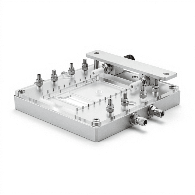
Reusable chip — designed for multiple experimental runs. Compatible with standard microfluidic tubing: steel pins (0.7 mm ID / 1.0 mm OD) and silicone tubing (0.8 mm ID / 1.9 mm OD). Available in bulk packs (5‑, 10‑, and 25‑unit) for lab-scale and consumable workflows.

Louise Corscadden, PhD
Director of Science · ConductScience
Ask Louise about PMMA Cell Culture Microfluidic Chip fit, setup, configuration, or quote prep.
Already working with us? Sign in to connect this with My Scientist.
Key Specifications
Full details →- Model fit
- 3 selectable configurations
- SKU family
- WHM-0016
- Sizing
- 25.0 x 15.0 x 3.0 cm
- Ordering
- Online checkout and quote request available
- Category
- Lab Equipment
- Build notes
- Confirm accessories, station layout, and support needs before purchase
The PMMA Cell Culture Microfluidic Chip provides a precisely engineered platform for controlled cell culture experiments within microenvironments. Fabricated from polymethyl methacrylate (PMMA), this chip features 100-micrometer deep channels that enable laminar flow conditions and controlled microenvironmental parameters for cellular studies. The transparent PMMA material offers optical clarity for real-time microscopic observation while maintaining chemical compatibility with standard cell culture media and reagents.
The 25mm x 15mm footprint provides sufficient culture area while maintaining compatibility with standard microscope stages and imaging systems. The shallow channel depth facilitates nutrient and oxygen diffusion to cultured cells while enabling precise control over shear stress and chemical gradients. This microfluidic platform supports applications ranging from basic cell behavior studies to complex organ-on-chip modeling systems.
How It Works
Microfluidic cell culture operates on the principle of controlled fluid dynamics at the microscale, where low Reynolds numbers ensure laminar flow conditions. The 100-micrometer channel depth creates an environment where surface tension and viscous forces dominate over inertial forces, enabling precise control over fluid mixing, shear stress, and chemical gradients. Cells cultured within these channels experience controlled microenvironmental conditions that can be maintained over extended periods.
The PMMA substrate provides optical transparency across the visible spectrum, enabling real-time fluorescence and brightfield microscopy. The material's surface properties support cell adhesion either directly or through standard surface treatments such as fibronectin or collagen coating. Chemical gradients are established through controlled perfusion at multiple inlet ports, while waste removal occurs through designated outlet channels, maintaining steady-state culture conditions.
The shallow channel geometry facilitates rapid equilibration of dissolved gases and nutrients while minimizing diffusion distances. This architecture supports both adherent cell culture on the channel bottom and suspension cell studies within the flowing medium, depending on experimental requirements.
Features & Benefits
Pack Size
- 5-Pack
- 10-Pack
- 25-Pack
Weight
- 0.03 kg
Dimensions
- L: 25.0 mm
- W: 15.0 mm
- H: 3.0 mm
| Feature | This Product | Typical Alternative | Advantage |
|---|---|---|---|
| Material Type | PMMA (polymethyl methacrylate) | Glass or PDMS substrates are common in microfluidics | Combines optical clarity of glass with the manufacturing flexibility and lower cost of polymer materials |
| Channel Depth | 100 micrometers | Channel depths vary widely from 10-500 micrometers | Optimized depth accommodates single cell layers while maintaining efficient nutrient and gas exchange |
| Platform Dimensions | 25mm x 15mm footprint | Microfluidic chips range from microscope slide size to larger custom formats | Standard size provides adequate culture area while fitting most microscope stages and experimental setups |
| Weight | 0.03 kg lightweight design | Glass chips typically weigh more due to material density | Facilitates handling and reduces mechanical stress on microscope stages during extended experiments |
This PMMA microfluidic chip provides an optimized balance of optical performance, biocompatibility, and practical handling characteristics. The standardized 100-micrometer channel depth and compact form factor address common requirements in cell culture microscopy applications.
| Model | SKU | Listed price | Status | Dimensions |
|---|---|---|---|---|
| 25-Pack | WHM-0016-25PK | Quote | Available | 25.0 x 15.0 x 3.0 cm |
| 10-Pack | WHM-0016-10PK | $1,490.00 | Available | 25.0 x 15.0 x 3.0 cm |
| 5-Pack | WHM-0016-5PK | Quote | Available | 25.0 x 15.0 x 3.0 cm |
Practical Tips
Pre-condition channels with culture medium for at least 1 hour before cell introduction to equilibrate surface properties and eliminate air bubbles.
Why: Ensures consistent cell adhesion and eliminates artifacts from surface conditioning during the experiment.
Clean channels immediately after use with enzymatic detergent followed by extensive rinsing to prevent protein buildup.
Why: Prevents cross-contamination and maintains optical clarity for subsequent experiments.
Verify actual flow rates using fluorescent tracer particles or dyes before each experiment to account for tubing resistance variations.
Why: Ensures accurate shear stress conditions and reproducible microenvironmental parameters across experiments.
Use phase contrast or DIC microscopy to visualize channel boundaries and verify uniform cell distribution before fluorescence imaging.
Why: Confirms proper cell loading and identifies potential flow pattern irregularities that could affect results.
If cells are not adhering properly, increase surface treatment incubation time or concentration, and verify medium pH and osmolality.
Why: PMMA surface properties can vary and may require optimization for specific cell types or experimental conditions.
Handle chips with powder-free gloves to prevent surface contamination and potential cytotoxic residues from reaching cultured cells.
Why: Maintains sterile conditions and prevents introduction of materials that could affect cell viability or behavior.
Setup Guide
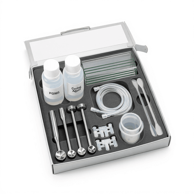
What’s in the Box
- PMMA Cell Culture Microfluidic Chip
- User manual with protocols (typical)
- Certificate of specifications (typical)
Warranty
ConductScience provides a one-year manufacturer warranty covering material defects and dimensional specifications. Technical support includes protocol guidance and troubleshooting assistance for microfluidic cell culture applications.
Compliance
References
Background reading relevant to this product:
What surface treatments are compatible with PMMA for cell adhesion?
PMMA surfaces can be treated with fibronectin, collagen, poly-L-lysine, or laminin for enhanced cell adhesion. Plasma treatment or UV-ozone exposure can also increase surface hydrophilicity and protein adsorption.
How do I prevent bubble formation in the microfluidic channels?
Pre-wet all tubing and connections with degassed medium, maintain consistent flow rates, and avoid rapid pressure changes. Use bubble traps in the inlet lines if necessary.
What microscopy techniques are suitable for this chip thickness?
The 3mm total thickness accommodates most standard microscopy techniques including brightfield, phase contrast, fluorescence, and confocal microscopy with working distances up to 2mm.
Can this chip be reused after cell culture experiments?
PMMA chips can be cleaned and reused if properly sterilized. Use enzymatic detergents followed by extensive rinsing and sterilization protocols between uses.
What flow rates are appropriate for cell culture applications?
Flow rates typically range from 0.1 to 10 μL/min depending on cell type and experimental requirements. Consult product datasheet for shear stress calculations at specific flow rates.
How does channel depth affect cell behavior?
The 100-micrometer depth provides sufficient space for most cell types while creating controlled microenvironmental conditions that can influence cell morphology, migration, and differentiation patterns.
Have a question about this product?
Have a question? Just ask.
Send it over and we'll email you a personalized answer — no call, no scheduling.
Prefer to talk it through?