Introduction
Parkinson’s disease (PD) is a common neurodegenerative disease characterized by the loss of 50% to 70% of the dopaminergic neurons in the substantia nigra pars compacta (SNc), loss of dopamine (DA) in the striatum, and the development of intracytoplasmic inclusions called Lewy bodies (LB), composed of α-synuclein and ubiquitin. α-synuclein has an important role in the pathogenesis of PD as it is found in all LBs. Parkinson’s disease is characterized by tremors, rigidity, bradykinesia, and postural instability; however, these manifestations could also be accompanied by non-motor symptoms such as olfactory deficits, neuropsychiatric disorders, and sleep disorders. Although the complete PD disease process is still not completely understood, various animal models have enabled a better understanding of its pathology, etiology, and molecular mechanisms.
Primarily, the animal models for PD were generated using neurotoxins such as 6-hydroxydopamine (6-OHDA) and 1-methyl-4-phenyl-1,2,3,6-tetrahydropyridine (MPTP) that resulting in acute neurodegeneration and death of dopaminergic neurons in the substantia nigra following intracranial 6-OHDA or intraperitoneal MPTP injection. Chronic administration of L-dopa to 6-OHDA-lesioned animals could create useful animal models to study L-dopa-induced dyskinesia. These models have been well-recognized and widely used in neuroscience research. Most of them use direct toxin injections into the SNpc or medial forebrain bundle (MFB), producing a rapid degeneration of DA neurons reflecting the late stages of PD. Contrastingly, the 6-OHDA model involves the administration of 6-OHDA in the striatum for the development of progressive and moderate lesions reflecting the early stages of the disease. Animal models are invaluable tools to gain knowledge of mechanisms involved in the pathogenesis of PD and to evaluate new therapeutic approaches. The recent development of non-invasive imaging methods in rodents further gives a unique opportunity to observe brain activity, neurotransmission, and neuroinflammation during the disease, and the effects of potential treatments (Blesa., Phani., Jackson-Lewis., & Przedborski., 2012).
Neurotoxic PD models (Blesa. & Przedborski., 2014)
6-Hydroxy dopamine (6-OHDA) model
6-Hydroxydopamine is a classical model for Parkinson’s disease study. 6-OHDA is a selective catecholaminergic neurotoxin that is used to generate lesions in the nigrostriatal DA neurons in test animals. Since 6-OHDA cannot cross the blood-brain barrier, its systemic administration fails to induce PD. So, this induction model requires the injection of 6-OHDA into the SNc, striatum, or medial forebrain bundle. The intrastriatal injection of this neurotoxin causes progressive retrograde neuronal degeneration in the SNc. As in PD, DA neurons degenerate, and the non-DA neurons are preserved. 6-OHDA triggers the formation of reactive oxygen species (ROS) to create oxidative stress after entering the neuron via the dopamine transporter, and consequently, neurotoxicity develops. Stereotaxic injection of 6-OHDA into the SNpc of the striatum induces neuronal cell death of the tyrosine hydroxylase (TH)-containing neurons in the test subject’s brain. After the neuron cell death, the dopamine levels in the striatum decrease. Although 6-OHDA injections do not induce the formation of Lewy bodies like those observed in PD, it reportedly interacts with the main protein of Lewy bodies: synuclein. 6-OHDA is an attractive neurotoxin for the initiation of the PD neurodegenerative process as it is a product of dopamine metabolism, and so is a useful model for the investigation of PD therapeutics.
1-Methyl-4-phenyl-1,2,3,6-tetrahydropyridine
The compound 1-methyl-4-phenyl-1,2,3,6-tetrahydropyridine (MPTP) also induces neurotoxicity in the nigrostriatal DA neurons in the brains of test animals. 1-methyl-4-phenyl-1,2,3,6-tetrahydropyridine(MPTP) is a neurotoxin precursor of 1-methyl-4-phenylpyridinium (MPP+), which interferes with the nigrostriatal DA pathway and causes significant loss of dopaminergic neurons in the striatum and SNc. MPTP was identified from the synthetic meperidine analog as a by-product. It is metabolized to its active metabolite 1-methyl-4-phenylpyridinium (MPP+) by monoamine oxidase type B. This active metabolite is taken up into the neurons with the help of the dopamine transporter, where it blocks the complex I site of the mitochondrial electron chain to initiate other intercellular reactions and multisystemic lesions, including the oxidative stress, inflammation, and energy failure. Rapid conversion of MPTP to MPP+ makes the chronic treatments serial acute insults. Therefore, a true chronic model requires continuous delivery of MPTP using the osmotic pumps. MPTP has an advantage over other neurotoxin models because of its lipophilic nature as it can cross the blood-brain barrier, therefore allowing easier administration, including systemic administration. MPTP has been widely used to model Parkinson’s disease in laboratory animals. Chronic MPTP-induced monkey models of PD have shown dopaminergic cell loss, α-synuclein aggregation and upregulation, and neuritic α-synuclein pathology. MPTP is the gold standard for toxin-based animal models of PD for replicating almost all of the hallmarks of the disease.
Rotenone
Rotenone is a natural compound that usually occurs in several plants and has been used as a broad-spectrum pesticide and insecticide. Rotenone is both an herbicide and an insecticide. In animals, rotenone could be administered by different routes. Oral administration causes little neurotoxicity. Chronic systemic administration with osmotic pumps has been commonly used as a delivery regimen, especially in the Lewis rat, which is more sensitive to rotenone than other rat strains. Intraperitoneal injections elicit behavioral and neurochemical deficits, but the mortality rate is higher. Intravenous administration causes damage to nigrostriatal DA neurons accompanied by α-synuclein aggregation, LB formation, oxidative stress, and gastrointestinal problems. It blocks the mitochondrial electron transport chain by inhibiting complex I to cause neurotoxicity. Rotenone also blocks mitosis and inhibits cell division and proliferation. In rats, chronic systemic exposure to rotenone causes many features resembling PD, including nigrostriatal DA degeneration. This model has reproduced almost all the features of PD, including the formation of intracellular inclusions resembling LB. It could be injected intraperitoneally, intravenously, or subcutaneously for systemic treatment of Parkinson’s disease. It could also be directly injected into the brain stereotaxically. Rotenone is highly lipophilic and could easily cross the blood-brain barrier.
Paraquat
Paraquat (N, N’-dimethyl-4-4-4′-bipiridinium) (PQ) is a herbicide that is commonly used in agriculture and exhibits a structural resemblance to MPP+. PQ potentiates oxidative stress to exert its deleterious effects. In particular, the hydrogen peroxide, superoxide radical, and hydroxyl radicals damage lipids, proteins, and nucleic acids. Following the systemic application PQ in mice, the animals exhibit reduced motor activity and a dose-dependent loss of striatal and midbrain SNpc neurons. Furthermore, it was also suggested that the damage done by PQ is caused by radicalized Parquat and facilitated by the glial cells. This implies that PQ behaves like MPP+ in causing its toxic effects. PQ’s importance to Parkinson’s disease research is its ability to induce α-synuclein aggregation in individual dopaminergic neurons in the SNpc and its ability to induce Lewy-bodies like structures in DA neurons. This suggests that the Paraquat model could be valuable for recapitulating all the events involved in PD pathogenesis.
Protocols
6-Hydroxydopamine lesion model (Vetel et al., 2019)
- Inject the rats with pargyline hydrochloride (50 mg/kg) intra-peritoneally twenty minutes before surgery to prevent 6-OHDA oxidation.
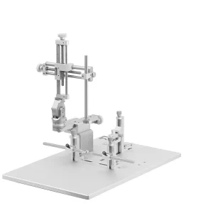
- Anesthetize the rats with isoflurane (4%, 500 mL/min), and place it on a stereotaxic frame.
- Maintain the anesthesia under isoflurane 2.5% (500 mL/min) during surgery.
- Monitor the body temperature and keep it as constant as possible (37°C) using a thermal probe.
- Inject 6-OHDA hydrochloride intrastriatal.
- Administer a total of 12 μg of 6-OHDA in three areas of the right striatum (2 mg/mL in 0.01% ascorbic acid dissolved in saline, pH 4.5, i.e., 4 μg in 2 μL for each area).
- Use Bregma stereotaxic coordinates: AP1 = +1.6 mm, L1 = -2.8 mm, P1 = -6 mm; AP2 = 0.0 mm, L2 = -4.1 mm, P2 = -5.5 mm; AP3 = -1.2 mm, L3 = -4.5 mm, P3 = -5.5 mm, according to the rat brain atlas of Paxinos and Watson (2009).
- Perfuse the 6-OHDA solution at the rate of 0.5 μL/minute.
- Keep the needle in place after each injection for 5 minutes to minimize solution backflow.
- Following surgery, inject the rats with buprenorphine (0.05 mg/kg subcutaneously) for post-operative pain.
MPTP rat model (Jackson-Lewis. & Przedborski., 2007)
Housing and acclimation
- Place the mice in a restricted room with a temperature of 22-270C and let them acclimate for 1 week before injection.
- Weigh, group, and code animals one day before starting the MPTP administration.
MPTP administration
- Choose a route of administration: subcutaneous or intraperitoneal. In addition, MPTP could either be injected (A) or infused (B).
- MPTP injection
- Determine the total volume of MPTP solution needed. It can be derived by adding the weights (in grams) of all mice to be injected with MPTP. For a 20 mg kg–1 dose of free base MPTP, the dose of MPTP. HCl is 20 mg kg–1 x 1.17 ¼ 23.4 mg kg-
- Weigh the necessary amount of MPTP. Given the recommended injection volume of 10 ml per 1 g body weight, corresponding to 10 ml kg–1, the dose of MPTP. HCl in milligrams per kilogram can be converted into a concentration of MPTP. HCl in milligrams per 10 ml. Then, multiply the total volume by the desired concentration of MPTP. HCl.
- Dissolve the calculated amount of MPTP. HCl in sterile saline (calculated amount). Cover the vial with parafilm and mix the solution.
- Inject the mice as per the dosing schedule worksheet, administering injections precisely 2 hours at the same time daily.
- MPTP infusion
- Determine the amount of MPTP. HCl is required for the experiment.
- Estimate the total volume of MPTP solution needed for the experiment by multiplying the total number of animals with the same body weight (e.g., 10 mice weighing 26 g) by 200 ml. According to this example, the total volume is 200 ml x 10 ¼ 2 ml.
- Fill the osmotic pump as follows;
- Weigh the empty pump with its flow moderator.
- Attach the filling tube to a tuberculin syringe and draw the room temperature solution up in the pump.
- Insert the filling tube into the opening at the top of the pump and gently push it to the bottom of the pump reservoir.
- Slowly fill the pump while holding it in a vertical position.
- Stop filling as soon as the solution appears at the outlet and carefully remove the filling tube. Wipe off the overflow with a tissue.
- Insert the flow moderator until the cap is flush with the pump top.
- Weigh the filled pump along with the flow moderator. The difference in the weights between the filled and empty pump gives the net weight of the loaded solution.
- Prepare a surgery area in the fume hood of the MPTP procedure room.
- Anesthetize the animals. Once the mouse is properly anesthetized, shave, and rinse the abdominal area with betadine.
- Using a scalpel, make a 1-1.5-cm long midline incision in the mid-abdomen.
- Using forceps, carefully tent the musculoperitoneal layer to prevent bowel damage and incise the peritoneal wall from beneath the cutaneous incision.
- Insert the filled pump into the peritoneal cavity.
- Suture the musculoperitoneal layer taking care to avoid perforating the bowel.
- Close the skin incision by placing 2-3 wound clips or sutures.
Note: Perform the following steps as per the required experimental protocol.
Rotenone model (Wrangel., et al., 2015)
Animal care
- Keep three to four animals per cage under controlled environmental conditions (temperature 22◦C, relative humidity 45-55%, 12 hours light/dark cycle).
- Feed all the animals with standard rat chow and water ad libitum.
Rotenone treatment
- Dissolve Rotenone in dimethylsulfoxide (DMSO) and dilute it using a sterile natural oil (middle chain triglycerides MCT; Miglyol 812) to make a final emulsion containing 2.5 mg rotenone/ml and 20 µl DMSO/ml.
- Treat the rats with chronic intraperitoneal (i.p.) injections of rotenone (2.5 mg/kg body weight once daily) for 60 days.
- Monitor the treated animals once daily.
Paraquat model (Cicchetti et al., 2005)
Toxin administration
- House the animals separately in a controlled-temperature environment maintained under a 12 hours light/dark cycle with free access to water and food.
- Inject the animals intraperitoneally with Paraquat (PQ dichloride hydrate (10 mg/kg), or a combination of PQ and maneb (MB) (30 mg/kg)) twice a week at the same time in the morning for 4 weeks.
- Monitor the animals daily for weight variation.
- Perform the subsequent protocol step as per the experimental needs.
Conclusion
Several animal models of Parkinson’s disease have been developed to study the pathogenesis and evaluate potential therapies of this disease. Toxic models have presented almost all the hallmarks of PD. Also, the toxic models are useful to screen potential drugs for symptomatic treatment of the disease. The limitation of the toxin models is that most of them display features of late stages of PD, whereas the genetic animal models use overexpression or knock-out technology to model earlier stages of the disease. The model should be chosen depending on the research questions required. These models provide useful study tools to investigate the pathogenesis and treatment regimens for Parkinson’s disease.
References
- C. Wrangel., K. Schwabe., N. John., K. J. Krauss., & Alam., M. (2015). The rotenone-induced rat model of Parkinson’s disease: behavioral and electrophysiological findings. Behav Brain Res, 279, 52-61.
- F. Cicchetti., N. Lapointe., A. Roberge-Tremblay., M. Saint-Pierre., L. Jimenez., W. B. Ficke., & Gross., E. R. (2005). Systemic exposure to paraquat and maneb models early Parkinson’s disease in young adult rats. Neurobiol Dis, 20(2), 360-371.
- J. Blesa., & Przedborski., S. (2014). Parkinson’s disease: animal models and dopaminergic cell vulnerability. Frontiers in neuroanatomy, 8.
- J. Blesa., S. Phani., V. Jackson-Lewis., & Przedborski., S. (2012). Classic and New Animal Models of Parkinson’s Disease. Journal of Biomedicine and Biotechnology.
- S. Vetel., S. Sérrière., J. Vercouillie., J. Vergote., G. Chicheri., B. J. Deloye., . . . Chalon., S. (2019). Extensive exploration of a novel rat model of Parkinson’s disease using partial 6-hydroxydopamine lesion of dopaminergic neurons suggests new therapeutic approaches. Synapse, 73(3).
- V. Jackson-Lewis., & Przedborski., S. (2007). Protocol for the MPTP mouse model of Parkinson’s disease. Nature protocols, 2, 141-151.


