ConductScience Pathology & Histology
Process. Section. Stain. Quantify.
Tissue processing, microtomy, slide preparation, and computer vision image analysis for research labs.
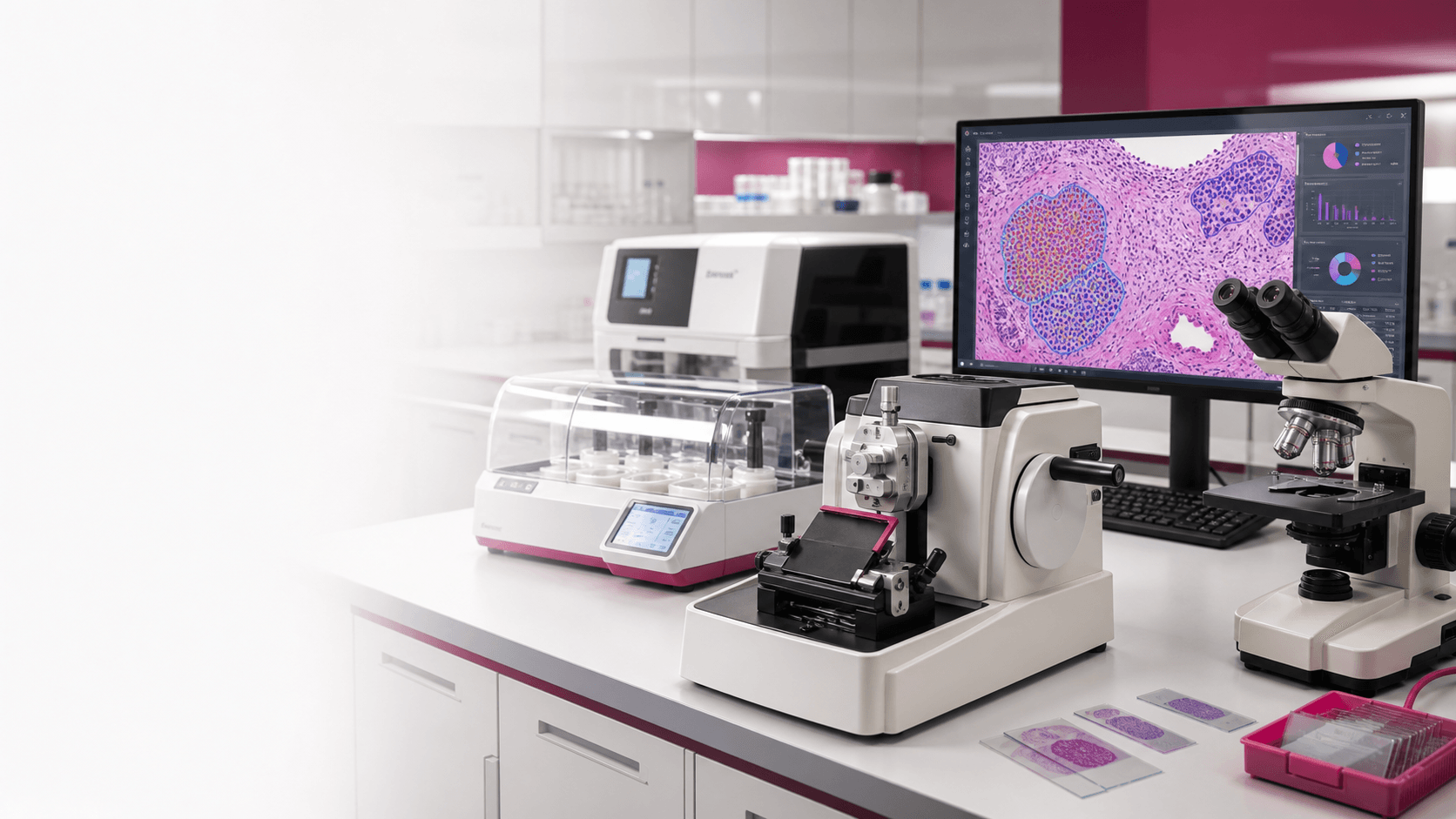
Microtome
Slide QC
ConductVision
7 steps
Complete tissue-to-data lane
8 hardware lines
Microtomy, processing, embedding, staining
1 software stack
ConductVision Image, Microscopy, Morphology
Research use
Reproducible. Auditable. No diagnostic claims.
Section 01 / Workflow Builder
Build the bench around the tissue, stain, and output.
Select the starting conditions. The planner returns the hardware lane, ConductVision lane, free tools, Lab Manager starter files, and a quote URL that carries the choices forward.
Section 02 / Workflow Overview
One workflow. Two lanes.
Hardware shapes the slide. ConductVision turns the slide into measurements. Start where you need to start. We fit the rest around it.
Lane A
Histology bench
01Process
Fix, dehydrate, infiltrate.
Instrument role
Tissue processors standardize fixation, dehydration, clearing, and paraffin infiltration before the sample reaches the cutting bench.
ConductVision role
This is the pre-analytics gate. The downstream image workflow records processing method, tissue type, and batch context against the slide set.
Output
Processed cassette batch with method context ready for embedding.
02Embed
Orient and block in paraffin.
Instrument role
Embedding stations, heated forceps, cold plates, and molds set orientation before sectioning quality is determined.
ConductVision role
Orientation notes and cassette IDs travel forward so review overlays can be tied back to preparation choices.
Output
Oriented paraffin blocks with traceable cassette IDs.
03Section
Microtomy and cryosectioning.
Instrument role
Manual, semi-automatic, rotary microtomes, and floor-standing cryostats create repeatable section thickness and ribbon quality.
ConductVision role
Section quality becomes the first visible QC signal before automated quantification starts.
Output
Mounted paraffin or frozen sections ready for staining.
04Stain
H&E, IHC, special stains.
Instrument role
Stainers, water baths, slide warmers, and coverslippers finish the slide and reduce manual variability.
ConductVision role
Stain type selects the matching analysis preset, including H&E nuclei, DAB percent area, fibrosis, or morphology review.
Output
Finished slides with stain-specific analysis intent.
Lane B
ConductVision analysis
05Image
Slide and field-level capture.
Instrument role
Microscopes and slide cameras provide the field, magnification, and illumination conditions for analysis.
ConductVision role
ConductVision Microscopy and Image organize capture, calibration, batch naming, and region selection.
Output
Captured slide fields with acquisition metadata.
06Quantify
Nuclei, DAB, fibrosis, area.
Instrument role
The hardware lane supplies consistent sections and stain quality so measurements are comparable across batches.
ConductVision role
ConductVision Image applies nuclei, DAB, percent-area, morphology, and batch-processing routines with visible overlays.
Output
Per-region measurements, overlays, QC flags, and review states.
07Export
CSV, overlays, methods, audit.
Instrument role
Bench instruments remain part of the exported method trail rather than being separated from the analysis result.
ConductVision role
Exports package CSV tables, overlay images, methods text, and audit-friendly batch summaries for research reporting.
Output
Research-ready data package with method context.
Section 03 / Two Buying Lanes
I need slides. I need answers.
Most labs need both. Configure each lane independently or as a bundle.
Lane A / Hardware-led
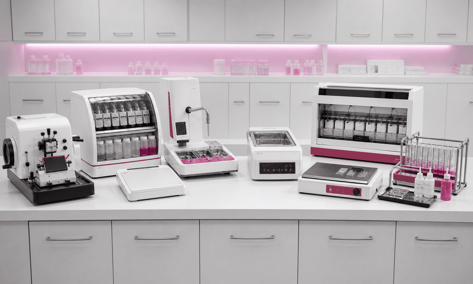
Histology Instruments
Reliable tissue-prep bench. Processing, microtomy, embedding, staining, slide finishing.
Open hub
Lane B / Software-led
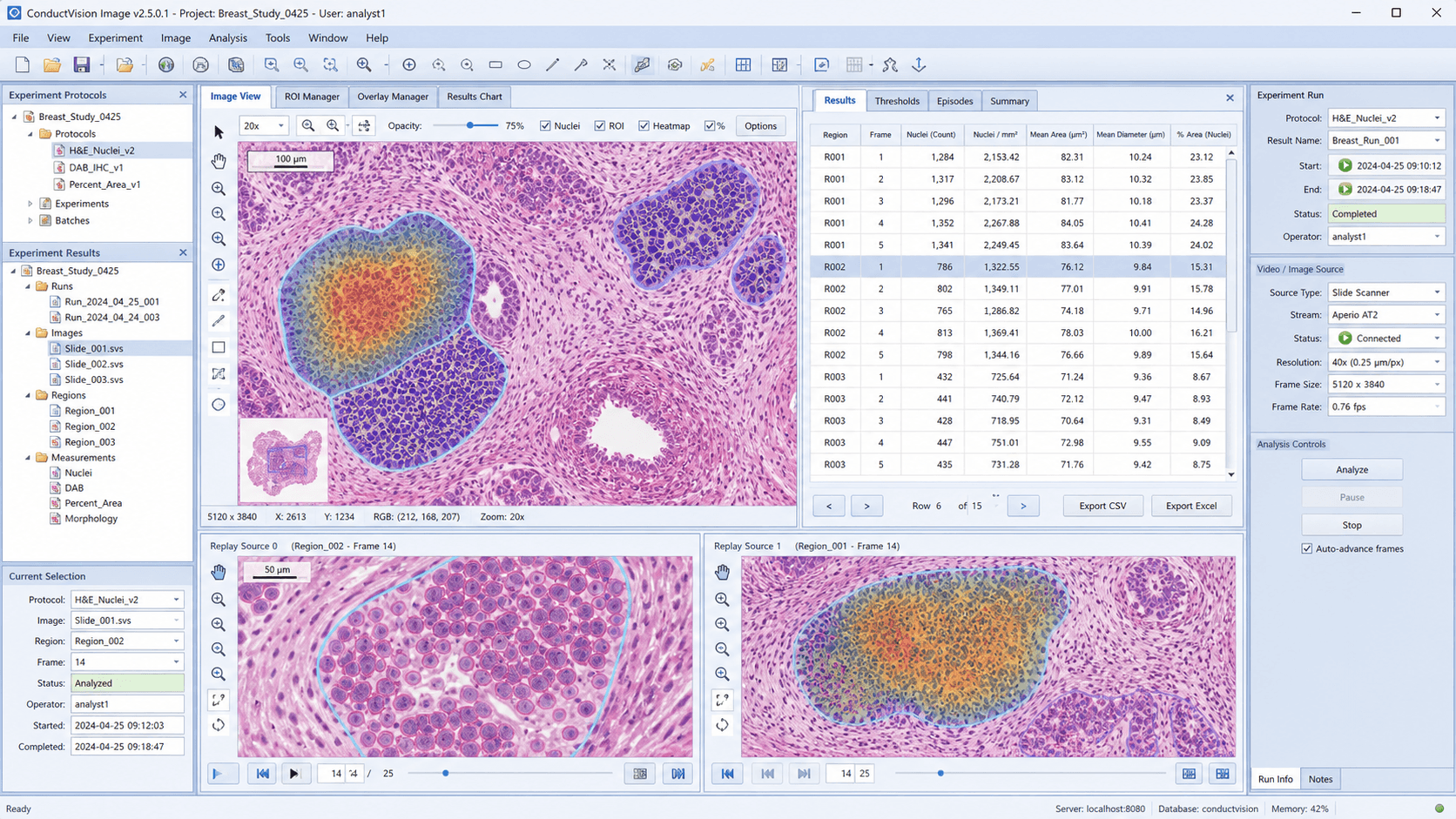
Pathology Image Analysis
Quantify H&E nuclei, IHC and DAB, tissue area, morphology, and section quality.
Open hub
Section 04 / Free Planning Tools
Calculators before quote requests.
Each tool should return a concrete planning output: a product path, a ConductVision package, a CSV where useful, and quote context that does not need to be retyped.
Microtome vs Cryostat Selector
Pick the right sectioning instrument for your tissue, section temperature, and downstream stain.
Inputs: Tissue, thickness, temperature, throughput
Output: Instrument class, why it fits, product links
Run toolTissue Processor Capacity Planner
Turn cassette volume, run frequency, and protocol length into a processor capacity recommendation.
Inputs: Cassettes per day, run schedule, protocol length
Output: Batch capacity, bottleneck warning, processor class
Run toolHistology Bench Budget Planner
Separate required bench items from optional automation and prepare a quote-ready configuration.
Inputs: New lab or upgrade, workflow steps, automation level
Output: Budget categories, required vs optional, CSV
Run toolH&E Staining Run Planner
Plan slides per run, racks, reagent refresh, coverslipping, and QC checks.
Inputs: Slides per run, racks, reagent schedule
Output: Run checklist, QC checks, stainer recommendation
Run toolPathology Image Analysis Planner
Map stain, image source, endpoint, ROI workflow, and batch size to a ConductVision package.
Inputs: Stain, image source, endpoint, ROI, batch
Output: CV package, overlays, CSV, methods text
Run toolSection 05 / Free Lab Manager Resource
Track this bench after the purchase decision.
Lab Manager is the free operating layer for inventory, equipment records, calibration windows, maintenance logs, budgets, and SOP documents. The page should give customers a starter pack they can import.
histology-equipment-starter.csv
Equipment records for microtomes, cryostats, processors, stainers, warmers, and coverslippers.
histology-reagents-consumables.csv
Starter inventory rows for blades, slides, cassettes, paraffin, stains, alcohols, and mounting media.
histology-maintenance-schedule.csv
Inspection, cleaning, calibration, and service cadence for the preparation bench.
histology-budget-categories.csv
Budget categories that separate required instruments, automation, consumables, and software.
histology-sop-document-checklist.csv
SOP, manual, certificate, and methods-document checklist for shared lab operation.
Section 06 / Purchasing Guides
Buying guidance at the decision point.
The page should not force customers to infer what matters. These guide cards explain the next buying decision and connect it to tools, products, and ConductVision outputs.
Setup
Buying your first histology bench
What to buy first, what can wait, and which items are required for reliable slide preparation.
Selection
Microtome vs cryostat
Choose paraffin sectioning, frozen sectioning, or a hybrid path based on tissue and endpoint.
Throughput
Tissue processor capacity and automation
Use cassette volume, protocol duration, and staffing constraints to decide when automation helps.
Quality
How slide preparation affects image analysis
Connect folds, chatter, tissue coverage, and stain uniformity to ConductVision QC and exports.
Section 07 / Buyer Matrix
Choose your workflow.
Start from the sample and stain. The buying path should show the hardware lane, ConductVision package, and output the lab can expect.
Use case
Routine H&E quantification
Sample and stain
Paraffin sections, H&E stain
Throughput
Single lab or small batch runs
Recommended setup
Microtome, water bath, slide warmer + ConductVision Image with H&E nuclei and histology QC presets
Expected output
Nuclei density, tissue coverage, QC flags, overlay PNGs, CSV
Use case
IHC and DAB analysis
Sample and stain
Paraffin sections, chromogenic IHC
Throughput
Study batches and repeat panels
Recommended setup
Tissue processor, embedding station, rotary microtome, stainer + DAB percent-positive area, intensity bins, ROI review
Expected output
Positive area, optical density, region summaries, methods text
Use case
Frozen section and neurohistology
Sample and stain
Fresh frozen tissue, brain sections, morphology stains
Throughput
Protocol-driven research cohorts
Recommended setup
Cryostat, slide warmer, microscope capture path + ConductVision Morphology plus Image QC and batch exports
Expected output
Morphometry tables, field overlays, section artifact review
Use case
High-throughput core lab
Sample and stain
Mixed tissues, H&E, IHC, special stains
Throughput
Multi-user queue and shared methods
Recommended setup
Processor, embedding, microtome, stainer, coverslipper + Locked protocols, batch reporting, review states, audit trail
Expected output
Per-slide metrics, batch consistency view, CSV and protocol JSON
Section 08 / Featured Hardware
Eight categories. One bench.
Each line is configurable as a standalone instrument or as part of a workflow bundle.
 microtomes
Microtomes
7 products. Manual, semi-auto, rotary.
 tissue processors
Tissue processors
11 products. Processing, embedding, cassette support.
 embedding systems
Embedding systems
Embedding station. Heated paraffin and cold plate.
 cryostats
Cryostats
1 product. Floor-standing frozen section microtome.
 water baths
Water baths
Flotation bath. Ribbon handling and slide transfer.
 slide warmers
Slide warmers
Slide dryer. Flat-bed warming and drying.
 stainers
Stainers
12 products. H&E, special staining, drying, coverslipping.
 coverslipping
Coverslipping
Automated coverslipper. Glass, film, mounting media.
Section 09 / ConductVision Layer
The software half of the lane.
ConductVision adds QC, quantification, and reporting on top of microscope fields or slide-scanner image exports. Use it as a histology gate, a pathology analysis engine, or both.
Feature 01
Histology QC
Fold detection, chatter, tissue coverage, stain uniformity, and slide-level review state.
Feature 02
H&E nuclei
Per-region nuclei count, density, tissue coverage, and visible overlays for routine H&E fields.
Feature 03
IHC and DAB
Percent-positive area, optical density, intensity bins, and ROI-level DAB summaries.
Feature 04
Section artifacts
Flag tears, folds, missing tissue, and inconsistent staining across batches.
Feature 05
Batch reporting
Per-slide metrics rolled into batch consistency views for shared methods and core facilities.
Feature 06
Exports
CSV tables, overlay PNGs, methods text, locked protocol JSON, and audit-friendly summaries.
Example analysis package
Overlay review
Annotated PNGs show nuclei, DAB-positive regions, tissue masks, artifacts, and reviewed ROIs.
Measurement tables
CSV exports include per-region counts, density, percent area, optical density, and slide identifiers.
Methods package
Methods text, locked protocol JSON, batch summary, and review state travel with the result set.
Section 10 / Workflow Bundles
Pre-configured starting points.
Three bundles cover the most common research benches. Each is quote-ready and configurable.
Bundle 01
Histology Quantification Starter
Sectioning bench plus ConductVision Image with H&E presets.
Best for
- Labs moving from manual slide review to repeatable H&E measurement.
Includes
- Manual microtome
- Water bath
- Slide warmer
- ConductVision Image H&E preset
Outputs
- Nuclei count and density
- Tissue coverage QC
- Overlay images and CSV tables
Optional add-ons
- Microscopy capture setup
- Additional H&E protocol templates
Bundle 02
Preclinical Pathology Quantification
A full sample-to-data lane for rodent studies, IHC and DAB.
Best for
- Preclinical teams running repeat animal cohorts with IHC endpoints.
Includes
- Tissue processor
- Embedding workstation
- Rotary microtome
- ConductVision Image IHC/DAB workflow
Outputs
- DAB percent-positive area
- Intensity bins
- ROI summaries and methods text
Optional add-ons
- Slide stainer
- Automated coverslipper
- Batch reporting setup
Bundle 03
Neurohistology Morphometry
Cryosection bench plus ConductVision Morphology and Image.
Best for
- Neuroscience labs sectioning frozen tissue and measuring morphology.
Includes
- Floor-standing cryostat
- Slide warmer
- ConductVision Morphology
- ConductVision Image QC
Outputs
- Morphometry measurements
- Section artifact flags
- Field overlays and CSV exports
Optional add-ons
- Microscopy workflow calibration
- Study-specific morphology templates
Section 11 / Recommended Next Step
Build a pathology and histology workflow quote.
Tell us what you are sectioning and what you need to measure. We come back with a hardware and ConductVision configuration that fits your bench, sample type, and throughput.