Southern blotting is a hybridization technique used for deoxyribose nucleic acid (DNA). The method was named after Edward M. Southern, who developed the technique in the 1970s. The method involves the transfer of DNA fragments from an electrophoresis gel to a membrane causing immobilization of the DNA fragments and bands are produced. After immobilization, the DNA fragments could be subjected to hybridization analysis, enabling the bands with sequence similarity to a labeled probe. Oligonucleotides, similar to the target sequence are designed. These oligonucleotides are chemically synthesized, radiolabeled, and then used for the cloned DNA fragments. Sequences hybridizing with the hybridization probe are further analyzed to obtain the full-length sequence of the gene of interest. Southern blotting could be used for homology-based cloning by exploring the amino acid sequence of the protein product of the gene of interest. Furthermore, southern blotting is used to identify methylated sites in particular genes. In addition to its use for DNA analysis, the immunologists have long used the southern blotting for gene identification in somatic rearrangements and transgene studies.
Principle
Southern blotting is based on the principle of separation of DNA fragments by gel electrophoresis followed by the identification by labeled probe hybridization. The DNA fragments are separated based on their size and charge during electrophoresis. The technique involves the transfer of electrophoresed DNA from the electrophoresis gel to a nitrocellulose membrane. The nitrocellulose filter is sandwiched between the gel and the stack of paper towels which draws the transfer buffer from the gel through capillary action. The desired DNA is detected using a labeled probe complementary to the desired DNA.
Apparatus & Equipment
The agarose gel for southern blotting is mounted on a filter paper dipped in a reservoir containing transfer buffer. The hybridization membrane is sandwiched between the gel and paper towels which draw the transfer buffer through the gel by capillary action. The DNA molecules are run on the membrane. Nitrocellulose or nylon serves as the membrane material. After transfer, the DNA is fixed onto the membrane. Then, the membrane is placed in a solution of labeled RNA, single-stranded DNA, or oligodeoxynucleotide complementary to the blot transferred DNA bands to be detected.
Training Protocol
Southern blotting onto nylon or nitrocellulose membrane (Brown, 2001)
- Digest the DNA using the restriction endonuclease(s), run in an agarose gel with appropriate DNA size markers, dye the nucleotide strands with ethidium bromide, and identify the band position by visualizing the membrane.
- Treat the gel with distilled water ∼10 gel volumes 0.25 M HCl for 30 minutes, again with distilled water ∼10 volumes denaturation solution (1.5 M NaCl/0.5 M NaOH) twice for 20 minutes, and then with distilled water ∼10 volumes neutralization solution (1.5 M NaCl/0.5 M Tris-Cl, pH 7.0) for 20 minutes for twice, with gentle shaking at room temperature.
- Assemble the transfer stack consisting of a sponge, Whatman 3MM paper, the gel, pieces of plastic wrap, a nylon membrane equilibrated in distilled water or nitrocellulose membrane, five pieces of Whatman 3MM paper, and a 4-cm stack of paper towels. As each layer is applied, wet it with 20× saline sodium citrate (SCC) and remove air bubbles if any by carefully rolling a 10-ml glass pipet over the surface.
- Place a glass plate on top of the stack and put a 0.2 to 0.4-kg weight on the top to hold the stack in place. Leave it overnight.
- Recover the membrane. Mark the positions of the wells on the membrane and cut one corner to mark the orientation.
- Rinse the membrane in 2× saline sodium citrate (SSC), then place it on a sheet of Whatman 3MM paper and let it dry thoroughly.
- Wrap nylon membrane in UV-transparent plastic wrap and place DNA-side-down on a UV transilluminator (254-nm wavelength).
Note: The nitrocellulose membranes should be placed between two sheets of Whatman 3MM paper and baked under vacuum at 80°C for 2 hours.
Mutation detection by southern blotting (Mellars. & Gomez., 2011)
DNA restriction
- Dispense 10 mg of deoxyribose nucleic acid in a 500 ml microtube.
- Add 4 mL of 10× restriction enzyme buffer. Make the final volume to 40 mL by adding the distilled water.
- Add 1mL of Bcl1 restriction enzyme.
- Vortex the mixture, pulse-spin, and incubate it at 50°C overnight.
Gel preparation and DNA separation
- Assemble a 25 cm long gel casting tray and gel comb.
- Make 300 mL of 0.5% agarose (300 mL 1× Tris-borate ethylene diamine tri-acetic acid (TBE) and 1.5 g agarose).
- To dissolve the agarose by heating in a microwave or on a hot plate.
- Let it cool to 55–60°C and add 25 mL ethidium bromide.
- Pour it into the casting tray, remove air bubbles, and let it set.
- Prepare the samples after incubation and add 1.5 mL of 10% sodium dodecyl sulfate (SDS) and 8 mL of 6× sucrose loading buffer.
- Prepare the molecular weight marker by adding 0.5 mg–10 mL of distilled water and 5 mL of loading buffer.
- Remove the gel comb, place it in the electrophoresis tank, and cover it with 1× TBE.
- Carefully load the samples into the gel and run the samples on the gel from negative to positive at 45 V, for 40 hours.
Southern blotting
Gel preparation
- Carefully visualize the gel on the transilluminator. Spot the area of interest by noting the sizes of the bands of the marker.
- Carefully inject a small amount of ink into the position of two of the bands of the marker nearest to the area of interest. Cut the top and bottom of the gel and isolate the targeted section.
- Transfer it to a sandwich box and immerse in 0.25 M HCl for 10-15 minutes, on an orbital shaker for depurination.
- Replace the HCl with 500 mL denaturing buffer (1.5 M NaCl, 0.5 M NaOH) and leave the sandwich box on an orbital shaker for 60 minutes at low speed at room temperature.
- Replace the denaturing buffer with 500 mL neutralizing buffer (0.5 M Tris–HCl, 1.5 M NaCl. pH 7.3, adjust with 10 M NaOH). Leave the box on an orbital shaker at low speed for 90 minutes at room temperature.
Blot assembling
- Place the casting tray upside down in a large plastic tray to support the gel. Lay three to four layers of Whatman filter paper over the casting tray for the wick.
- Pour 500 mL of 20× SSC on the filter paper and saturate the base of the large with it.
- After 90 minutes in neutralizing buffer, slide the gel onto a casting tray and transfer it to the prepared blotting base.
- Surround the gel with cling film to cover the exposed area of Whatman filter paper up to the gel edge.
- Add 450 mL distilled water to 50 mL of 20x SSC to make 2× SSC.
- Cut a piece of Hybond N membrane slightly larger than the gel. Soak it in 2× SSC and then layer it onto the gel.
Note: Make sure to remove entrapped air bubbles if any.
- Cut four sheets of Whatman filter paper slightly larger than the gel. Soak them in 2× SSC and layer them onto the blot.
- Place a dry piece of Whatman filter paper on the blot and then place a stack of tissue approx. 6 cm in height covering the whole gel. Then, cover this with a stack of paper towels.
- Place a glass plate and a 500 g weight on the stack. Leave the blot overnight.
- Remove the paper towels, tissues, and filter paper the following morning.
- Rinse the Hybond membrane briefly in 2× SSC and blot dry using the filter paper.
- Mark the membrane with the help of a soft-lead pencil.
- Fix the DNA on the membrane by placing it under UV light for 5 minutes.
Probe labeling
- Add 30 ng of the designed probe to 5 mL of the primer mix and denature for 5 minutes at 99°C and then keep the mixture on ice.
- Add labeling buffer, containing the dNTPs, with 2.5 mL of 32P and 2 mL of Klenow fragment polymerase.
- Incubate the mixture to allow incorporation of the radiolabel for 10 minutes to 60 minutes at 37°C.
Hybridization
- Turn ON the hybridization oven 30 minutes before use and heat to 65°C.
- Warm 20 mL of 2× SSC in a water bath.
- Warm 10 mL of PerfectHyb Plus in a water bath.
- Roll the blot up with the DNA side inside and the pencil mark to the outside.
- Place the blot in a hybridization bottle and add 20 mL warmed 2× SSC.
- Unroll the blot on the bottle wall by tilting and rolling the bottle. Make sure no air bubble enters the blot.
- Pour the SSC and 10 mL of pre-warmed PerfectHyb Plus buffer.
- Tightly cap the bottle and place it in the oven.
- Pre-hybridize for 30 minutes up to a maximum of 4 hours.
- Denature the labeled probe by heating it to 100°C for 5 minutes and then placing it on ice.
- Warm 10 mL of PerfectHyb Plus buffer to 65°C.
- After pre-hybridization, remove the bottle from the oven and pour off the pre-hybridization buffer.
- Pour the probe solution in 10 mL of warmed buffer and immediately add to the blot.
- Replace the bottle in the oven and rotate at 65°C overnight.
Blot washing
- Pour 250 mL of washing buffer in a flask and warm to 65°C.
- Remove the bottle from the oven and discard the excess buffer.
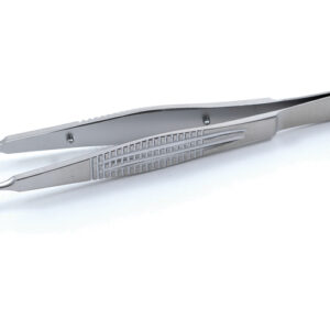
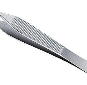
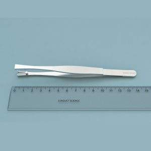
- Detach the membrane with tweezers and place it in a plastic sandwich box.
- Add half of the buffer, seal the box, and place in a shaking water bath at 65°C for 15 minutes. Keep the remaining buffer at 65°C.
- Pour off the wash buffer and add the remaining. Wash for a further 15 minutes.
- Pour off the buffer and dry the blot using the Whatman filter paper.
- Run the Geiger counter over the blot.
- Air-dry the blot and wrap in cling film.
Autoradiography
- Tape the blot onto an X-ray film and pace another film on the top to make a sandwich autoradiograph in a darkroom.
- Place at -70°C for 48 hours before developing the first film.
- If bands are weak, replace the cassette in the freezer for 24-48 hours before the second film.
Applications
Characterization of human herpesvirus strains (Aubin. et al., 1991)
Southern blotting is a valuable technique for genome analysis and characterization. In the study, eight human herpesvirus 6 (HHV-6) strains were analyzed using the Southern blot and polymerase chain reaction. Nucleic acids from infected cells were digested by restriction enzymes and hybridized with cloned BamH1 fragments. In parallel, the DNA was amplified using the polymerase chain reaction employing primers derived from the strain SIE nucleotide sequence. The results indicated that all the strains were closely related to one another. However, marked differences in restriction patterns were observed in two groups, typified by strains SIE and HST, respectively. The findings of the study suggest that the HHV-6 species exhibit genetic polymorphism, and the sequences could be used as useful markers for the diagnosis and molecular epidemiology of HHV-6 infections.
Tracking of mouse cell lineage (Lo., Coulling., & Kirby., 1987)
The analysis of cell lineage has provided detailed insights into the underlying mechanisms of pattern formation, determination, and differentiation of cells in developmental systems. The feasibility of DNA microinjection for mouse embryos lineage studies was determined. For this, tissues from three transgenic mice mosaic with an exogenously introduced mouse β-major globin gene were analyzed using the Southern-blotting and in situ hybridization. The results presented early segregation of cells in the somatic and germ cell lineages. The in-situ hybridization data suggested that cells of the transformed lineages are finely dispersed which indicates an extensive cell-cell mixing during mouse development. Whereas the kidney cells had a patchy distribution, the transformed cells in the villi were aggregated, and the positive and negative seminiferous tubules were segregated in the testis. The study suggested a clonal basis for the organ development stages for kidney, intestine, and testis. The Southern blotting and in-situ hybridization allowed the direct quantification and localization of lineage descendants derived from the founder cells (marked or transformed).
Analysis of Epstein-Barr viral genomes in lymphoid malignancy (Ohshima. et al., 1990)
Southern blot analysis was used for the analysis of Epstein-Barr virus (EBV) genome and its comparison with non-neoplastic lymphadenopathic and healthy thymus tissues. In specimens marked positive by the Southern blot and PCR, in-situ hybridization studies were performed. By Southern blot analysis, it was found that 26% Hodgkin’s disease samples, 5.6% B-cell lymphomas (5.6%) and 11% T-cell lymphomas had EBV DNA. While in some samples, EBV DNA was not detected. The results indicate that the presence of EBV DNA is not related to lymphoid malignancy; however, the enhancement of the DNA could lead to neoplastic conditions.
Precautions
- Wear gloves and a lab coat while handling the materials for Southern blotting.
- Make sure no air bubbles got entrapped in gel, membrane, and Whatman filter paper as they may cause the DNA to run irregularly.
- Monitor the agarose while heating, as it is liable to boil quickly.
- Gently remove the gel comb to avoid well collapse.
- Slowly release the sample in the wells for a smooth transfer.
- Once the nitrocellulose membrane is placed, do not move it as the DNA transfer may already have taken place.
Strengths and Limitations
- Southern blotting is a suitable technique to analyze long stretches of DNA such as the multikilobase restriction fragments.
- The expression levels and expression profiles are better detected as compared to the other genomic analyses methods.
- Southern blotting has a wide array of applications in the fields of DNA fingerprinting, including paternity and maternity testing, forensic studies, and personal identification.
- The method is cheaper than genome sequencing and is useful to quantify the amount of DNA as well.
- The method may require a large quantity of high-quality DNA as compared to other genome analyzing techniques.
References
- Brown, T. (2001). Southern blotting. Curr Protoc Immunol, Chapter 10:Unit 10.
- Mellars., & Gomez., k. (2011). Mutation detection by Southern blotting. Methods Mol Biol, 688, 281-291.
- Ohshima., M. Kikuchi., F. Eguchi., Y. Masuda., Y. Sumiyoshi., H. Mohtai., . . . Kimura., N. (1990). Analysis of Epstein-Barr viral genomes in lymphoid malignancy using Southern blotting, polymerase chain reaction and in situ hybridization. Virchows Arch B Cell Pathol Incl Mol Pathol, 59(6), 383-90.
- J. Aubin., H. Collandre., D. Candotti., D. Ingrand., C. Rouzioux., M. Burgard., . . . Agut., H. (1991). Several groups among human herpesvirus 6 strains can be distinguished by Southern blotting and polymerase chain reaction. J Clin Microbiol, 29(2), 367-72.
- C. Lo., M. Coulling., & Kirby., C. (1987). Tracking of mouse cell lineage using microinjected DNA sequences: analyses using genomic Southern blotting and tissue-section in situ hybridizations. Differentiation, 35(1), 37-44.
