Object Detection, Segmentation, or Density Map
Three ways to count. The right one depends on your cells.
ConductVision Image ships three analysis modes, selectable per protocol and switchable without restarting. Separable cells with clear boundaries want object detection. Overlapping cells where shape matters want segmentation. Cultures so dense that individual boundaries disappear want a density map, where the count is the integral of the map rather than a sum of objects.
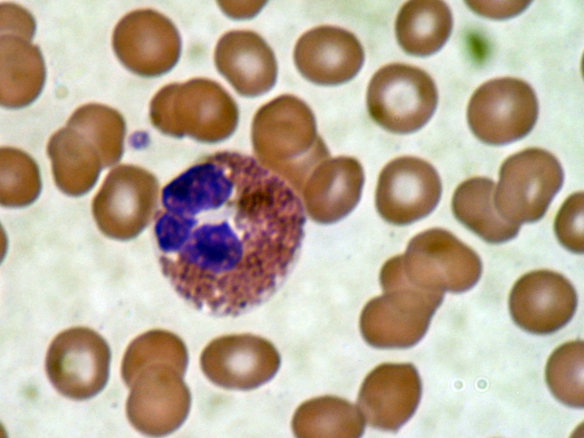
Which mode fits your sample
This is the software’s own guidance: each mode states when it is best, what it outputs, and where it is used.
Object Detection
- Best when: cells separable
- Output: count + locations
- Blood smears
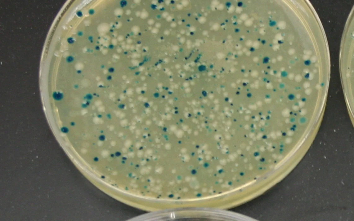
- Bacteria colonies
- Yeast cells
Segmentation
- Best when: cells overlap, shape matters
- Output: individual cell masks
- Accurate counts + morphology
Histology- Fluorescence microscopy
Organoids
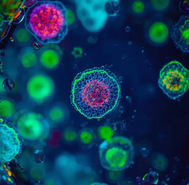
Density Map
- Best when: extremely dense, boundaries unclear
- Output: density heatmap
- Count = integral of map
- Colony counting
- Tissue sections
- High-density cultures
Tuning & recount
- Max Flow
- Min Cell Prob
- Min Cell Size
- Recount without re-running
How It Works
Pick a mode
Choose object detection, segmentation, or density map on the protocol.
Set parameters
Max Flow, Min Cell Prob, and Min Cell Size control what counts as an object.
Preview
Check detection on a single image before committing the batch.
Recount
Adjust parameters and recount from the existing result, no full re-run.

Not sure which mode fits your cells?
Send a representative image and we will tell you which mode to run, and why.
Request a Demo