Works With Your Microscope
Tell the engine your optics; it selects the model.
Microscopes differ in noise statistics, contrast, resolution and anisotropy, and background structure. Selecting your microscopy type helps the system choose the right AI model for the image you actually captured. Brightfield, fluorescence, phase contrast, and differential interference contrast are all first-class optical contrasts, with widefield and two-photon handled explicitly. You can switch types without restarting the application.
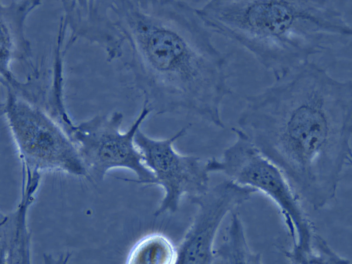
Supported imaging modes
Every type below is a selectable option on the protocol, and it steers which AI model runs.
Optical contrast
- Brightfield (H&E, IHC)
Fluorescence- Phase Contrast
- Differential Interference Contrast
Fluorescence
WidefieldTwo-photon
Other
- Other / custom optics
Module-specific
- Plate type (colony)
- Microspheres image type
How It Works
Select optics
Pick the optical contrast and fluorescence mode you imaged with.
Model selected
The system uses your microscopy type to guide AI model selection.
Preview
Confirm detection looks right on a representative image.
Run
Process a single image or the whole folder with the type locked in.


Imaging on something unusual?
Send a sample image from your scope and we will confirm the right model before you buy.
Request a Demo