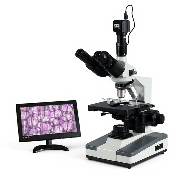
Build-in Camera Biological Microscope
Digital biological microscope with integrated camera system providing 1.3-3.0 megapixel imaging, achromatic objectives (4X-100X), and USB2.0 connectivity for direct image capture and analysis.

Louise Corscadden, PhD
Director of Science · ConductScience
Ask Louise about Build-in Camera Biological Microscope fit, setup, configuration, or quote prep.
Already working with us? Sign in to connect this with My Scientist.
Key Specifications
Full details →- Model fit
- Configured during quote
- SKU family
- BIO-0519
- Sizing
- 49.2 x 39.6 x 26.2 cm
- Ordering
- Online checkout and quote request available
- Category
- Microscopy
- Build notes
- Confirm accessories, station layout, and support needs before purchase
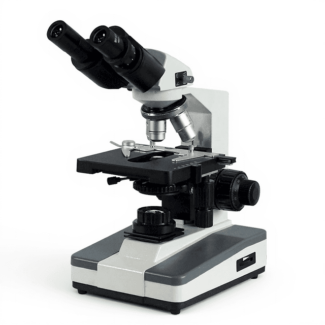
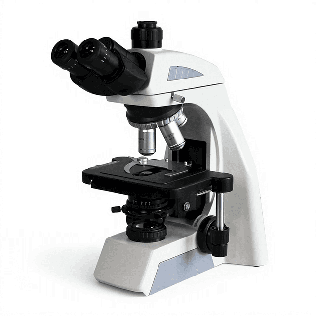
The Build-in Camera Biological Microscope (BIO-0519) is a digital microscopy system combining traditional optical microscopy with integrated digital imaging capabilities. The system features a built-in camera with selectable resolutions from 1.3 to 3.0 megapixels, providing direct USB2.0 connectivity for real-time image capture and analysis. The microscope incorporates achromatic objectives (4X, 10X, 40X, 100X) with WF10X/18 eyepieces, delivering an 18mm field of view for biological specimen observation.
The integrated Scope Image 9.0 software package enables direct image acquisition, measurement, and documentation on Windows-based systems. This configuration eliminates the need for separate camera attachments while maintaining the optical performance required for routine biological microscopy applications. The system supports both visual observation through eyepieces and simultaneous digital documentation, streamlining workflow in laboratory and educational environments.
How It Works
The microscope operates on conventional brightfield illumination principles, where transmitted light passes through the specimen and is focused by the objective lens system onto the image plane. The achromatic objective lenses correct for chromatic aberration across the visible spectrum, providing sharp imaging with minimal color fringing. The 18mm field of view is achieved through the WF10X/18 eyepiece design, optimizing the balance between magnification and observable area.
The integrated camera system captures the optical image formed at the eyepiece level through a beam-splitting or direct imaging pathway. The CMOS or CCD sensor converts the optical image into digital format at selectable resolutions from 1.3 to 3.0 megapixels. USB2.0 interface provides real-time data transfer to the host computer, enabling live viewing and image capture through the Scope Image 9.0 software platform.
Digital image processing occurs within the software environment, allowing for measurement tools, annotation, and file export in standard formats. The system maintains optical performance equivalent to conventional microscopes while adding digital documentation capabilities without requiring external camera attachments or complex alignment procedures.
Features & Benefits
Automation Level
- semi-automated
√
- √
Valid Pixel
- 1280*1024(1.3M Pixel)
1600*1200(2.0M Pixel)
- ●
2048*1036(3.0M Pixel)
- ●
2052*1944(5.0M Pixel)
- /
Output Mode
- USB2.0
Operation System
- WINDOWS7/8 2000/XP/VISTA
Software
- Scope image 9.0
Range of Viewing Field
- ; 18mm(BMB-300M)
Eyepiece
- WF10X/18
P16X/11
- ●
Extra Wide Filed Eyepiece EW10X/20 with Diopter Ad
- /
Objective
- Achromatic Objective 4X, 10X, 40X, 100X
Infinite Plan Achromatic Objective 4X, 10X, 40X, 1
- /
Brand
- ConductScience
Research Domain
- Cell Biology
- Clinical Diagnostics
- Developmental Biology
- Histopathology
- Materials Science
- Microbiology
Weight
- 7.5 kg
Dimensions
- L: 49.2 mm
- W: 39.6 mm
- H: 26.2 mm
| Feature | This Product | Typical Alternative | Advantage |
|---|---|---|---|
| Camera Integration | Built-in camera system with direct optical path | External camera attachments requiring separate mounting and alignment | Eliminates alignment variables and maintains consistent optical performance without additional setup complexity. |
| Resolution Options | Selectable 1.3M, 2.0M, and 3.0M pixel resolutions | Fixed resolution cameras requiring hardware changes for different applications | Allows optimization of image quality versus file size based on specific documentation requirements. |
| Software Integration | Dedicated Scope Image 9.0 software with measurement tools | Generic imaging software with limited microscopy-specific features | Provides calibrated measurement capabilities and microscopy-optimized workflow tools. |
| Connectivity | USB2.0 interface for direct computer connection | Varies by model, may require additional interface cards or converters | Ensures broad compatibility with standard computer systems without additional hardware requirements. |
| Field of View | 18mm field of view with WF10X/18 eyepieces | Standard eyepieces often provide smaller field of view | Wider observation area improves specimen survey efficiency and reduces repositioning during examination. |
This microscope integrates digital imaging capabilities directly into the optical design, providing consistent performance and simplified workflow compared to systems requiring separate camera attachments. The selectable resolution options and dedicated software package offer flexibility for various documentation requirements while maintaining the optical quality of conventional biological microscopy.
Practical Tips
Calibrate measurement tools in the software using a stage micrometer at each objective magnification to ensure dimensional accuracy.
Why: Each objective has different magnification factors that must be compensated for accurate measurements.
Clean camera sensor and optical elements monthly using appropriate lens cleaning materials and techniques to maintain image quality.
Why: Dust and contamination on internal optics cannot be bypassed and will appear in all captured images.
Use the lowest camera resolution adequate for your application to reduce file sizes and processing time while maintaining required image detail.
Why: Higher resolutions create larger files that may slow software performance without providing additional useful information for routine applications.
Establish consistent illumination settings and document them for each type of specimen to maintain reproducible imaging conditions.
Why: Inconsistent illumination affects image contrast and color balance, making comparisons between specimens unreliable.
If live image appears dark or unclear, check that the correct camera resolution is selected and USB connection is secure before adjusting optical settings.
Why: Software configuration issues often present as apparent optical problems but can be resolved without touching the microscope hardware.
Allow adequate ventilation around the microscope and computer to prevent overheating during extended imaging sessions.
Why: Continuous operation of the camera sensor and processing hardware generates heat that can affect system performance and longevity.
Setup Guide
What’s in the Box
- Build-in Camera Biological Microscope main unit
- Achromatic objectives: 4X, 10X, 40X, 100X
- WF10X/18 eyepieces (pair)
- USB2.0 connection cable
- Scope Image 9.0 software installation media
- Power adapter and cord
- User manual and software documentation (typical)
- Dust covers for optics (typical)
Warranty
ConductScience provides a standard one-year manufacturer warranty covering optical and electronic components, with technical support for software installation and operation. Extended service plans may be available for institutional users requiring on-site calibration services.
Compliance
What is the maximum resolution available with the integrated camera system?
The camera offers selectable resolutions up to 3.0 megapixels (2048×1536 pixels), with intermediate options of 2.0M (1600×1200) and 1.3M (1280×1024) pixels depending on application requirements.
Can the system capture images while simultaneously using the eyepieces for visual observation?
Yes, the integrated camera design allows simultaneous eyepiece viewing and digital image capture, enabling collaborative observation and real-time documentation during microscopy sessions.
What file formats does the Scope Image 9.0 software support for image export?
Consult the software documentation for specific supported formats. The software typically supports standard imaging formats for compatibility with image analysis and presentation applications.
Are the objectives parfocal for rapid magnification changes?
The achromatic objective set (4X, 10X, 40X, 100X) is designed for parfocal operation, allowing magnification changes with minimal refocusing between objectives.
What are the illumination requirements and can LED systems be used?
Consult product datasheet for specific illumination specifications. The system works with standard transmitted light sources appropriate for brightfield microscopy applications.
How does image quality compare to separate camera attachments on research-grade microscopes?
The integrated camera eliminates alignment variables and optical path losses associated with external attachments, providing consistent image quality optimized for the specific optical design of this system.
What computer specifications are recommended for optimal software performance?
The system requires Windows 7/8/2000/XP/Vista compatibility with USB2.0 ports. Consult software documentation for RAM and processor recommendations based on intended image processing workflows.
Can measurements and annotations be calibrated for different objective magnifications?
The Scope Image 9.0 software includes measurement tools that can be calibrated for accurate dimensional measurements. Consult software documentation for specific calibration procedures and accuracy specifications.
Have a question about this product?
Have a question? Just ask.
Send it over and we'll email you a personalized answer — no call, no scheduling.
Prefer to talk it through?
Accessories
Enhance your setup with compatible accessories


Replacement Parts & Consumables


Related Products