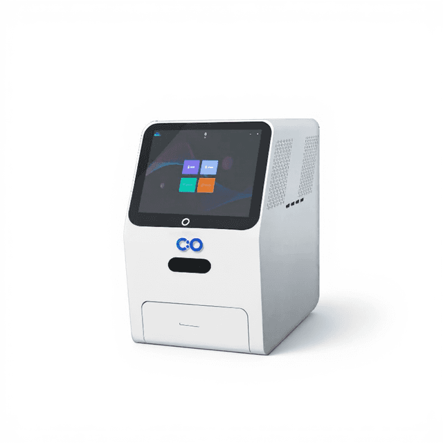
Gel Imaging System W1000 Plus
Gel imaging system for nucleic acid gel documentation, gel excision, and protein gel imaging.
6.3 MP color camera; 240×240 mm field of view; 310 nm transmitted / 254 nm / 365 nm LED UV sources; motorized zoom lens; built-in UV laser protection shield (OD > 7) for safe in-instrument gel cutting.

Louise Corscadden, PhD
Director of Science · ConductScience
Ask Louise about Gel Imaging System W1000 Plus fit, setup, configuration, or quote prep.
Already working with us? Sign in to connect this with My Scientist.
Key Specifications
Full details →- Model fit
- Configured during quote
- SKU family
- CS-W1000 Plus
- Sizing
- Model-specific dimensions confirmed from the selected configuration
- Ordering
- Online checkout and quote request available
- Category
- Gel & Clarity Testing
- Build notes
- Confirm accessories, station layout, and support needs before purchase
The W1000 Plus Gel Imaging System is designed for nucleic acid gel documentation, nucleic acid band excision, and protein gel imaging. A 6.3 MP color camera paired with a motorized zoom lens covers gel formats up to 240×240 mm. Three independent LED UV light sources — a 310 nm transmitted array for uniform illumination across the full gel surface, plus 254 nm and 365 nm side-mounted sources — are compatible with EtBr, SYBR Green, SerRed, SerBlue, GoldView, and other common nucleic acid stains. The built-in UV laser protection shield (cutoff depth >OD7) enables safe gel band excision directly inside the instrument. An optional dual blue-white transilluminator accessory provides switchable blue/white transmitted light for blue-light excitable stains, with three-level power adjustment for each source.
Key Features
- 6.3 MP color camera (3072×2048, 2.4×2.4 µm pixels) for natural-color gel imaging
- Motorized zoom lens (8–48 mm, F1.0–F16) with coarse, fine, and ultra-fine adjustment
- 240×240 mm maximum effective imaging area — accommodates large-format gels
- Three UV light sources: 310 nm transmitted LED array + 254 nm and 365 nm side-mounted LED
- Optional 8-bit (256 gray levels) or 16-bit (65,535 levels) grayscale output
- Built-in UV laser protection shield (OD > 7) for safe in-instrument gel excision
- Fully light-sealed dark box; door sensor automatically controls the bright-field LED
- Auto exposure for one-step imaging without manual parameter adjustment
- Optional dual blue-white transilluminator (switchable, 3-level power per source)
- 10.4-inch touchscreen running Windows 10; 16 GB RAM, 512 GB SSD, Wi-Fi/Bluetooth
Applications
- Nucleic acid gel imaging (EtBr, SYBR Green, SerRed, SerBlue, GoldView)
- Nucleic acid gel band excision and purification
- PCR product verification and DNA sizing
- Protein gel imaging (Coomassie blue, silver stain)
- RNA gel electrophoresis documentation
Technical Specifications
| Parameter | Specification |
|---|---|
| Dimensions (W×D×H) | 400×371×700 mm |
| Camera | |
| Camera Type | Color Camera |
| Pixel Resolution | 6.3 Megapixels |
| Resolution | 3072×2048 |
| Pixel Size | 2.4×2.4 µm |
| Target Size | 1/1.8" (7.37×4.92 mm) |
| Full Well Capacity | 10.4 ke⁻ |
| Sensitivity | 760 mV |
| Readout Noise | 2.14 e⁻ |
| Dark Current | 0.15 mV |
| Signal-to-Noise Ratio | 40.2 dB |
| Exposure Time | 17 µs – 15 s |
| Binning Mode | 1×1, 2×2, 3×3 |
| Grayscale | 8-bit (256 levels) or 16-bit (65,535 levels), selectable |
| Cooling | Not applicable (color camera) |
| Lens | |
| Type | Motorized zoom lens |
| Focal Length | 8–48 mm |
| Aperture | F1.0–F16 |
| Close-up Lens | 2× |
| Filter | 590/60 nm |
| Light Sources | |
| Bright-Field | Downward-facing LED white light, symmetrically distributed on both sides |
| Transmitted UV | 310 nm LED array with uniform transmitted illumination |
| Side UV | 254 nm / 365 nm LED, symmetrically distributed on both sides |
| Optional | Dual blue-white transilluminator — switchable blue/white, 3-level cyclic power adjustment for each source |
| Dark Box | |
| Light Isolation | Fully light-sealed; isolates environmental light |
| Door Control | Door sensor controls bright-field LED on/off |
| Gel Cutting | Extractable UV source + UV protective board (OD > 7) |
| Imaging Area | |
| Maximum Effective Field of View | 240×240 mm |
| Software | |
| Camera Settings | Contrast, exposure time, gain adjustment |
| Lens Adjustment | Coarse / fine / ultra-fine; zoom, focus, aperture |
| Imaging Modes | Nucleic acid gel, protein gel, gel cutting |
| Auto Exposure | Yes |
| Image Tools | Open/Save, crop, flip, rotate, mark, restore, print |
| System | |
| Industrial Computer | 10.4-inch display (1024×768), Windows 10, 16 GB RAM, 512 GB SSD, Bluetooth/Wi-Fi |
| External Interfaces | USB 3.0 × 2 |
| Operating Voltage | 90–132 VAC / 180–264 VAC (selectable via switch), 47–63 Hz |
| Power Consumption | ≤300 W |
| Net Weight | 30.65 kg |
Ordering Information
| Cat. No. | Description |
|---|---|
| CS-W1000 Plus | Gel Imaging System W1000 Plus — Set |
Also Available
Need chemiluminescence Western blot imaging in addition to gel documentation? Explore the W5000 Plus — a combined chemiluminescence and gel imaging system. For dedicated ECL Western blot detection, see the W2000 Plus. For advanced fluorescence Western blot imaging, see the W6000.
No Q&A yet. Have a question? Ask below.
Have a question about this product?
Have a question? Just ask.
Send it over and we'll email you a personalized answer — no call, no scheduling.
Prefer to talk it through?