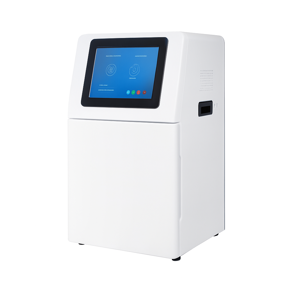
Multifunctional Fluorescence Imaging System
Multifunctional fluorescence imaging system integrating fluorescence imaging, chemiluminescence imaging, tissue fluorescence imaging, and gel imaging.
Three top-mounted fluorescence excitation sources (470 nm, 530 nm, 650 nm) for multiplex fluorescent WB; proprietary image inversion for standard pre-stained ladders; deep-cooled 9 MP monochrome camera; motorized filter wheel; 155×155 mm WB / 215×215 mm gel field of view.

Louise Corscadden, PhD
Director of Science · ConductScience
Ask Louise about Multifunctional Fluorescence Imaging System fit, setup, configuration, or quote prep.
Already working with us? Sign in to connect this with My Scientist.
Key Specifications
Full details →- Model fit
- Configured during quote
- SKU family
- CS-W6000
- Sizing
- Model-specific dimensions confirmed from the selected configuration
- Ordering
- Online checkout and quote request available
- Category
- Gel & Clarity Testing
- Build notes
- Confirm accessories, station layout, and support needs before purchase
The W6000 Multifunctional Fluorescence Imaging System integrates four core imaging modules — fluorescence, chemiluminescence, tissue fluorescence, and gel imaging — into a single platform. Three top-mounted fluorescence excitation sources at 470 nm, 530 nm, and 650 nm enable simultaneous detection of multiple target proteins on a single membrane using fluorescently labeled secondary antibodies. A proprietary image inversion function allows standard pre-stained protein ladders to be used directly in fluorescent WB experiments: bright ladder bands captured in brightfield are fully resolved under darkfield after inversion processing, eliminating the need for fluorescence-conjugated ladder substitutes. A deep-cooled monochrome camera (−40°C relative to ambient) and motorized filter wheel serve both the fluorescence and chemiluminescence channels, while integrated 310/254/365 nm UV LED sources and motorized lens handle nucleic acid and protein gel documentation in the same instrument.
Key Features
- Three fluorescence excitation channels — 470 nm, 530 nm, 650 nm — top-mounted, symmetrically positioned
- Multiplex fluorescent WB: detect multiple target proteins simultaneously on a single membrane
- Image inversion function: use standard pre-stained protein ladders directly in fluorescent WB without fluorescence-conjugated ladder substitutes
- Deep-cooled 9 MP monochrome camera: −40°C relative to ambient; dark current 0.0003 e⁻/s/pixel at −15°C
- Motorized filter wheel switches between fluorescence, chemiluminescence, and gel imaging modes
- Chemiluminescence WB: 0.1 s fine-increment exposure adjustment; time accumulation; one-click ladder removal
- Tissue fluorescence imaging: autofluorescence detection with real-time frame selection for quantitative analysis
- Cell and microbial fluorescence: rapid assessment in culture plates and bacterial agar plates
- Nucleic acid gel imaging (310 nm transmitted + 254/365 nm side UV); compatible with EB, SYBR Green, SerRed, SerBlue, GoldView
- Gel band excision with UV laser safety shield (OD > 7); protein gel imaging in color and grayscale
Applications
- Multiplex fluorescent Western blot imaging
- ECL chemiluminescence Western blot detection
- Tissue section autofluorescence imaging
- Fluorescent protein expression imaging in cell culture plates
- Microbial fluorescence assays on bacterial agar plates
- Nucleic acid gel imaging (EtBr, SYBR Green, SerRed, SerBlue, GoldView)
- Protein gel imaging (Coomassie blue, silver stain)
- Nucleic acid gel band excision and downstream purification
Technical Specifications
| Parameter | Specification |
|---|---|
| Dimensions (W×D×H) | 400×371×700 mm |
| Camera | |
| Camera Type | Monochrome Camera |
| Pixel Resolution | 9 Megapixels |
| Resolution | 3000×3000 |
| Pixel Size | 3.76×3.76 µm |
| Target Size | 1" (11.28×11.28 mm) |
| Full Well Capacity | 16.5 ke⁻ (HCG), 50.5 ke⁻ (LCG) |
| Sensitivity | 877 mV @ 1/30 s |
| Readout Noise | 1.24 e⁻ (HCG), 3.22 e⁻ (LCG) |
| Dark Current | 0.0003 e⁻/s/pixel at −15°C |
| Signal-to-Noise Ratio | 42.2 dB (HCG), 47 dB (LCG) |
| Exposure Time | 0.1 ms – 1 h |
| Binning Mode | 1×1, 2×2, 3×3 |
| Grayscale | 16-bit (65,536 gray levels) |
| Cooling | −40°C relative to ambient temperature |
| Lens | |
| Type | Motorized Lens |
| Focal Length | 17 mm |
| Aperture | F0.95–F16 |
| Light Sources | |
| Bright-Field | Episcopic LED white light, symmetrically distributed on both sides |
| Transmitted UV | 310 nm LED array with uniform transmitted illumination |
| Side UV | Episcopic 254 nm / 365 nm LED UV, symmetrically distributed on both sides |
| Fluorescence | Episcopic fluorescence light sources: 470 nm, 530 nm, 650 nm — symmetrically distributed on top |
| Optional | Blue/white dual light source — switchable, 3 power levels per source |
| Dark Box | |
| Light-Tightness | Fully light-tight; isolates ambient light |
| Door Sensor | Controls brightfield and 310 nm UV LED sources |
| Filter Wheel | Switches filters according to mode — chemiluminescence, gel, and fluorescence applications |
| Gel Cutting | Extractable UV source; UV laser safety shield (OD > 7) |
| Imaging Area | |
| Blotting Membrane | 155×155 mm effective field of view |
| Protein Gel | 155×155 mm effective field of view |
| Nucleic Acid Gel | 215×215 mm effective field of view |
| Fluorescent Membrane | 136×136 mm effective field of view |
| Software | |
| Auto Exposure | Smart exposure with automatic binning; combined with timeline imaging and time accumulation for one-operation optimal imaging |
| Exposure Modes | High Quality (highest image quality) / Standard (balanced) / High Sensitivity (fastest) |
| Real-time Imaging | Yes — live signal preview with overexposure indication |
| Temporal Imaging | Yes — retrieve any frame within the recorded exposure window |
| Time Accumulation | Yes — extend exposure after initial run |
| Marker Removal | One-click removal of non-specific marker signal bands |
| System | |
| Industrial Computer | 10.4-inch display (1024×768), Windows 10, 16 GB RAM, 512 GB SSD, Bluetooth/Wi-Fi |
| External Interfaces | USB 3.0 × 2 |
| Operating Voltage | 90–132 VAC / 180–264 VAC (selectable via switch), 47–63 Hz |
| Power Consumption | ≤300 W |
| Net Weight | 35 kg |
Ordering Information
| Cat. No. | Description |
|---|---|
| CS-W6000 | Multifunctional Fluorescence Imaging System — Set |
Also Available
For chemiluminescence-only Western blot imaging, see the W2000 Plus. For combined chemiluminescence WB and gel documentation without fluorescence channels, see the W5000 Plus. For dual-camera ECL imaging with true-color pre-stained marker capture, explore the W3000 Plus.
No Q&A yet. Have a question? Ask below.
Have a question about this product?
Have a question? Just ask.
Send it over and we'll email you a personalized answer — no call, no scheduling.
Prefer to talk it through?