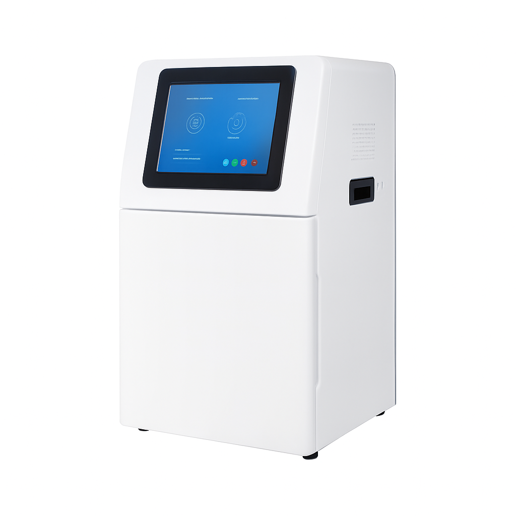
Multiplex Fluorescence Imaging System W3000 Plus
Dual-camera chemiluminescence imaging system for Western blot detection.
Dedicated deep-cooled 9 MP monochrome camera for high-sensitivity ECL band detection plus an independent 45 MP color camera for true-color pre-stained ladder imaging. One-click marker restoration eliminates non-specific antibody cross-reactivity. 136×136 mm field of view (expandable to 200×200 mm).

Louise Corscadden, PhD
Director of Science · ConductScience
Ask Louise about Multiplex Fluorescence Imaging System W3000 Plus fit, setup, configuration, or quote prep.
Already working with us? Sign in to connect this with My Scientist.
Key Specifications
Full details →- Model fit
- Configured during quote
- SKU family
- CS-W3000 Plus
- Sizing
- Model-specific dimensions confirmed from the selected configuration
- Ordering
- Online checkout and quote request available
- Category
- Gel & Clarity Testing
- Build notes
- Confirm accessories, station layout, and support needs before purchase
The W3000 Plus Chemiluminescence Imaging System combines two independent optical channels: a deep-cooled monochrome camera for ECL Western blot band detection and a 45 MP color camera for true-color pre-stained ladder imaging. The monochrome channel records the chemiluminescent signal with 16-bit fidelity and full-range sensitivity, while the color channel captures the marker in natural color — eliminating the color-shift artifacts and non-specific cross-reactivity that occur in single-camera systems where the same sensor handles both signals. A one-click marker restoration function removes non-specific antibody binding signals from the marker and restores the true band pattern. The complete time-lapse exposure record allows any frame to be selected in 0.1 s increments after the experiment is complete, and underexposed samples can receive additional exposure without discarding the initial acquisition.
Key Features
- Dual-camera design: independent monochrome ECL channel + color marker channel
- Monochrome camera: 9 MP, deep-cooled −40°C relative to ambient; dark current 0.0003 e⁻/s/pixel at −15°C
- Color camera: 45 MP, 2.315×2.315 µm pixels, 1.4" sensor (18.93×13 mm)
- One-click marker restoration: removes non-specific antibody signals; recovers true marker band colors
- 16-bit time-lapse acquisition for monochrome channel; any frame selectable in 0.1 s increments
- Time accumulation: extend exposure on top of the original run without data loss
- Three exposure modes: High Quality / Standard / High Sensitivity
- Simultaneous imaging of 10 or more membranes
- 136×136 mm working area (expandable to 200×200 mm)
- 10.4-inch Windows 10 display; 16 GB RAM, 512 GB SSD, Wi-Fi/Bluetooth; USB 3.0 × 2
Applications
- ECL chemiluminescence Western blot detection with co-registered color ladder
- True-color pre-stained protein ladder documentation
- High-throughput multi-membrane WB workflows
- Experiments requiring accurate molecular weight co-localization
- Quantitative band density analysis
Technical Specifications
| Parameter | Camera 1 (Monochrome — ECL) | Camera 2 (Color — Marker) |
|---|---|---|
| Dimensions (W×D×H) | 400×371×700 mm | |
| Camera Type | Black and White Camera | Color Camera |
| Pixel Resolution | 9 million pixels | 45 million pixels |
| Resolution | 2992×3000 | 2992×3000 (as stated; see note) |
| Pixel Size | 3.76×3.76 µm | 2.315×2.315 µm |
| Target Size | 1" (11.28×11.28 mm) | 1.4" (18.93×13 mm) |
| Full Well Capacity | 16.5 ke⁻ (HCG), 50.5 ke⁻ (LCG) | 10.8 ke⁻ |
| Sensitivity | 877 mV @ 1/30 s | 419 mV |
| Readout Noise | 1.24 e⁻ (HCG), 3.22 e⁻ (LCG) | 2.12 e⁻ |
| Dark Current | 0.0003 e⁻/s/pixel at −15°C | 0.12 mV |
| Signal-to-Noise Ratio | 42.2 dB (HCG), 47 dB (LCG) | 40.3 dB |
| Exposure Time | 0.1 ms – 1 h | 17 µs – 15 s |
| Binning Mode | 1×1, 2×2, 3×3 | 1×1, 2×2, 3×3 |
| Grayscale | 16-bit (65,536 levels) | 8-bit (256 gray levels) |
| Cooling | Relative to ambient temperature −40°C | — |
Note: Camera 2 resolution is listed as "2992×3000" on the source specification page.
Based on sensor dimensions (18.93×13 mm) and pixel pitch (2.315 µm), the physical
pixel count is approximately 45 MP. These two values may reflect different reporting
conventions. Confirm with the product datasheet for your application.
| Parameter | Specification |
|---|---|
| Lens (Camera 1) | |
| Type | Prime lens |
| Focal Length | 17 mm |
| Aperture | F0.95–F16 |
| Light Sources | |
| Bright-Field | Downward-facing LED white light, symmetrically distributed on both sides |
| Dark Box | |
| Light Isolation | Fully light-sealed; isolates environmental light |
| Door Control | Door sensor controls bright-field LED on/off |
| Imaging Area | |
| Effective Field of View | 136×136 mm (expandable to 200×200 mm) |
| Software | |
| Exposure Modes | High Quality / Standard / High Sensitivity |
| Auto Exposure | Intelligent exposure with automatic binning |
| Real-time Imaging | Yes — live signal preview with overexposure indication |
| Time-lapse Imaging | Yes — retrieve any frame within the recorded exposure window |
| Time Accumulation | Yes — extend exposure after initial run |
| Optional Settings | Color Marker / Black & White Marker; Overexposure Prompt / No Overexposure Prompt |
| System | |
| Industrial Computer | 10.4-inch display (1024×768), Windows 10, 16 GB RAM, 512 GB SSD, Bluetooth/Wi-Fi |
| External Interfaces | USB 3.0 × 2 |
| Operating Voltage | 90–132 VAC / 180–264 VAC (selectable via switch), 47–63 Hz |
| Power Consumption | ≤200 W |
| Net Weight | 23.45 kg |
Ordering Information
| Cat. No. | Description |
|---|---|
| CS-W3000 Plus | Multiplex Fluorescence Imaging System W3000 Plus — Set |
Also Available
Need a simpler chemiluminescence-only system? See the W2000 Plus. For combined WB and gel imaging in one instrument, explore the W5000 Plus. For advanced fluorescence Western blot imaging (up to 6-plex), see the W6000.
No Q&A yet. Have a question? Ask below.
Have a question about this product?
Have a question? Just ask.
Send it over and we'll email you a personalized answer — no call, no scheduling.
Prefer to talk it through?