Microfluidic Flow Cytometry Chip
Microfluidic chip for single-cell analysis using hydrodynamic focusing, enabling flow cytometry applications in a compact, microscale format.

Louise Corscadden, PhD
Director of Science · ConductScience
Ask Louise about Microfluidic Flow Cytometry Chip fit, setup, configuration, or quote prep.
Already working with us? Sign in to connect this with My Scientist.
Key Specifications
Full details →- Model fit
- 3 selectable configurations
- SKU family
- WHM-0027
- Sizing
- 25.0 x 15.0 x 3.0 cm
- Ordering
- Online checkout and quote request available
- Category
- Lab Equipment
- Build notes
- Confirm accessories, station layout, and support needs before purchase
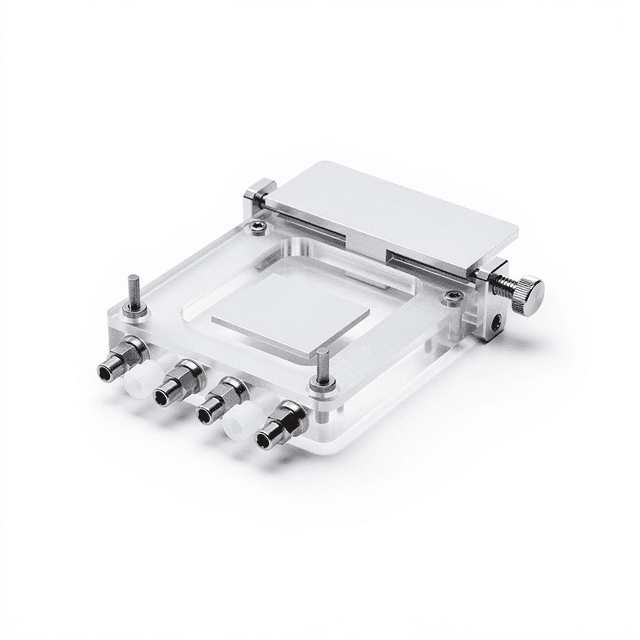
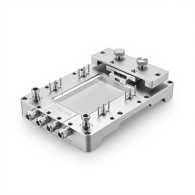
The Microfluidic Flow Cytometry Chip is a precision-engineered device that enables single-cell analysis through hydrodynamic focusing in a microfluidic environment. This chip integrates the principles of flow cytometry with microfluidics technology, allowing researchers to analyze individual cells or particles in a controlled microscale flow stream. The device uses hydrodynamic focusing to position cells in the center of a fluid stream, enabling precise optical interrogation for cell counting, sizing, and characterization.
Designed for applications requiring single-cell resolution, this microfluidic chip provides researchers with a platform for studying cellular heterogeneity, cell viability, and population dynamics. The compact 25mm x 15mm x 3mm form factor makes it compatible with standard microscopy setups and portable analysis systems. The chip's design facilitates laminar flow conditions essential for accurate single-cell measurements, supporting a wide range of cell biology and analytical applications where traditional flow cytometry may be impractical or cost-prohibitive.
How It Works
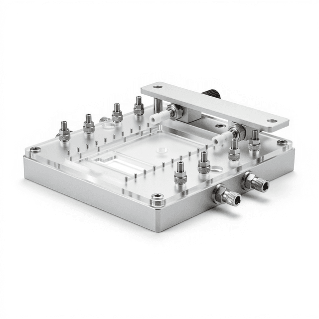
The microfluidic flow cytometry chip operates on the principle of hydrodynamic focusing, where sample fluid containing cells is introduced into the center of a sheath flow stream. The chip architecture creates laminar flow conditions that position cells in single file within the interrogation zone. As cells pass through the focused stream, they can be analyzed using optical detection methods including fluorescence, light scattering, or brightfield imaging.
The hydrodynamic focusing mechanism relies on the velocity profile differences between the sample and sheath flows. The sheath fluid, flowing at higher velocity along the channel walls, compresses the sample stream into a narrow core where individual cells are separated and aligned. This ensures each cell passes through the detection zone individually, enabling accurate single-cell measurements without cell aggregation or coincidence errors.
The chip's microfabricated channels provide precise flow control and consistent optical path lengths, critical for quantitative measurements. The transparent materials used in construction allow for optical access from multiple angles, supporting various detection modalities including forward scatter, side scatter, and fluorescence detection at multiple wavelengths.
Features & Benefits
Channel Width
- 100 um
- 150 um
- 200 um
- 50 um
Bonding Type
- PDMS-Glass
- PDMS-PDMS
Pack Size
- 5-Pack
- 10-Pack
- 25-Pack
Weight
- 0.03 kg
Dimensions
- L: 25.0 mm
- W: 15.0 mm
- H: 3.0 mm
| Feature | This Product | Typical Alternative | Advantage |
|---|---|---|---|
| Channel Pattern | Flow-focusing junction with downstream serpentine channel — 3 ports | Sheath fluid confinement narrows sample stream for single-particle analysis | |
| Channel Depth | 50 μm (fixed) | Standard across all configurations in this product line | |
| Channel Width Options | 50, 100, 150, or 200 μm (8 configurations total) | Match focusing width to target cell or particle diameter | |
| Bonding Options | PDMS-to-PDMS or PDMS-to-glass | Glass bonding recommended for fluorescence and bright-field imaging | |
| Manufacturer Part Numbers | 3.2.002.00.033 through 3.2.002.00.040 (Model: WHBZ-RC) | Odd numbers = PDMS-PDMS; even numbers = PDMS-glass | |
| Chip Dimensions | 25 × 15 × 3 mm chip on 25.4 × 76.2 mm glass slide | Standard microscope slide format | |
| Included Accessories | Stainless steel tubes (0.7 × 1.0 × 15 mm), silicone tubing (0.8 × 1.9 mm) | 3 connection points: 1 sample inlet + 2 sheath inlets (or 1 outlet) |
Flow-focusing PDMS chip for hydrodynamic sample confinement, cytometry, and monodisperse droplet generation. Serpentine downstream section extends residence time. 8 configurations: 4 widths × 2 bonding types.
| Model | SKU | Listed price | Status | Dimensions |
|---|---|---|---|---|
| 25-Pack | WHM-0027-200-um-pdms-glass-25-pack | Quote | Available | 25.0 x 15.0 x 3.0 cm |
| 10-Pack | WHM-0027-200-um-pdms-glass-10-pack | Quote | Available | 25.0 x 15.0 x 3.0 cm |
| 5-Pack | WHM-0027-200-um-pdms-glass-5-pack | Quote | Available | 25.0 x 15.0 x 3.0 cm |
| 25-Pack | WHM-0027-200-um-pdms-pdms-25-pack | Quote | Available | 25.0 x 15.0 x 3.0 cm |
| 10-Pack | WHM-0027-200-um-pdms-pdms-10-pack | Quote | Available | 25.0 x 15.0 x 3.0 cm |
| 5-Pack | WHM-0027-200-um-pdms-pdms-5-pack | Quote | Available | 25.0 x 15.0 x 3.0 cm |
| 25-Pack | WHM-0027-150-um-pdms-glass-25-pack | Quote | Available | 25.0 x 15.0 x 3.0 cm |
| 10-Pack | WHM-0027-150-um-pdms-glass-10-pack | Quote | Available | 25.0 x 15.0 x 3.0 cm |
| 5-Pack | WHM-0027-150-um-pdms-glass-5-pack | Quote | Available | 25.0 x 15.0 x 3.0 cm |
| 25-Pack | WHM-0027-150-um-pdms-pdms-25-pack | Quote | Available | 25.0 x 15.0 x 3.0 cm |
| 10-Pack | WHM-0027-150-um-pdms-pdms-10-pack | Quote | Available | 25.0 x 15.0 x 3.0 cm |
| 5-Pack | WHM-0027-150-um-pdms-pdms-5-pack | Quote | Available | 25.0 x 15.0 x 3.0 cm |
| 25-Pack | WHM-0027-100-um-pdms-glass-25-pack | Quote | Available | 25.0 x 15.0 x 3.0 cm |
| 10-Pack | WHM-0027-100-um-pdms-glass-10-pack | Quote | Available | 25.0 x 15.0 x 3.0 cm |
| 5-Pack | WHM-0027-100-um-pdms-glass-5-pack | Quote | Available | 25.0 x 15.0 x 3.0 cm |
| 25-Pack | WHM-0027-100-um-pdms-pdms-25-pack | Quote | Available | 25.0 x 15.0 x 3.0 cm |
| 10-Pack | WHM-0027-100-um-pdms-pdms-10-pack | Quote | Available | 25.0 x 15.0 x 3.0 cm |
| 5-Pack | WHM-0027-100-um-pdms-pdms-5-pack | Quote | Available | 25.0 x 15.0 x 3.0 cm |
| 25-Pack | WHM-0027-50-um-pdms-glass-25-pack | Quote | Available | 25.0 x 15.0 x 3.0 cm |
| 10-Pack | WHM-0027-50-um-pdms-glass-10-pack | Quote | Available | 25.0 x 15.0 x 3.0 cm |
| 5-Pack | WHM-0027-50-um-pdms-glass-5-pack | Quote | Available | 25.0 x 15.0 x 3.0 cm |
| 25-Pack | WHM-0027-50-um-pdms-pdms-25-pack | Quote | Available | 25.0 x 15.0 x 3.0 cm |
| 10-Pack | WHM-0027-50-um-pdms-pdms-10-pack | $1,490.00 | Available | 25.0 x 15.0 x 3.0 cm |
| 5-Pack | WHM-0027-50-um-pdms-pdms-5-pack | Quote | Available | 25.0 x 15.0 x 3.0 cm |
Practical Tips
Use fluorescent calibration beads of known size and intensity before each experiment to establish detection parameters and verify system performance.
Why: Ensures measurement accuracy and provides reference standards for quantitative analysis.
Flush channels with 10% bleach solution followed by sterile water after each use to prevent bacterial growth and cross-contamination.
Why: Maintains channel integrity and prevents biofilm formation that could affect flow patterns.
Filter all samples and buffers through 0.22 μm filters before introduction to prevent channel clogging from debris or aggregates.
Why: Prevents blockages that can disrupt hydrodynamic focusing and compromise data quality.
If cells appear to cluster or flow irregularly, reduce sample concentration or increase sheath flow rate to improve focusing.
Why: Proper hydrodynamic focusing is essential for single-cell resolution and accurate counting.
Monitor the focused stream width throughout the experiment to ensure consistent hydrodynamic focusing conditions.
Why: Changes in focusing quality directly impact measurement precision and reproducibility.
Always wear appropriate personal protective equipment and handle biological samples according to institutional biosafety protocols.
Why: Protects laboratory personnel from potential biological hazards in cell samples.
Maintain sample temperature at 4°C during analysis to preserve cell viability and reduce metabolic activity.
Why: Prevents cell deterioration and maintains consistent optical properties during extended analysis periods.
Establish flow rate stability for at least 5 minutes before beginning data collection to ensure steady-state conditions.
Why: Flow variations can affect cell positioning and lead to measurement artifacts or false readings.
Setup Guide
What’s in the Box
- Microfluidic flow cytometry chip
- User manual with operating protocols
- Quality control certificate (typical)
- Protective storage case (typical)
Warranty
ConductScience provides a standard 1-year manufacturer warranty covering defects in materials and workmanship. Technical support includes application guidance and troubleshooting assistance for optimal performance.
Compliance
References
Background reading relevant to this product:
What cell concentrations work best with this microfluidic chip?
Optimal cell concentrations range from 10^6 to 10^7 cells/mL to ensure single-cell detection while maintaining reasonable throughput. Higher concentrations may cause cell aggregation or coincidence errors.
How do I establish proper hydrodynamic focusing?
Maintain a sheath-to-sample flow rate ratio between 10:1 and 100:1, with typical sheath flow rates of 10-100 μL/min. Monitor the focused stream width under microscopy to verify proper cell alignment.
What detection methods are compatible with this chip?
The chip supports fluorescence detection, light scattering measurements, and brightfield imaging. Multiple detection angles are possible due to the transparent chip material and open optical access.
How do I prevent channel clogging during operation?
Filter samples through 40-70 μm filters before introduction, maintain consistent flow rates, and flush channels with buffer between samples. Avoid air bubble introduction which can trap particles.
What is the typical throughput for cell analysis?
Throughput depends on flow rates and detection method, typically ranging from 100 to 10,000 cells per minute. Higher flow rates increase throughput but may compromise detection accuracy.
Can this chip be reused for multiple experiments?
Yes, with proper cleaning protocols between uses. Flush thoroughly with appropriate cleaning solutions and buffers, and verify channel integrity under microscopy before reuse.
What microscope objectives work best for detection?
High numerical aperture objectives (0.4-1.4 NA) provide optimal resolution for single-cell detection. The 3mm chip height accommodates standard objectives with appropriate working distances.
How does this compare to conventional flow cytometry?
Offers lower throughput than conventional systems but requires much smaller sample volumes and provides direct optical access for imaging. Better suited for precious samples or when imaging capability is required.
Have a question about this product?
Have a question? Just ask.
Send it over and we'll email you a personalized answer — no call, no scheduling.
Prefer to talk it through?
Accessories
Enhance your setup with compatible accessories