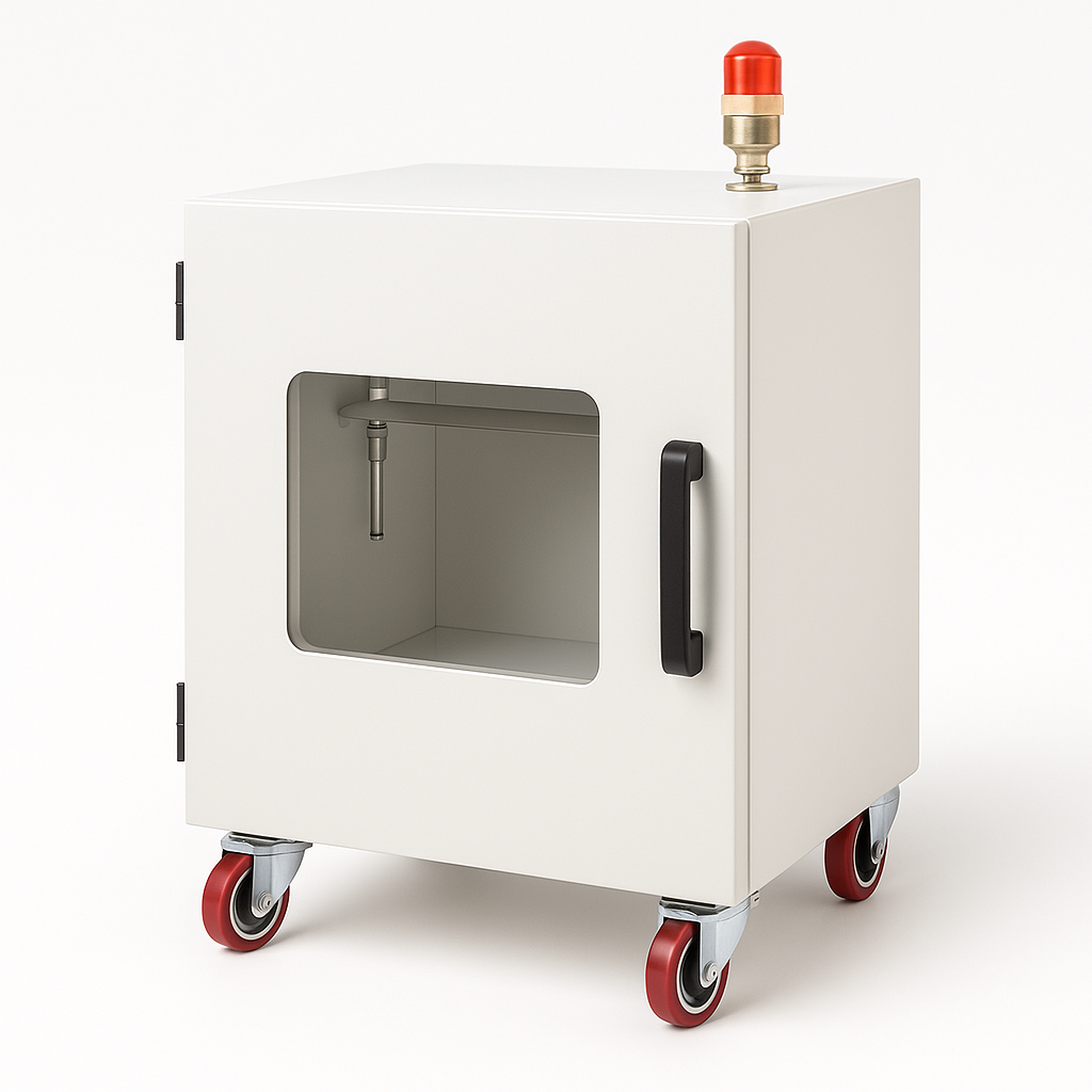
Seed X-Ray Inspection System
Non-destructive seed X-ray systems for fullness, vigor, and pest detection.
16-bit digital imaging, 3–4 s acquisition. Two models for any throughput. Request a quote.

Louise Corscadden, PhD
Director of Science · ConductScience
Ask Louise about Seed X-Ray Inspection System fit, setup, configuration, or quote prep.
Already working with us? Sign in to connect this with My Scientist.
Key Specifications
Full details →- Model fit
- Configured during quote
- SKU family
- AGR-0010
- Sizing
- Model-specific dimensions confirmed from the selected configuration
- Ordering
- Online checkout and quote request available
- Category
- Agriculture & Food Science
- Build notes
- Confirm accessories, station layout, and support needs before purchase
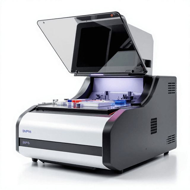
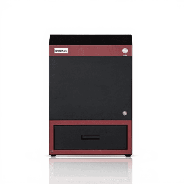
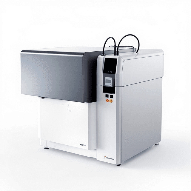
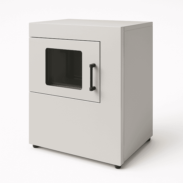
The HY-1600T and HY-1800T Seed X-Ray Machines are non-destructive digital radiography systems designed for microscopic examination of seeds and small plant specimens. Using real-time X-ray imaging, they reveal internal seed structures without cutting or sectioning—allowing researchers to assess fullness, detect empty or deformed grains, evaluate vigor and maturity, and identify internal insect infestations within intact fruits and seeds.
Both models share a 16-bit A/D conversion pipeline and a 4.0 LP/mm spatial resolution, delivering high-contrast digital images in as little as 3–4 seconds per acquisition. The HY-1600T is configured for smaller-scale or single-accession workflows, with a 130 × 160 mm imaging field of view and a 125 µm pixel pitch, while the HY-1800T scales up to a 350 × 430 mm field of view with a 125–140 µm pixel pitch and a higher tube current range (1–10 mA), accommodating bulk sample trays and specimens up to 600 mm thick. Both models support a customizable imaging area to match specific study designs.
The integrated software package enables one-click image enhancements—including contrast enhancement, edge enhancement, false color display, local enhancement, and inverted color—alongside quantitative seed measurements (length, width, circumference, area, and angle). A standard dual-layer adjustable shelf system lets operators select the imaging plane that best suits sample size and field-of-view requirements. A foot-switch remote trigger and a lead-shielded enclosure engineered to meet national X-ray leakage standards under zero-distance conditions keep operators safe during routine acquisition sessions. The system includes a computer as standard, and images can be archived, recalled, or printed at any time.
Because chamber size, imaging field, and tube current requirements vary by seed type and throughput, the HY-1600T and HY-1800T are configured to your study protocol. Contact our team to request a quote and discuss the configuration that fits your research workflow.
Models
- HY-1600T
- HY-1800T
A/D Conversion
- 16 Bits
- 16 Bits
Spatial Resolution
- 4.0 LP/mm
- 4.0 LP/mm
Tube Voltage
- 40–60 kV
- 40–60 kV
Pixel Pitch
- 125 µm
- 140 µm
X-Ray Tube Current
- 0.5–1.0 mA
- 1–10 mA
Maximum Specimen Thickness
- 300 mm
- 600 mm
Digital Imaging Field of View
- 130 × 160 mm (customizable)
- 350 × 430 mm (customizable)
Dimensions (W × D × H)
- 710 × 530 × 350 mm
- 1350 × 800 × 710 mm
Weight
- 110 kg
- 350 kg
What types of seed quality parameters can these X-ray systems evaluate?
The systems are designed to assess seed fullness, identify empty or deformed grains, determine vigor and maturity stage, and detect internal insect infestations within intact seeds or fruits—all without destructive sample preparation.
What is the difference between the HY-1600T and the HY-1800T?
The HY-1600T offers a 130 × 160 mm imaging field of view, a 125 µm pixel pitch, a tube current range of 0.5–1.0 mA, and accommodates specimens up to 300 mm thick, making it well suited to smaller sample sets. The HY-1800T expands to a 350 × 430 mm imaging field, a 140 µm pixel pitch, a tube current range of 1–10 mA, and handles specimens up to 600 mm thick, supporting larger bulk trays or bigger specimens. Both models share the same tube voltage range (40–60 kV), 16-bit A/D conversion, and 4.0 LP/mm spatial resolution.
How fast does the system acquire an X-ray image?
A single seed X-ray imaging acquisition can be completed in as little as 3–4 seconds, enabling efficient screening of multiple accessions or large seed lots in a single session.
What image analysis tools are included in the software?
The bundled software supports one-click image processing operations including contrast enhancement, edge enhancement, inverted color display, false color display, and local enhancement. It also provides quantitative measurement tools for seed length, width, circumference, area, and angle. Images can be saved, recalled, and printed at any time via a connected printer—no film processing is required.
How is operator safety addressed during X-ray acquisition?
The system includes a lead-shielded enclosure engineered so that X-ray leakage at zero distance meets national safety standards. A foot-switch remote trigger allows the operator to remain in the data acquisition area while initiating exposures, keeping them physically separated from the X-ray source during imaging.
Have a question about this product?
Have a question? Just ask.
Send it over and we'll email you a personalized answer — no call, no scheduling.
Prefer to talk it through?
Frequently Bought Together