
Bacteria Enumeration: Methods for Counting Bacteria
Direct and indirect counting, viable and total counts, and the three techniques labs rely on most — plate counts, turbidimetry, and direct microscopy.
What Is Bacteria Enumeration?
Bacteria enumeration is the process of determining the number of bacteria in a sample. Researchers count or compare bacterial populations for many reasons, and it is routine work in food, water, and dairy microbiology labs.[1]
Knowing how many microbes are present in milk, curd, buttermilk, or water tells a lab whether a product or water source is hygienic and safe to consume. Because bacteria are invisible to the naked eye, counting them relies on microscopy and on high-throughput protocols that make large populations tractable.
This guide covers the categories of bacterial counting and the techniques used to count bacteria in food and water samples, along with the advantages and limitations of each.
Counting colonies from a plate image?
Plate counts and CFU tallies are slow and error-prone by hand. Image-analysis software counts colonies directly from a photo of the plate — reproducibly and with an auditable record.
Explore ConductVision Colony Counting →Categories of Bacteria Enumeration
Bacteria enumeration falls into four categories.[2]
- Direct cell count. Counts bacteria in a liquid medium or colonies on a plate directly under the microscope using counting chambers.
- Indirect cell count. Estimates bacterial numbers rather than counting each cell — for example, inferring concentration from colonies via the plate count method.
- Viable cell count. Counts only bacteria that are metabolically active and actively dividing in the culture medium.
- Total cell count. Counts all cells, both metabolically active and inactive or dead.[2]
These categories combine in four ways depending on the experiment:[2]
- Direct/Viable: the standard plate count — repeated dilutions determine viable cells; used for cellular vaccines and milk.
- Indirect/Viable: statistical inference from growth patterns; used for soil, laboratory cultures, and water.
- Direct/Total: dyes and fluorescent stains make cells visible under a fluorescence microscope; used for aquatic samples.[2]
- Indirect/Total: spectroscopy estimates numbers from light transmitted through the culture; used for food samples.[2]
Standard Plate Count (Viable Counts)
Bacteria can number in the millions per sample, so serial dilution is used to bring counts into a countable range. Diluted aliquots are plated on appropriate media, incubated, and the resulting colonies are counted — a method also called plate count or colony count.[3]
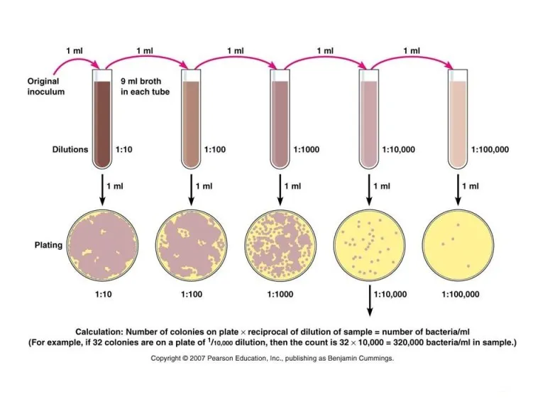
Streak-plate and pour-plate methods support the standard plate count. Plates must not be overcrowded: statistically, only plates with 30–300 colonies are valid to count.[3]
Because it is hard to guarantee that each colony grew from a single cell, results are reported as Colony Forming Units (CFUs) rather than exact cell numbers:[3]
CFUs per ml = number of colonies (30–300 plate) × dilution factor of the plate counted.
Counting the 30–300 colonies on each plate is exactly the step that image-analysis software such as ConductVision Colony Counting automates, removing manual tally error and keeping an image record of the plate.
Advantages
- Feasible for milk, water, foods, soils, and cultures.
- Relatively simple and more sensitive than turbidimetry.
- Allows positive identification of the organism counted.
- Easy to count small numbers of bacteria.
Limitations
- Time-consuming — dilution, plating, incubation, and media prep each take fixed time.[3]
- Only living cells form colonies, so only they are counted.
- Clumps or chains can form a single colony, biasing results.
- Only organisms that grow under the provided conditions are counted.
Turbidimetric Measurement
Turbidity is the loss of transmitted-light intensity caused by suspended particles scattering the beam.[5] A colloidal bacterial suspension blocks and reflects light in proportion to cell concentration, so absorbance estimates the number of bacteria — a fast method well suited to counting many cultures.
Because turbidity readings must first be calibrated against actual cell numbers, labs pair it with the standard plate count:[5]
- Measure turbidity across known concentrations of the species in culture.
- Use standard plate count to establish viable cells per ml for those samples.
- Plot a standard curve of optical density against living-organism counts.
Once the curve exists, later turbidity readings convert directly to viable counts without repeating the slow plate count.
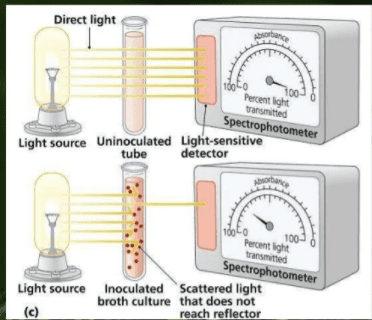
A spectrophotometer or colorimeter measures turbidity: cultures in a cuvette interfere with a light beam, and the instrument output is converted to cell numbers. Before reading, the spectrophotometer is zeroed to 100% transmittance (0% absorbance) using uninoculated medium, and readings are converted with:[1]
Absorbance (O.D.) = 2 − log(% Transmittance)
Wavelength depends on solution color — for example, 420 nm for white, 540 nm for yellow, and 600–625 nm for yellow-to-brown solutions.[1]
Advantages
- Faster than the standard plate count.
- Non-destructive — samples are not spoiled.
Limitations
- Requires a high cell density — around 100 million cells per ml.
- Very dense cultures mask the light and produce inaccurate readings.
Direct Microscopic Count
Direct microscopic counting quantifies bacteria in water, food, milk, and air samples.[3] A measured sample volume is spread over a defined slide area, representative fields are counted, and the average is scaled by the volume-area factor.[3]
Petroff-Hausser and Levy counting chambers are common. The Petroff-Hausser chamber is a thick slide with a central chamber 0.02 mm (1/50 mm) deep and an etched grid with improved Neubauer rulings.
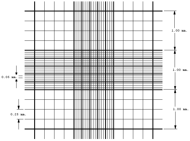
A single drop is applied with a Pasteur pipette and cells are counted directly across 10–20 high-power fields. The per-field average gives bacteria per ml of the original sample:[3]
cells/ml = (total cells counted × 2.0×10⁷ × dilution factor) / number of small squares counted
Using Fluorescent Dyes
Direct counts can also use fluorescent dyes that stain all bacteria, a single species, or a specific cell component — for example CTC, auramine, acridine orange, and rhodamine. Acridine orange is the most common: a known volume is stained, filtered through a 0.22 µm filter that traps the bacteria, and examined under a fluorescence microscope. Counting bacteria in a defined filter area gives the original concentration.
Advantages
- Rapid, simple, and easy.
- Morphology is visible while counting.
- Even dense suspensions can be counted after dilution.
Limitations
- Does not distinguish living from dead cells.
- Cannot count viable cells specifically.
- Small cells can be missed.
- Unsuitable for low-density suspensions (<107 cells per ml).
Conclusion
Bacteria enumeration determines the number of bacterial cells in a sample, split into four categories — direct, indirect, viable, and total — that combine to fit the experiment. The most common techniques are the standard plate count, turbidimetry, and direct microscopic count.
These methods are essential in food and beverage industries, where bacterial counts confirm that a sample is safe and uncontaminated, and they extend to agriculture and processing. Each technique has real limitations, which continues to motivate faster, more accurate enumeration methods — including image-analysis software that counts colonies straight from a plate photo.
References
- Bacteria enumeration. SlideShare.
- What Is Enumeration in Microbiology? Sciencing.
- Isolation and Enumeration of Bacteria. Microbe Online.
- Pour Plate Method: Procedure, Uses, (Dis)Advantages. Microbe Online.
- Bacterial Enumeration: Definition, Methods & Example. Study.com.
Count Colonies from a Photo of the Plate
ConductVision Colony Counting detects and tallies colonies directly in your plate images — reproducible CFU counts with an auditable record, no manual clicking.