ConductVision Disease and Toxicity Models
Neurotoxicity testing in brain organoids
A ConductVision pilot tracks the size and shape of brain organoids around the clock after a test exposure, and measures readouts inside cells in your stained sections.
Readouts
What valproic acid, ethanol and opioids changed
| Readout | Level | Image needed | Evidence today |
|---|---|---|---|
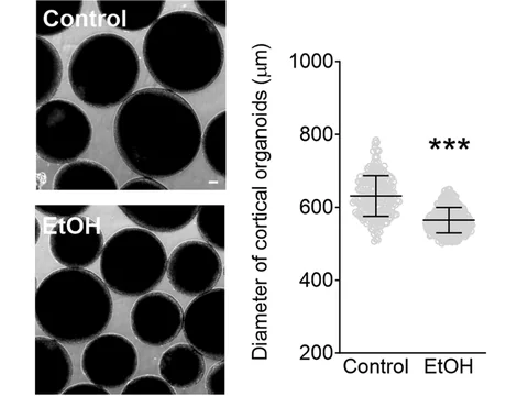
| Size, growth, shape and fragmentation | Whole organoid | Camera, label-free, every 5 to 15 minutes | At 250 and 500 µM, valproic acid made brain organoids smaller by day 42.1 Ethanol-exposed cortical organoids had a smaller diameter than controls at two months (210 to 333 organoids per condition).2 Methadone halted the growth of human cortical organoids in a dose-dependent manner, and after prolonged exposure the organoids disintegrated.3 Chronic fentanyl did not change the size of midbrain organoids over 77 days.4 |
| Progenitor cells and layers | Inside cells | Microscope, immunostained sections | Valproic acid at 250 and 500 µM thinned the layer of SOX2-positive progenitors.1 In forebrain organoids grown from human embryonic stem cells, ethanol disrupted radial glial processes and reduced the generation of HOPX-positive outer radial glial cells.5 |
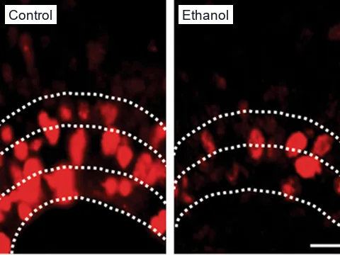
| Dividing cells | Inside cells | Microscope, Ki67 or EdU staining of sections | After valproic acid, progenitor proliferation fell in a dose-dependent manner.1 Ethanol-exposed organoids had fewer Ki67-positive dividing cells than controls at two months, and by three months the difference had narrowed.2 |
| Cell death markers | Inside cells | Microscope, cleaved caspase 3 staining of sections | Staining for cleaved caspase 3 showed more cell death in ethanol-exposed organoids at two months.2 In the forebrain organoids, ethanol increased apoptosis in Nestin-positive radial glial cells.5 |
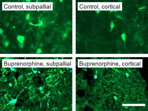
| Interneuron migration | Inside cells | Microscope, fluorescent reporter in sections | More GFP-labeled interneurons migrated into cortical spheroids after buprenorphine than after control or oxycodone.6 |
| Network activity | Not imaging | Multi-electrode array | Multi-electrode array recordings showed impaired network formation and activity in ethanol-exposed organoids.2 Methadone suppressed spontaneous action potential firing in a dose-dependent manner.3 A trend toward higher activity in buprenorphine-exposed spheroids was not statistically significant.6 |
| Gene expression and cell types | Not imaging | Sequencing | Bulk and single-cell sequencing found changes after methadone, oxycodone, buprenorphine and fentanyl.4,7,8 |
Glass and colleagues grew cortical organoids from one stem cell line in three laboratories, and size varied by site at every time point.9 They judged organoid size unsuitable for pooling across sites.9 That is why a pilot compares each organoid with controls grown and imaged alongside it.
Developmental neurotoxicity
From animal guideline studies to human cell batteries
Guideline studies of developmental neurotoxicity, such as OECD Test Guideline 426, expose rodent offspring from implantation on gestation day 6 through lactation to postnatal day 21.10 Such studies are resource-intensive in time, cost and animals, and a limited number of substances have been tested in them.10 They also say little about which cellular processes a chemical disrupts.11
Blum and colleagues built a battery of ten assays on human neural cells at different developmental stages and screened 120 test compounds.12 The 120 included 28 known toxicants and 17 negative controls.12 The battery gave alerts for 24 of the toxicants and for none of the controls.12 Carstens and colleagues analyzed 92 chemicals screened in all 57 endpoints of a cell-based battery, including 53 putative positives and 13 negatives.11 Hierarchical clustering by potency, with cytotoxicity included, classified positive chemicals with 93% sensitivity.11
In brain organoids, a lab can follow a test compound's effects at two levels: the whole organoid and the cells inside it.
Developmental neurotoxicity
Valproic acid, in children and in organoids
In a Danish population study of 655,615 children, the 508 exposed to valproate before birth had a 4.42% risk of autism spectrum disorder, significantly increased even after adjusting for maternal epilepsy.13
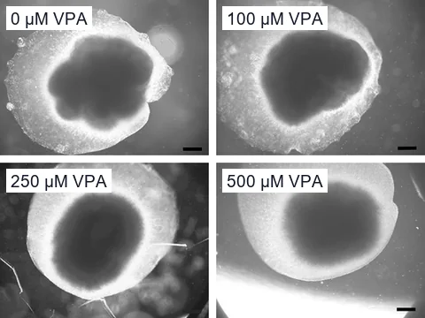
Zang and colleagues exposed brain organoids grown from human embryonic stem cells to 100, 250 or 500 µM valproic acid from day 16.1 By days 42 and 56, organoids at 250 or 500 µM had a smaller surface area and diameter than controls, and 100 µM made no detectable difference.1
A camera pilot records the same size measures for each visible organoid.
Developmental neurotoxicity
The progenitor layer thinned
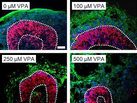
In sections stained for SOX2 and DCX, the layer of SOX2-positive progenitors was thinner at 250 and 500 µM on days 28, 42 and 56.1 Valproic acid also reduced the production of outer radial glia-like cells, progenitors that contribute to neocortical expansion.1
In a pilot, ConductVision measures layers and cell counts like these in your stained sections and checks them against your own measurements on a subset of the images.
Our analysis
Size differences our outlines detect between lines
Size is the readout a camera pilot records, so we tested whether our automatic outlines detect size differences between organoid lines. We outlined every organoid automatically in Schröter and colleagues' public dataset of 1,407 microscope images at about 3 µm per pixel, from a healthy line and three disease lines of 16 organoids each.14 On our automatic outlines, organoids from the TUBA1A and tyrosine hydroxylase deficiency lines were larger than the healthy line on all five size measures, such as area and diameter. Mean projected area was 16 percent larger in the TUBA1A line and 38 percent larger in the tyrosine hydroxylase deficiency line. Rank-biserial correlations ran from 0.95 to 1.00, where 1.00 means the two lines did not overlap at all. Adjusted q values were below 0.0001. Every difference held when any one organoid was left out.
Prenatal alcohol exposure
Ethanol exposure in cortical organoids
Prenatal alcohol exposure is the foremost preventable cause of intellectual disability.2 Among first-graders in 4 US communities, conservative estimates of fetal alcohol spectrum disorder prevalence ranged from 1.1% to 5.0%.15
Adams and colleagues added ethanol to the medium of cortical organoids grown from three human iPSC lines, at 100 mM for seven days, starting when the organoids were about 4 weeks old.2 Within a couple of hours, the concentration settled at about 20 mM.2
Prenatal alcohol exposure
Dividing cells need stained sections
Readouts inside cells, such as Ki67-positive dividing cells, need sections of fixed organoids stained and imaged under a microscope.
Prenatal opioid exposure
Few opioid effects showed in whole organoids
We reviewed five published organoid and spheroid studies of opioids, and only one, a methadone study, reported a whole-organoid change a camera could see.3,4,6,7,8 Most effects were inside cells or in gene expression, which need a microscope or sequencing to find.4,6,7,8
Kim and colleagues grew human midbrain organoids with fentanyl from the first day of neuronal induction.4 Over 77 days of culture, the organoids' gross morphology and size did not differ from controls.4 The authors suggest growth in both groups may have reached an upper limit without a supporting hydrogel scaffold and vasculature.4 Single-cell sequencing of 25,510 cells showed that chronic fentanyl arrested neuronal subtype specification.4
Prenatal opioid exposure
More interneurons migrated after buprenorphine
Nieto-Estévez and colleagues grew cortical and subpallial spheroids, labeled interneurons in the subpallial spheroids with GFP and fused the two, so interneurons could migrate into the cortical side.6 Spheroids exposed to 2 ng/ml buprenorphine from day 50 for 10 days sent more labeled interneurons into the cortical side.6
Prenatal opioid exposure
Sequencing found changes after all four drugs
Dwivedi and colleagues exposed cortical organoids to 1 µM methadone for 50 days, and bulk RNA sequencing found 2,124 genes changed, including genes for synapses, extracellular matrix and cilia.7 Ho and colleagues sequenced the nuclei of 25,787 cells from forebrain organoids of three male donors.8 Buprenorphine changed transcription regulation in glial cells, and oxycodone induced type I interferon signaling in many cell types.8 In Kim and colleagues' midbrain organoids at day 77, mature dopaminergic neurons made up 14.3% of cells in controls and 8.6% after fentanyl.4
These results come from sequencing, outside what ConductVision measures.
Pilots
Two ways to measure your models
- Whole organoid
24/7 camera pilot
In the pilot, cameras image your plates every 5 to 15 minutes. ConductVision tracks each visible organoid's size, growth and shape, and logs each fusion, fragmentation or structural collapse.
Plan a camera pilot - Inside cells
Analysis of your microscope images
In a pilot, ConductVision measures readouts inside cells, such as Ki67-stained dividing cells, in your lab's microscope images, and checks them against your counts.
Plan an image analysis pilot
References
- Zang Z, Yin H, Du Z, et al. (2022). Valproic acid exposure decreases neurogenic potential of outer radial glia in human brain organoids. Frontiers in Molecular Neuroscience.
- Adams JW, Negraes PD, Truong J, et al. (2023). Impact of alcohol exposure on neural development and network formation in human cortical organoids. Molecular Psychiatry.
- Yao H, Wu W, Cerf I, et al. (2020). Methadone interrupts neural growth and function in human cortical organoids. Stem Cell Research.
- Kim HS, Xiao Y, Chen X, et al. (2024). Chronic Opioid Treatment Arrests Neurodevelopment and Alters Synaptic Activity in Human Midbrain Organoids. Advanced Science.
- Lü L, Yuan F, Fan H, et al. (2023). Ethanol exposure disrupted the formation of radial glial processes and impaired the generation and migration of outer radial glial cells in forebrain organoids derived from human embryonic stem cells. Experimental Neurology.
- Nieto-Estévez V, Donegan JJ, McMahon CL, et al. (2022). Buprenorphine Exposure Alters the Development and Migration of Interneurons in the Cortex. Frontiers in Molecular Neuroscience.
- Dwivedi I, Caldwell AB, Zhou D, et al. (2023). Methadone alters transcriptional programs associated with synapse formation in human cortical organoids. Translational Psychiatry.
- Ho MF, Zhang C, Moon I, et al. (2024). Single cell transcriptomics reveals distinct transcriptional responses to oxycodone and buprenorphine by iPSC-derived brain organoids from patients with opioid use disorder. Molecular Psychiatry.
- Glass MR, Waxman EA, Yamashita S, et al. (2024). Cross-site reproducibility of human cortical organoids reveals consistent cell type composition and architecture. Stem Cell Reports.
- Tal T, Myhre O, Fritsche E, et al. (2024). New approach methods to assess developmental and adult neurotoxicity for regulatory use: a PARC work package 5 project. Frontiers in Toxicology.
- Carstens KE, Carpenter AF, Martin MM, et al. (2022). Integrating Data From In Vitro New Approach Methodologies for Developmental Neurotoxicity. Toxicological Sciences.
- Blum J, Masjosthusmann S, Bartmann K, et al. (2023). Establishment of a human cell-based in vitro battery to assess developmental neurotoxicity hazard of chemicals. Chemosphere.
- Christensen J, Grønborg TK, Sørensen MJ, et al. (2013). Prenatal Valproate Exposure and Risk of Autism Spectrum Disorders and Childhood Autism. JAMA.
- Schröter J, Deininger L, Lupse B, et al. (2024). A large and diverse brain organoid dataset of 1,400 cross-laboratory images of 64 trackable brain organoids. Scientific Data.
- May PA, Chambers CD, Kalberg WO, et al. (2018). Prevalence of Fetal Alcohol Spectrum Disorders in 4 US Communities. JAMA.
Plan a neurotoxicity pilot
Tell us your organoid model, the compounds or exposures you study and the readouts you need.