ConductVision Disease and Toxicity Models
Disease and toxicity models in cells and organoids
For disease modeling and toxicity testing, a ConductVision pilot measures whole organoids from camera images taken around the clock, and cells from your own microscope images.
Readouts
What each readout needs, and the evidence today
A camera images whole organoids without dyes or fixation. Readouts inside cells need a microscope image of fixed or fluorescently labeled cells, and some readouts need electrodes or sequencing instead.
| Readout | Level | Image needed | Evidence today |
|---|---|---|---|
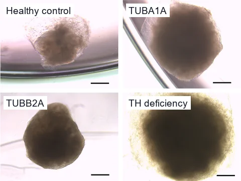
| Size, growth, shape and fragmentation | Whole organoid | Camera, label-free, every 5 to 15 minutes | In Schröter and colleagues' dataset, four iPSC lines of brain organoids, a healthy control and three disease lines, grew in clearly distinct patterns.1 We outlined each organoid automatically in their microscope images at about 3 µm per pixel.1 On our automatic outlines, two disease lines, TUBA1A and tyrosine hydroxylase deficiency, measured larger than the healthy line on all five size measures. Ethanol-exposed cortical organoids had a smaller diameter at two months than controls.2 |
| Progenitor cells and layers | Inside cells | Microscope, immunostained sections | At 250 and 500 µM, the two higher doses tested, valproic acid thinned the layer of SOX2-positive progenitors in brain organoids.3Developmental neurotoxicity |
| Dividing cells | Inside cells | Microscope, Ki67 or EdU staining of sections | Ethanol-exposed cortical organoids had fewer Ki67-positive dividing cells at two months.2Prenatal alcohol exposure |
| Cell death markers | Inside cells | Microscope, cleaved caspase 3 staining of sections | Staining for cleaved caspase 3 showed more cell death in ethanol-exposed cortical organoids at two months.2Prenatal alcohol exposure |
| Nuclear RNA foci | Inside cells | Microscope, RNA FISH | In cortical organoids grown from DM1 stem cells, CUG repeat RNA foci appeared early in maturation.4 Neurons and astrocytes in C9orf72 neuromuscular organoids carried RNA foci.5Repeat expansion disorders |
| MBNL1 sequestered in RNA foci | Inside cells | Microscope, RNA FISH with MBNL1 immunostaining | In DM1 fibroblasts, MBNL1 colocalized with RNA foci, though some antibodies showed it more clearly than others.6Myotonic dystrophy |
| Dipeptide-repeat proteins | Inside cells | Microscope, immunostained sections | Deep-layer neurons in C9orf72 brain organoid slices accumulated the dipeptide-repeat protein poly(GA).7C9orf72 ALS/FTD |
| DNA damage foci | Inside cells | Microscope, γ-H2AX immunostaining | C9orf72 brain organoid slices showed γ-H2AX DNA damage foci in deep-layer neurons.7 We scored our spot detector on 90 γ-H2AX images of irradiated human lymphocytes that were not used to set it up. It found 75 percent of the foci experts had marked, and 84 percent of its detections were marked foci (F1 0.79).Nuclear and RNA foci analysis |
| Pyknotic nuclei | Inside cells | Microscope, nuclear stain | C9orf72 brain organoid slices had more pyknotic neurons than controls.7C9orf72 ALS/FTD |
| Network activity | Not imaging | Multi-electrode array | Multi-electrode array recordings showed impaired network formation and activity in ethanol-exposed cortical organoids.2Prenatal alcohol exposure |
| Gene expression and cell types | Not imaging | Sequencing | Measured by sequencing, outside what ConductVision measures. |
Glass and colleagues grew cortical organoids from one stem cell line in three laboratories, and size varied by site at every time point.8 They judged organoid size unsuitable for pooling across sites.8 That is why a pilot compares each organoid with controls grown and imaged alongside it.
How we measure, and how wellModels
Models, toxicity testing and methods
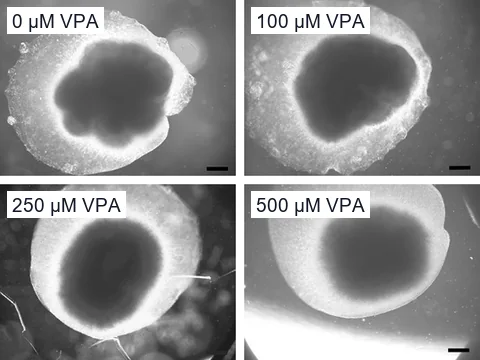
Adapted from Zang et al. 2022 · CC BY 4.0 Neurotoxicity testing in brain organoids
Valproic acid and ethanol made brain organoids smaller and changed the progenitor and dividing cells inside them.2,3
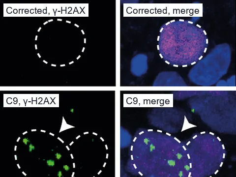
Adapted from Szebényi et al. 2021 · CC BY 4.0 Repeat expansion disorders
Nuclear RNA foci, the MBNL1 they sequester and repeat proteins in myotonic dystrophy and C9orf72 ALS/FTD models.
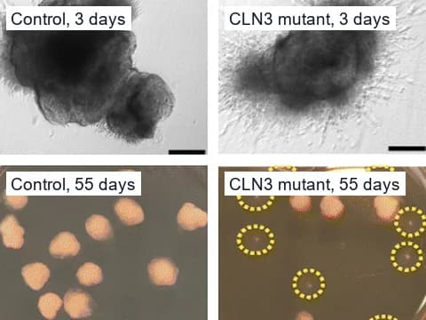
Adapted from Gomez-Giro et al. 2019 · CC BY 4.0 Brain and neural organoids
Size, growth, shape and outline of each brain organoid, recorded around the clock in a camera pilot.
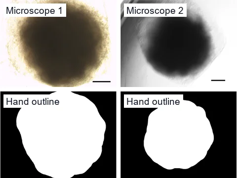
Adapted from Schröter et al. 2024 · CC BY 4.0 Image analysis methods and validation
How closely our automatic outlines matched outlines drawn by hand in brain organoid images, and what is not yet validated.
Pilots
Two ways to measure your models
- Whole organoid
24/7 camera pilot
In the pilot, cameras image your plates every 5 to 15 minutes. ConductVision tracks each visible organoid's size, growth and shape, and logs each fusion, fragmentation or structural collapse.
Plan a camera pilot - Inside cells
Analysis of your microscope images
In a pilot, ConductVision measures readouts inside cells, such as Ki67-stained dividing cells, in your lab's microscope images, and checks them against your counts.
Plan an image analysis pilot
Questions labs ask
Can a camera see inside the cells of an organoid?
No. A camera measures whole organoids, such as their size, growth, shape and fragmentation. Readouts inside cells, such as dividing cells, need a microscope image.
Can you analyze the microscope images my lab already has?
Yes, for readouts inside cells. In a pilot, ConductVision measures them in your fixed or fluorescent images and reports how closely its counts match yours on a subset of those images.
Will a pilot tell me which organoids died?
Not from camera images alone. In a pilot, ConductVision labels organoids dead or viable only after checking those labels against your own viability assay.
References
- Schröter J, Deininger L, Lupse B, et al. (2024). A large and diverse brain organoid dataset of 1,400 cross-laboratory images of 64 trackable brain organoids. Scientific Data.
- Adams JW, Negraes PD, Truong J, et al. (2023). Impact of alcohol exposure on neural development and network formation in human cortical organoids. Molecular Psychiatry.
- Zang Z, Yin H, Du Z, et al. (2022). Valproic acid exposure decreases neurogenic potential of outer radial glia in human brain organoids. Frontiers in Molecular Neuroscience.
- Morelli KH, Jin W, Shathe S, et al. (2022). MECP2-related pathways are dysregulated in a cortical organoid model of myotonic dystrophy. Science Translational Medicine.
- Gao C, Shi Q, Pan X, et al. (2024). Neuromuscular organoids model spinal neuromuscular pathologies in C9orf72 amyotrophic lateral sclerosis. Cell Reports.
- Aoki Y, Ohki A, Yanaizu M, et al. (2025). Comparative Analysis of MBNL1 Antibodies: Characterization of Recognition Sites and Detection of RNA Foci Colocalization. Genes.
- Szebényi K, Wenger LMD, Sun Y, et al. (2021). Human ALS/FTD brain organoid slice cultures display distinct early astrocyte and targetable neuronal pathology. Nature Neuroscience.
- Glass MR, Waxman EA, Yamashita S, et al. (2024). Cross-site reproducibility of human cortical organoids reveals consistent cell type composition and architecture. Stem Cell Reports.
- Gomez-Giro G, Arias-Fuenzalida J, Jarazo J, et al. (2019). Synapse alterations precede neuronal damage and storage pathology in a human cerebral organoid model of CLN3-juvenile neuronal ceroid lipofuscinosis. Acta Neuropathologica Communications.
Plan a disease or toxicity model pilot
Tell us your cell or organoid model, the disease or exposure you study and the readouts you need.