Microcirculation Microscope
Specialized optical microscope system with 380X magnification and HD color camera for real-time microcirculation visualization and blood flow analysis in research applications.

Louise Corscadden, PhD
Director of Science · ConductScience
Ask Louise about Microcirculation Microscope fit, setup, configuration, or quote prep.
Already working with us? Sign in to connect this with My Scientist.
Key Specifications
Full details →- Model fit
- Configured during quote
- SKU family
- BIO-0492
- Sizing
- 38.0 x 27.0 x 34.0 cm
- Ordering
- Online checkout and quote request available
- Category
- Microscopy
- Build notes
- Confirm accessories, station layout, and support needs before purchase
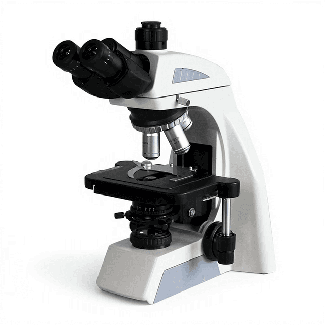
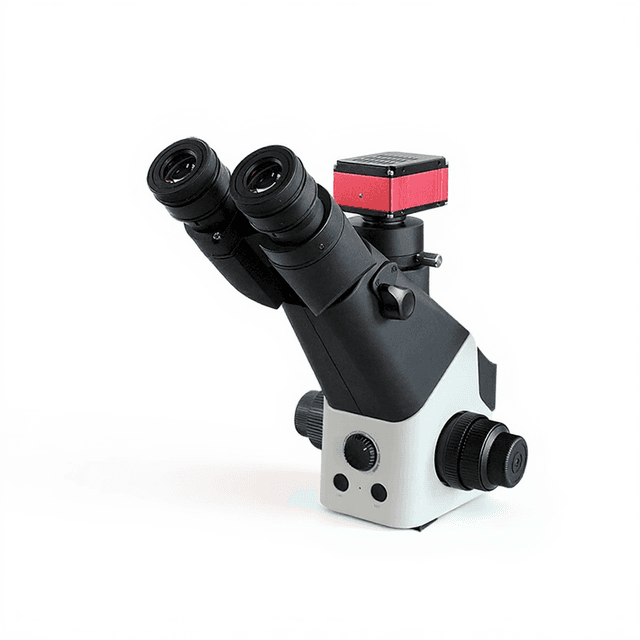
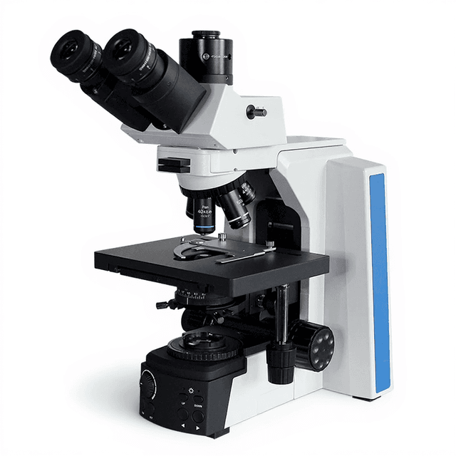
The Microcirculation Microscope is a specialized optical imaging system designed for real-time visualization and analysis of microvascular structures and blood flow dynamics. This 380X magnification system incorporates a color camera with HD LCD display for direct observation and documentation of microcirculatory patterns in tissue samples and experimental preparations.
The instrument features a 10X objective with coaxial coarse and fine focusing mechanisms, providing precise Z-axis control with mechanical upper and lower limits. The 360° rotating stage with X-Y axis movement (30×80mm range) enables comprehensive sample positioning and analysis. LED illumination with high brightness and low heat generation ensures optimal visualization conditions while minimizing thermal effects on living preparations.
How It Works
The Microcirculation Microscope operates on transmitted light optical microscopy principles, utilizing LED illumination to provide uniform, high-intensity lighting with minimal heat generation. Light passes through the specimen and is collected by the 10X objective lens, which focuses the image onto a color camera sensor system.
The coaxial focusing mechanism allows for precise control of the focal plane through both coarse and fine adjustments along the Z-axis. Mechanical limits prevent over-adjustment and protect both the objective and specimen. The integrated color camera captures the magnified image and displays it in real-time on the HD LCD screen, enabling immediate visualization and digital documentation.
The 360° rotating stage with X-Y axis movement provides comprehensive specimen positioning capabilities across a 30×80mm working area. This mechanical stage system ensures stable specimen mounting while allowing for systematic scanning and analysis of different regions within the sample.
Features & Benefits
Automation Level
- manual
WXH-10
- WXH-12
Magnification
- 380X
480X
- 580X
Display Size(mm)
- 161*121
204*153
- 185*240
Objective
- 10X
Focus System
- Coaxial coarse/slightly adjusted, Z axis movement, Mechanical upper and lower limits
Stage
- 360°revolve, X, Y axis movement, moving range 30*80mm
lllumination
- LED illumination with high brightness and lower consumption, new luminaire with lower temperature an
Camera
- Color camera system, HD LCD display
Standard Accessory
- Aluminum box, Display screen, Power cable, Asphalt, Manual, Flip chart, Microcirculation flattery
Package Size
- 270*340*380mm
Brand
- ConductScience
Research Domain
- Cardiovascular
- Cell Biology
- Clinical Diagnostics
- Materials Science
- Microbiology
- Pharmaceutical QC
Power/Voltage
- AC100~230V, 50/60Hz
Weight
- 5.5kg
Weight
- 5.5 kg
Dimensions
- L: 38.0 mm
- W: 27.0 mm
- H: 34.0 mm
| Feature | This Product | Typical Alternative | Advantage |
|---|---|---|---|
| Magnification Range | 380X optical magnification with 10X objective | Entry-level systems often provide 100-400X range with multiple objectives | Fixed magnification optimized for microcirculation studies eliminates objective changing and maintains consistent image quality |
| Display System | Integrated HD LCD color display (161×121mm) | Traditional microscopes rely on eyepiece observation | Direct screen visualization enables documentation and collaborative observation without eyepiece sharing |
| Illumination Technology | LED illumination with high brightness and low heat generation | Many basic systems use halogen bulbs with higher heat output | LED technology extends lamp life and reduces thermal effects on living specimens during extended observation |
| Stage Movement | 360° rotation with X-Y axis movement across 30×80mm range | Basic mechanical stages offer limited movement range | Comprehensive positioning capability enables systematic scanning of larger specimen areas |
| Focusing System | Coaxial coarse and fine adjustment with mechanical limits | Separate coarse and fine focus controls without limit protection | Mechanical limits prevent over-adjustment protecting both objective and specimens from damage |
| Portability | 5.5kg total weight with aluminum carrying case | Laboratory microscopes typically require permanent installation | Portable design enables field applications and flexible laboratory deployment |
This microscope system integrates HD display technology with LED illumination and comprehensive stage movement capabilities in a portable configuration. The system focuses on microcirculation applications with optimized magnification and thermal management features for extended observation periods.
Practical Tips
Verify optical alignment and focus accuracy using a calibrated test slide before beginning critical observations.
Why: Ensures measurement accuracy and consistent image quality across different specimens.
Clean LED illumination optics and objective lens monthly with appropriate optical cleaning solutions.
Why: Maintains optimal light transmission and prevents image degradation from dust or contamination.
Allow 10-15 minutes warm-up time for LED illumination system stabilization before critical imaging.
Why: LED output stabilizes with temperature, ensuring consistent illumination intensity throughout observation sessions.
If stage movement becomes stiff, check for debris in the mechanical tracks and lubricate as specified in the manual.
Why: Smooth stage operation is essential for precise specimen positioning and prevents sample damage.
Adjust LED brightness to prevent oversaturation while maintaining adequate contrast for your specimen type.
Why: Proper illumination balance optimizes image contrast and prevents loss of detail in bright or dark specimen areas.
Use the mechanical focus limits to prevent objective-specimen contact during initial focusing procedures.
Why: Protects expensive optical components and prevents specimen damage during routine operation.
Store the system in its aluminum case with objective lens covered when not in use for extended periods.
Why: Protects optical surfaces from environmental contamination and maintains system readiness for field deployment.
Setup Guide
What’s in the Box
- Microcirculation Microscope main unit
- HD LCD display screen
- Power cable
- Aluminum carrying case
- User manual
- Flip chart documentation
- Microcirculation accessories
- Asphalt sample preparation materials
Warranty
ConductScience provides a standard one-year manufacturer warranty covering defects in materials and workmanship. Technical support and application guidance are available throughout the warranty period.
Compliance
What is the working distance of the 10X objective and stage clearance?
The system provides 30×80mm X-Y stage movement range with 360° rotation capability. For specific working distance specifications, consult the product datasheet.
Can the camera system capture and store digital images?
The system includes a color camera with HD LCD display for real-time visualization. For digital image capture and storage capabilities, consult the product datasheet.
What specimen types are compatible with this microscope configuration?
The system is designed for transmitted light microscopy applications including tissue sections, cell cultures, and solution-based preparations that fit within the stage working area.
How does the LED illumination system compare to halogen lighting?
LED illumination provides high brightness with significantly lower heat generation and power consumption compared to traditional halogen systems, extending lamp life and reducing thermal effects on specimens.
What maintenance is required for optimal performance?
Regular cleaning of optical surfaces, periodic calibration of the focusing mechanism, and LED system inspection are recommended. Mechanical stage lubrication may be needed based on usage frequency.
Is the system compatible with additional objectives or magnifications?
The system is configured with a 10X objective providing 380X total magnification. For compatibility with alternative objectives or magnification options, consult the product datasheet.
What are the power requirements and consumption specifications?
The system operates on AC100-230V, 50/60Hz universal power supply. LED illumination provides high efficiency with lower power consumption compared to traditional lighting systems.
Have a question about this product?
Have a question? Just ask.
Send it over and we'll email you a personalized answer — no call, no scheduling.
Prefer to talk it through?
Accessories
Enhance your setup with compatible accessories

Frequently Bought Together
Related Products