Genetics of Drosophila
Drosophila has 4 pairs of chromosomes and the size of its genome is estimated to be approx 180 Mb―the euchromatin (transcriptionally active region ) is about 120 Mb. Out of 4 pairs of chromosomes, one is sex chromosome (acrocentric X chromosomes and metacentric Y chromosomes) and three are autosomes.
Other than mitotic chromosomes, Drosophila has “Polytene chromosomes-giant chromosomes.” This chromosome is formed by the process of “endoreduplication-multiple round of DNA replication, without cell division.” The polytene chromosomes are commonly found in salivary glands and most commonly isolated from larval salivary glands.
How does the genetics of Drosophila aid in the experiments?
A lot of information has been decoded about the Drosophila genome. Some genetic characteristics of drosophila are known as “genetic tools”, which provide essential aid in various researches including genetics, biotechnology, drug discovery, etc.
1. Classical genetics
It has been almost 100 years since the use of Drosophila in various researches. These meticulous studies have contributed a lot to the understanding of heredity and gene activity, construction of recombination maps, and the relation between sex linkage and inheritance of sex chromosomes.
Mutagenesis studies on Drosophila led to the construction of genetic maps, which was also facilitated by the studies of banding patterns of polytene chromosomes found in salivary glands of Drosophila larva.
2. Transposable elements
The P elements carry the gene for transposase activity and are responsible for hybrid dysgenesis―series of mutations as a result of crossing male with P element and a female without P element which leads to sterility and chromosomal aberration―in Drosophila. Its use in introducing cloned DNA in Drosophila was discovered way back in 1982.
The P elements are now an essential tool in genetics for gene tagging, gene disruption, chromosome engineering, and inducible gene expression. Moreover, the mutation, by the introduction of P elements into a gene, allows the molecular identification of the affected gene.
The controlled activity of transposase aids scientists to insert P elements at the desired location and generate a large number of mutations required for their studies.
3. The Drosophila genome
The whole genome of Drosophila was sequenced in 2000. Researchers have found that Drosophila has approximately 70% of orthologous genes associated with human diseases.
The availability of a chunk of information about Drosophila genomes aid researchers (as a model organism) for human disease research and drug discovery and it also avoids the labor of performing a number of molecular manipulations of Drosophila DNA.
Experimental methods to study Drosophila in labs
The study of Drosophila in labs serves many purposes which include the understanding of neurons, developmental processes, mutational studies, gene regulation, and signaling. A number of methods are used to study the different processes in different experiments. In this section, you will get a brief of cytogenetic methods used to study different parts/processes of Drosophila and its culturing in labs.
Other than the mentioned cytogenetic experiments in this section, the two other, most commonly used bioengineered, techniques used to study the Drosophila are:
- The GAL4/UAS system: Used to study gene expression and function in organisms.
- The FLP/ FRT system: Site-directed recombination technology.
1. Culturing Drosophila in labs
Equipment:
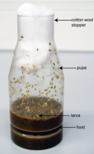
The Drosophila culturing does not require standard equipment. The great scientist T. H. Morgan used glass milk bottles to culture Drosophila for his experiments! Now, laboratories use bottles and vials to culture Drosophila.
Bottles are used to maintain a large population and culturing vials are used to maintain a small population and make crosses. Generally, glass bottles are preferred but autoclaved plastic bottles can also work well. Moreover, the size of small vials ranges from 96 mm by 25 mm to larger vials. To cover the bottles, plugs are used which can be cotton or foam plugs. However, cotton plugs are mostly preferred.
Media:
The preparation of media involves two ways: cooking the media or using ready-made and dehydrated media. The latter is most preferred, which avoids the labor of cooking the media, and is easier and quicker than the former. However, rehydration is required to use readymade media. To ensure the complete rehydration and culture process, follow the given procedure:
Rehydration Procedure of ready-made media:
- Add ⅕ to ⅖ volume of dry media to the bottle/vial.
- Add water to completely moisten the media.
- Let the vial rest for a few minutes and add water until it seems hydrated.
- The surface of completely hydrated media looks shiny without any spaces in the media.
- Allow the media to warm to room temperature. The optimum temperature to grow flies is at 25 ℃ with 60 % humidity.
- Add several grains of yeast on the surface of the media after the media is hydrated.
- Transfer the flies in the vial/bottles and plug it.
- Flies should be transferred to different clean vials/bottles to maintain the culture.
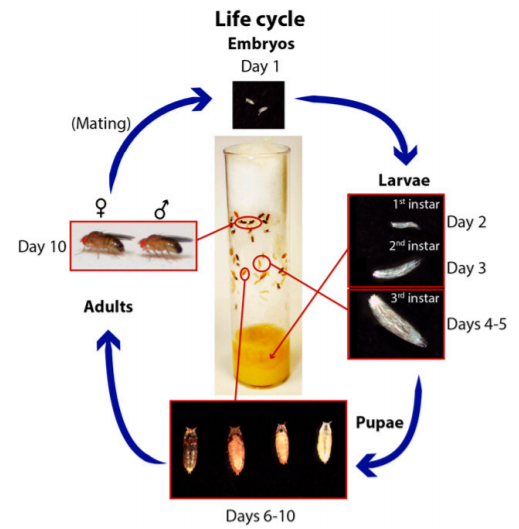
NOTE: Change the food always after or between 10-14 days.
2. Orcein staining to identify Polytene chromosomes
Procedure:
- Squash Preparation
- Select 3rd instar larva from the culture.
- Place them in a container having PBS and 0.8% saline.
- Put the larva on the slide containing a drop of 45% acetic acid and dissect the larva.
- Remove the salivary gland and allow it to sit in 45% acetic acid for 2-3 minutes.
- Stain the gland with the lactic–acetic-orcein stain for 5 minutes.
- Take a clean slide and put a drop of 1: 2 : 3 fixative. Transfer the gland to this slide.
- Put a coverslip over the gland and by using bibulous paper score a zigzag pattern over the coverslip to shear the chromosomes into fragments.
- Seal the edges of the coverslip and observe it in the microscope.
- Identifying chromosomes
- To identify the chromosome, resolve the banding patterns of the telomeric region and look for some landmarks such as puffs, constriction, and banded regions.
3. In-situ hybridization to polytene chromosomes
Procedure:
- Polytene Chromosome Preparation
- Dissect the salivary glands of the Drosophila in a drop of 0.7% NaCl and then transfer to 45 % acetic acid.
- Transfer the glands to the fixative for 3 minutes.
- Spread chromosomes by tapping the coverslip with a pencil in a circular motion.
- Cover the slide in blotting paper and leave overnight for better fixation.
- Freeze the slide in liquid nitrogen and flip off the coverslip.
- Place the slide in 70% ethanol for 5 min and air dry. The chromosomes can be stored desiccated at room temperature.
- Denaturation of Chromosome
- Two methods of denaturation are available: Alkali denaturation (involves the use of freshly prepared NaOH)and Heat denaturation (involves the use of Tris HCl, pH 7.5, and then transfer to 70 % and 96% ethanol).
- Use the denatured slide on the same day.
- Preparation of Labeled probes
- Prepare probes using DNA amplified by the polymerase chain reaction (PCR) in the presence of biotinylated nucleotides.
- Hybridization
- Pipette probe onto the chromosomes and cover it with a siliconized coverslip.
- Overnight incubate the slide in a box lined with moist tissue at 58 ℃.
- Remove the slide from the box and put it in 2X SSC. Wash the slide at 58 ℃ for 1 hour, with three changes of the solution.
- Signal Detection
- Wash the slides in PBS and PBS-TX for 2 minutes.
- Make a 1:250 dilution of streptavidin–horseradish peroxidase conjugate in the buffer and add to the chromosomes.
- Place the slide in a humid box and incubate at 37°C for 30 minutes.
- Wash the slide in PBS and PBS-TX.
- Place the DAB solution onto the chromosomes.
- Incubate at room temperature for 10-15 minutes.
- Rinse the slides in PBS.
- Observe the slide under the Phase Contrast microscope.
4. Immunostaining to study Larval brains
Procedure:
- Fixation of Mitotic Chromosomes
- Transfer the 3rd instar larva in a few drops of physiological solution (0.7% NaCl in distilled water) and dissect out its brain.
- Transfer the brain to a few drops of hypotonic solution (0.5% Trisodium citrate dihydrate in distilled water) for 8 minutes.
- Transfer the brain to a fixative solution (2% formaldehyde, 45% acetic acid in distilled water) for 1-8 minutes.
- Transfer four fixed brains to 4 drops of fixative solution and cover it with a siliconized coverslip.
- Put another clean slide on the coverslip, invert the sandwich, and squash for 1 minute between blotting paper.
- Freeze in liquid nitrogen, flip off the coverslip, and immerse the slide in 1X PBS at room temperature.
- Fluorescent immunostaining on Fixed chromosomes
- Put the slide in 1X PBS with dried nonfat milk and incubate in it for 30 minutes.
- Clean the slides in 1X PBS for 3 minutes.
- Put the primary antibody solution on the mitotic preparation and incubate for 1 hour at room temperature.
- Wash the slides in PBS for 5 minutes (three times).
- Put the secondary antibody solution on the mitotic preparation and incubate it for 1 hour at room temperature.
- Wash the slides in 1X PBS (three times) for 5 min in the dark.
- DAP1 staining and mounting
- Stain the slides in the DAP1 staining solution at room temperature for 4 minutes.
- Wash the slides in 1X PBS for 30 seconds.
- Mount the slide in a drop of antifade solution.
- Analyze the slides using an epifluorescence microscope (The slide can also be used to perform the FISH technique).
Note: To grasp the principle and theory of Cytogenetic techniques you can refer to “Cytogenetics: An advanced technique to color Chromosomes and Molecular Cytogenetics: in situ Hybridization-based technology”.


