Automatic Staining and Coverslipping Machine
Fully automated histopathology workstation that performs staining and coverslipping of 120 slides with robotic precision, touchscreen control, and customizable protocols.

Louise Corscadden, PhD
Director of Science · ConductScience
Ask Louise about Automatic Staining and Coverslipping Machine fit, setup, configuration, or quote prep.
Already working with us? Sign in to connect this with My Scientist.
Key Specifications
Full details →- Model fit
- 3 selectable configurations
- SKU family
- RF-RD-3000A
- Sizing
- 34.0 x 25.0 x 20.0 cm
- Ordering
- Online checkout and quote request available
- Category
- Pathology & Histology
- Build notes
- Confirm accessories, station layout, and support needs before purchase
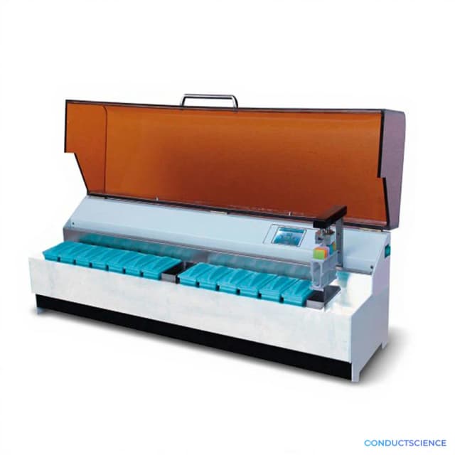
The RF-RD-3000A Automatic Staining and Coverslipping Machine is a fully automated histopathology workstation designed for high-throughput slide processing. This system handles both stained and unstained slides, performing automated staining protocols followed by precise coverslip application with controlled pressure to prevent air bubble formation. The machine accommodates 120 slides in a six-layer configuration (20 slides per layer) and processes standard 26×75mm microscope slides with 24×50mm coverslips.
The workstation features an intuitive touchscreen interface for protocol selection and monitoring, robotic arm transfer system for precise slide handling, and automated mounting medium dispensing. The system supports both predefined and custom staining protocols, making it suitable for routine H&E staining, special stains, and immunohistochemistry applications. Upon completion, fully prepared slides are delivered to an output tray ready for microscopic examination.
How It Works
The RD-3000A operates through a precision-controlled, multi-step automated process. Slides are loaded into the system's six-layer carousel that accommodates up to 120 slides total. A robotic arm transfer system moves slides through sequential processing stations, beginning with the staining module where automated reagent applications, rinses, and dehydration steps occur according to programmed protocols.
Following staining completion, slides advance to the coverslipping station where automated drop dispensing applies mounting medium in precise volumes. The coverslip application mechanism uses controlled pressure to ensure proper adhesion while preventing air bubble formation. The pressure-controlled system maintains consistent coverslip placement across all samples, regardless of tissue thickness variations.
The touchscreen interface allows operators to select predefined protocols or customize parameters for specific staining requirements. The system monitors each processing step and delivers completed slides to an output tray, maintaining sample identification throughout the workflow.
Features & Benefits
pa_equipment-type
- 4954
pa_automation-level
- fully-automated
pa_pathology-application
- 4971
pa_slide-capacity
- 5008
pa_staining-protocols
- 5011
pa_reagent-stations
- 5022
pa_processing-speed
- 4991
slide_capacity
- 60 slides per run
automation_level
- fully automated
staining_capability
- stained or unstained slides
coverslipping_capability
- automatic coverslip application with controlled pressure
interface_type
- intuitive touchscreen interface
protocol_flexibility
- predefined or custom staining and coverslipping protocols
mounting_medium_dispensing
- automated drop dispensing
air_bubble_prevention
- controlled pressure mechanism
slide_transfer_method
- robotic arm transfer system
reagent_application
- automated reagent applications, rinses, and dehydration steps
output_method
- output tray for fully prepared slides
Set parts
- One set
- Stainer
- Slide cover
Capacity
- 60 slides per run
Display Type
- Touchscreen
Research Domain
- Cancer Research
- Developmental Biology
- Histopathology
- Immunology
- Neuroscience
- Pharmaceutical QC
Weight
- 6.61 kg
Dimensions
- L: 34.0 mm
- W: 25.0 mm
- H: 20.0 mm
| Feature | This Product | Typical Alternative | Advantage |
|---|---|---|---|
| Slide Capacity | 120 slides in six-layer configuration | Manual systems process 10-20 slides per batch | Higher throughput reduces operator time and increases laboratory efficiency for large sample volumes. |
| Automation Level | Fully automated with robotic slide transfer | Semi-automated systems require manual slide handling between steps | Eliminates manual intervention and reduces risk of slide damage or contamination during processing. |
| Protocol Flexibility | Customizable protocols via touchscreen interface | Fixed protocols with limited parameter adjustment | Accommodates diverse staining requirements and allows protocol optimization for specific research needs. |
| Coverslip Application | Controlled pressure mechanism prevents air bubbles | Manual application prone to air bubble formation | Ensures consistent slide quality and reduces need for slide reprocessing due to application defects. |
| Dual Processing Capability | Handles both stained and unstained slides | Separate instruments for staining and coverslipping | Single workstation solution reduces equipment footprint and workflow complexity. |
The RF-RD-3000A combines high-capacity processing, full automation, and protocol flexibility in a single workstation. The controlled pressure coverslipping and dual-capability design provide comprehensive slide preparation with consistent quality control.
| Model | SKU | Listed price | Status | Dimensions |
|---|---|---|---|---|
| Slide cover | RF-RD-3000A | $31,000.00 | Available | 34.0 x 25.0 x 20.0 cm |
| Stainer | RF-RD-3000A | $30,000.00 | Available | 34.0 x 25.0 x 20.0 cm |
| One set | RF-RD-3000A | $57,000.00 | Available | 34.0 x 25.0 x 20.0 cm |
Practical Tips
Verify coverslip placement accuracy using test slides before processing valuable samples.
Why: Ensures proper alignment and pressure settings for optimal coverslip adhesion.
Clean reagent lines and dispensing nozzles weekly to prevent cross-contamination and ensure consistent staining quality.
Why: Prevents reagent carryover and maintains optimal fluid flow throughout the system.
Load slides with consistent tissue thickness within each batch to optimize coverslip application pressure.
Why: Uniform tissue thickness ensures consistent coverslip placement and prevents air bubble formation.
If coverslips appear loose or contain bubbles, verify mounting medium viscosity and dispenser calibration.
Why: Improper medium consistency or volume affects coverslip adhesion and optical clarity.
Document protocol parameters and reagent lot numbers for each run to ensure reproducibility.
Why: Maintains traceability and enables consistent results across different processing sessions.
Ensure adequate ventilation around the instrument when processing samples with volatile reagents.
Why: Protects operators from chemical exposure and maintains safe laboratory conditions.
Organize slides by staining protocol before loading to minimize setup time and reduce processing errors.
Why: Streamlines workflow and prevents protocol mix-ups during batch processing.
Setup Guide
What’s in the Box
- Automatic Staining and Coverslipping Machine main unit
- Stainer module
- Slide cover accessories
- Power cord
- User manual and protocol guide
- Installation hardware (typical)
- Reagent containers (typical)
- Calibration slides (typical)
Warranty
ConductScience provides a standard one-year manufacturer warranty covering parts and labor, with technical support for installation, calibration, and troubleshooting assistance.
Compliance
What slide dimensions are compatible with this system?
The system processes standard 26×75mm microscope slides with 24×50mm (±10mm variation) coverslips, which accommodates most routine histopathology applications.
Can the system handle both stained and unstained slides?
Yes, the workstation processes both stained and unstained slides, allowing laboratories to use it for complete staining workflows or coverslipping-only operations.
How many slides can be processed in a single run?
The system accommodates 120 slides total in a six-layer configuration with 20 slides per layer, enabling high-throughput batch processing.
Are custom staining protocols supported?
The touchscreen interface allows both predefined protocol selection and custom protocol development, accommodating specific laboratory requirements and specialized staining techniques.
How does the system prevent air bubbles during coverslipping?
The controlled pressure mechanism ensures consistent coverslip application while preventing air bubble formation, maintaining slide quality regardless of tissue thickness variations.
What maintenance is required for optimal performance?
Regular maintenance includes cleaning reagent lines, calibrating dispensing systems, and replacing consumable components according to the maintenance schedule provided in the user manual.
Can the system track slide identification throughout processing?
The system maintains sample identification throughout the automated workflow, ensuring processed slides can be properly matched to their original samples.
Have a question about this product?
Have a question? Just ask.
Send it over and we'll email you a personalized answer — no call, no scheduling.
Prefer to talk it through?
Accessories
Enhance your setup with compatible accessories