Specifications
| Parameter |
Details |
| Section thickness range |
0.5gm—100gm adjustable
0.Sqm-5gm, in 0.5gm increment
5gm-20gm, in 1gm increments
20gm-50gm, in 2gm increments
50gm-100gm, in Sqm increments |
| Trimming thickness range |
1 ym—600ym adjustable
0ym-50ym, in Sym increments
50ym-100ym, in 1 Oym increments
100ym-600ym, in 50ym increments |
| Specimen retraction |
0-90 μm adjustable
0-60 μm increment :5 μm
60-90 μm increment :10 μm |
| Maximum specimens size |
55*60 mm |
| Horizontal stroke |
≥22 mm |
| Vertical stroke |
≥65 mm |
| Freezing chamber temperature |
-10 ℃ to -60 ℃ |
| Specimen head rotary angle |
X- axis , Y-axis :12 ° Z-axis :360 ° |
| Minimum freezing shelf temperature |
- 45 ℃ |
| Freezing shelf_ Number of freezing station |
26 |
| Minimum Peltier shelf temperature |
- 65 ℃ |
| Peltier_Number of freezing station |
2 |
| Peltier_working time to reach - 60 ℃ |
15 min |
| Compressor type |
Double Danfoss compressor |
| Speed of fast-forward and back-forward |
0.9mm/s, 0.45mm/s |
| Maximum specimen size: |
55mm x 80mm |
Features
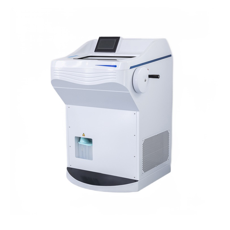
- The specimen retraction function prevents specimens from accidental blade damage. This function can be turned on or off.
- The unit can be defrosted manually or by using a controlled time setting.
- A high-quality LED screen displays all user specification data.
- The unit features high-brightness, low-power LED lights to provide stable and bright illumination.
- It includes a UV sterilization function and is equipped with a cleaner to quickly collect wastes in the chamber.
- The handwheel has 16 positions, ensuring safety.
- The cryostat is equipped with two original German Danfoss brand compressors, providing special cooling for the chamber, freezing shelf, knife blade, and specifications.
- A closed freezer cover plate ensures that cold in the cooling station is not lost.
- The fully enclosed structure simplifies and makes cleaning up sample waste more convenient, giving a more refined appearance.
Key features include:
- Fully enclosed design for the freezing chamber interior parts.
- Frame cross-ball vertical section design with the transmission part placed in the freezer chamber. The vertical stroke can be 72mm, allowing a 22mm horizontal stroke for specimens, enabling cutting of larger samples.
- The machine adopts a frame suspension structure, with the chassis shell serving only the role of protection and occlusion, ensuring stable operation during long-distance transport and precise slicing down to 1μm.
- A large-size quick-freezing shelf allows the preparation of 26 specimens simultaneously, with two holes for semiconductor refrigeration.
Specimen retraction function: prevents specimens from accidental blade damage. This function can be turned off.
The unit can be defrosted manually or by a controlled time setting.


Introduction
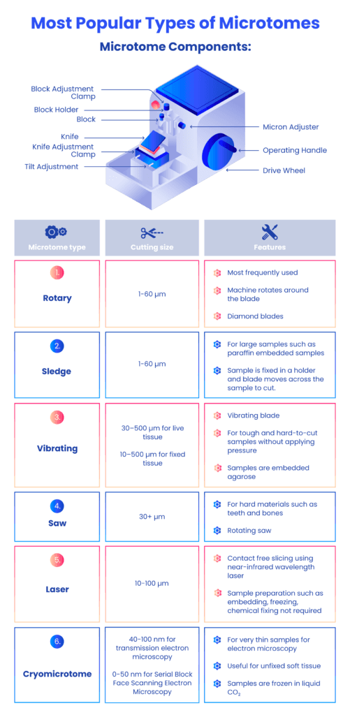
A cryostat microtome is a specialized instrument used for cutting thin sections of frozen specimens, typically for histology and pathology research. The main parts of a cryostat microtome include:
- Freezing Chamber:
- The compartment where specimens are frozen to a temperature low enough for sectioning.
- Microtome Sectioning Mechanism:
- The cutting mechanism responsible for slicing thin sections from the frozen specimen. It includes a blade holder, advance/retract mechanism, and controls for adjusting section thickness.
- Specimen Holder:
- A platform or clamp that holds the frozen specimen securely during the cutting process.
- Temperature Control System:
- Components that regulate and maintain the low temperature inside the freezing chamber. This often includes a refrigeration system, temperature controls, and sensors.
- Knife Holder and Blade:
- The knife holder supports the microtome blade, which is a sharp cutting tool that slices through the frozen specimen.
- Defrosting Mechanism:
- A system for removing ice buildup or frost from the microtome components, allowing for smooth operation.
- Display and Controls:
- User interface for setting and monitoring parameters such as temperature, cutting thickness, and other relevant settings.
- Illumination System:
- Light source to illuminate the specimen during the sectioning process, aiding visibility.
- Waste Collection System:
- A mechanism to collect and dispose of waste generated during the sectioning process.
- Vacuum System:
- Some cryostat microtomes include a vacuum system to assist in holding the specimen in place and removing debris.
- Storage Compartments:
- Spaces for storing additional blades, tools, and accessories.
Types of rotary Microtome
- Manual Cryostat Microtome:
- Requires manual operation for setting cutting parameters, advancing the specimen, and other adjustments. It is suitable for users who prefer hands-on control over the sectioning process.
- Semi-Automatic Cryostat Microtome:
- Combines manual control with some automated features, such as motorized advance/retract mechanisms or automated temperature control. This type offers a balance between manual flexibility and automated convenience.
- Fully Automatic Cryostat Microtome:
- Provides a high level of automation for various cutting parameters, including section thickness and specimen advance. This type is suitable for high-throughput applications where consistent and precise sectioning is crucial.
- Freezing Microtome:
- Specialized for cutting sections from frozen specimens. It includes a freezing chamber to maintain low temperatures during sectioning.
- Infrared Cryostat Microtome:
- Uses infrared technology to assist in the rapid freezing of specimens, providing efficient and precise sectioning capabilities.
- Digital Cryostat Microtome:
- Integrates advanced digital controls and interfaces for setting and monitoring cutting parameters. It often includes digital displays and programmable settings for improved precision.
- Cryostat Microtome with UV Disinfection:
- Equipped with a UV sterilization function to disinfect the cutting area and minimize the risk of contamination during sectioning.
- Clinical Cryostat Microtome:
- Specifically designed for clinical pathology applications, offering features tailored to the needs of diagnostic laboratories.
- Research Cryostat Microtome:
- Geared towards research laboratories, these models may have advanced features and capabilities suitable for a wide range of scientific investigations.
Strengths and Limitations
Strengths:
- Precision Sectioning: One of the key strengths is its ability to provide high-precision sectioning of frozen tissue samples. This is crucial in histology and pathology applications where thin and accurate sections are required for analysis.
- Cryogenic Cooling: The incorporation of cryogenic cooling ensures that the samples remain frozen during the sectioning process. This is particularly advantageous for preserving the integrity of delicate structures within the tissue.
- Versatility: Cryostat Rotary Freezing Microtomes are versatile instruments that can handle a variety of sample types, including soft and hard tissues. This makes them suitable for a wide range of research and diagnostic applications.
- Speed: Compared to traditional microtomes, cryostat rotary microtomes can provide quicker results due to the rapid freezing and sectioning process.
- Reduced Artifacts: The frozen sectioning process helps reduce artifacts that may be introduced by other methods, allowing for more accurate analysis of tissue structures.
Limitations:
- Cost: Cryostat Rotary Freezing Microtomes can be relatively expensive to purchase and maintain, making them less accessible for some research labs or smaller institutions with budget constraints.
- Complex Operation: Operating a cryostat microtome may require specialized training due to its complex design and the need to manage cryogenic temperatures. This can limit its use to trained personnel.
- Sample Size Restrictions: The size of the samples that can be accommodated may be limited by the dimensions of the cryostat chamber. Larger specimens may need to be processed differently.
- Maintenance Requirements: Cryostat microtomes require regular maintenance, including defrosting and cleaning, to ensure optimal performance. Failure to maintain the instrument properly can lead to issues with sectioning quality.
Summary
- Specimen retraction function for preventing accidental blade damage, with the option to turn it on or off.
- Manual or controlled time setting for defrosting the unit.
- High-quality LED screen displaying user specification data.
- UV sterilization function and waste collection cleaner for chamber hygiene.
- 16-position handwheel for enhanced safety during operation.
- Equipped with two original German Danfoss compressors for specialized cooling of various components.
- Fully enclosed design for the freezing chamber interior, simplifying cleaning and providing a refined appearance.
- Frame cross-ball vertical section design with a large stroke for cutting larger samples.
- Frame suspension structure ensures stable operation during transport, achieving precise slicing down to 1μm.
- Quick-freezing shelf for simultaneous preparation of 26 specimens, featuring semiconductor refrigeration.
- Various types of cryostat microtomes available, including manual, semi-automatic, fully automatic, freezing, infrared, digital, and those with UV disinfection.
- Clinical and research-specific cryostat microtomes catering to different laboratory needs.
References
- Tosney, K. W., & Landmesser, L. T. (1986). Neurites and growth cones in the chick embryo. Enhanced tissue preservation and visualization of HRP-labeled subpopulations in serial 25-microns plastic sections cut on a rotary microtome. Journal of Histochemistry & Cytochemistry, 34(7), 953–957. https://doi.org/10.1177/34.7.3519758
- Sharma, R. (2022). Microtomy. Retrieved February 5, 2022, from Slideshare.net website: https://www.slideshare.net/candysharma777/microtomy-67236029
- Frimiano, E M S, N, C. N., A, V. D., Sales, A., Mendes, & A, N. A. (2022). Histological study of the liver of the lizard Tropidurus torquatusWied 1820, (Squamata: Tropiduridae)(AU). Braz. J. Morphol. Sci, 165–170. Retrieved from https://pesquisa.bvsalud.org/portal/resource/pt/lil-644137
- Muhd-Farouk, H., Amin-Safwan, A., Mahsol, H., & Ikhwanuddin, M. (2017). Histological Characteristics on the Testes of Mud Spiny Lobster, Panulirus polyphagus (Herbst, 1793). Pakistan Journal of Biological Sciences: PJBS, 20(7), 365-371. https://doi.org/10.3923/pjbs.2017.365.371
- Sy, J., & Ang, L.-C. (2018). Microtomy: Cutting Formalin-Fixed, Paraffin-Embedded Sections. Methods in Molecular Biology, 269–278. https://doi.org/10.1007/978-1-4939-8935-5_23


