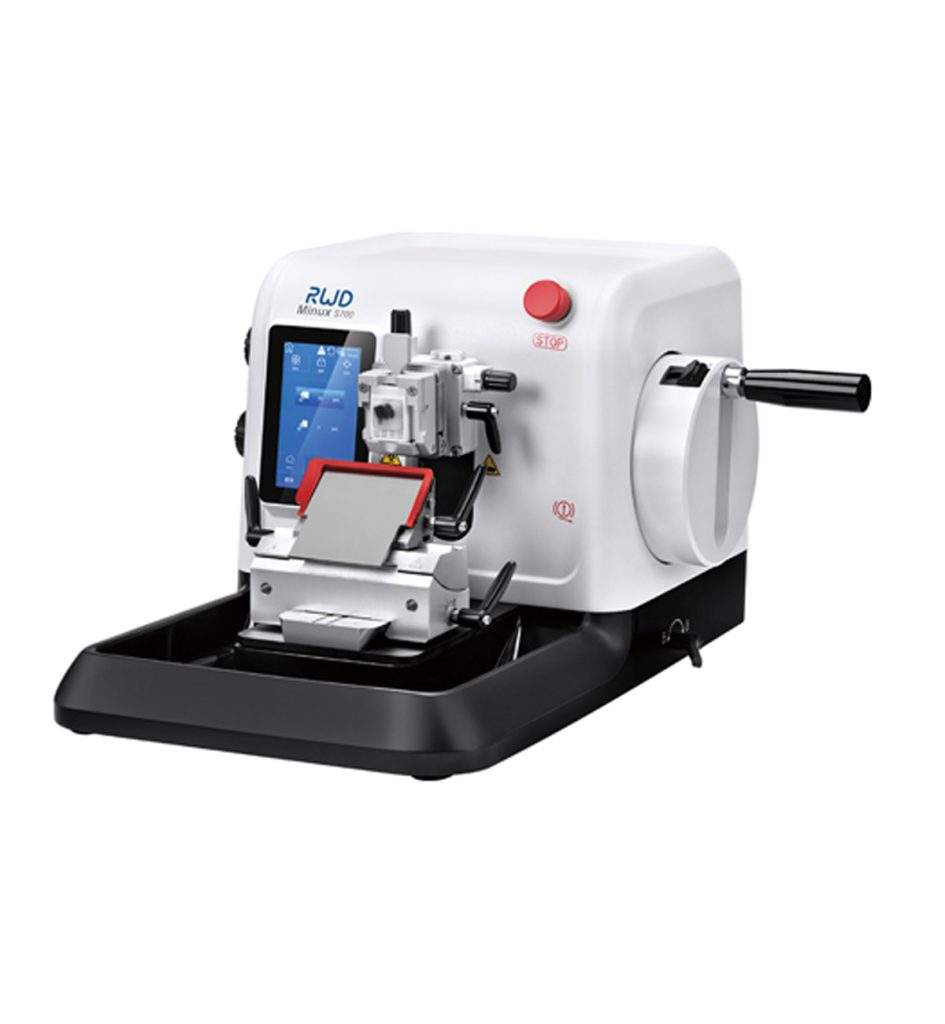
Specifications
|
S700 Semi-Auto microtome |
S700A Fully-auto microtome |
| Section thickness range: 0.5 - 100 μm |
√ |
√ |
| Trimming section thickness range: 1 - 800 μm |
√ |
√ |
| Electric coarse feed: 0 - 1800μm/s adjustable |
√ |
√ |
| Specimen retraction: 0 - 250μm adjustable |
√ |
√ |
| Horizontal feed: 28mm |
√ |
√ |
| Vertical stroke: 70mm |
√ |
√ |
| Specimen orientation: X/Y axis 8° |
√ |
√ |
| Maximum specimen size: 60*50*40mm |
√ |
√ |
| W x Dx H: 470*553*305mm |
√ |
√ |
| Machine Weight |
28 KG |
36.4 KG |
| Rock mode |
√ |
√ |
| Position memory function |
√ |
√ |
| Fully auto and manual section mode switch |
|
√ |
| Emergency stop button |
|
√ |
| Cut window |
|
√ |
| Auto section speed adjustable |
|
√ |
| 4 auto section modes: Cont., Int., Single, Multi. |
|
√ |
| Pedals (optional ) |
|
√ |
Standard configuration
|
S700 Semi-Auto Microtome |
S700A Fully-Auto Microtome |
| Host of Microtome |
√ |
√ |
| Blade holder assembly |
√ |
√ |
| Universal clamp |
√ |
√ |
| Tool set |
√ |
√ |
| Easy of cleaning section waste tray (Large) |
√ |
√ |
| User manual |
√ |
√ |
| Power cable |
√ |
√ |
| Dust cover |
√ |
√ |
| Dummy plug |
|
√ |
Features
Precision
-
- Top-level cross guide rails and precision screw rods, together with a five-phase high-resolution sampling motor and a horizontal sampling slider made of aviation aluminum, form a high-precision sampling system to ensure the quality of slices.
- Precision motor and encoder ensure stable and smooth automatic slicing.
-
- Precise tool holder lateral movement. The position of the same sample changes very little after changing the knife edge by moving laterally, and the sample can be cut within 5 knives.
- The knife holder base has scales for easy positioning of the knife holder.
Safety
- Handwheel double locking system
-
- At the same time, it is equipped with a double locking system of handwheel lock and any position lock and has its own beep sound, which significantly reduces the risk of hand injury during the sample block replacement operation, allowing you to change samples with peace of mind.
Knife guards and retractors
- Knife guards and retractors
-
- The knife guard covers the full length of the blade, avoiding the potential risk of injury during operation.
- When changing the blade, the ejector easily pushes out the blade for safe removal.
- Electronic brake and emergency stop button
-
- The electronic brake with stable performance keeps the handwheel in a stable state (after the automatic slicing is completed) to protect the safety of the operator.
- To quickly eliminate the danger, press the red emergency stop button to immediately stop the automatic sectioning.
Efficiency
-
- With the patented visual pointer mark, the angle adjustment data is intuitive and visible, when adjusting the tissue angle of different inclined planes, the direction of the sample head is accurately controlled to avoid waste of tissue samples.
- Quickly adjust multiple devices to the same angle to keep the same sample at the same angle when slicing different devices, reducing sample waste caused by rough trimming.
-
- The side injection knob is integrated with the fuselage, and the ergonomic design is more comfortable to operate.
- The injection speed can be freely adjusted between 0-1800um/s, and the operation is more efficient.
- It adopts military-grade components, which are strong and durable, with an active life of more than 20 million times.
-
- Large-capacity waste tray with magnetic suction function for easy disassembly and installation.
- Anti-static, non-waxing, easy to clean, and the cleaning time is greatly shortened.
Intelligence
-
- No need to spend time practicing slicing techniques, the machine automatically cuts out high-quality slices.
Automatic sectioning is stable and can maintain sectioning consistency. Whole-cut specimens such as embryos, brains, etc., the amount of sectioning are large and the task is heavy. Using automatic sectioning can relieve your sectioning pressure.
- Four automatic slicing modes to meet the needs of different slicing scenarios. The window cutting function ensures the quality of slices and improves the efficiency of automatic slices.
-
- The 5-inch touch screen has a history recording system, which is convenient for information retrospective tracking and quick problem-solving.

Introduction
A Rotary microtome cuts the sections of biological specimens into thin slices for use in microscopy. It gets its name because of the rotary action of the handwheel used for slicing samples. It is used to prepare thin layers of bone, mineral, teeth, and hair with section thicknesses ranging from 1-micron to 60 microns. For hard materials that use synthetic resins, they can slice up to 0.5 microns.
Parts of Rotary Microtome
The knife holder base holds the holder in place on the microtome stage. It can be moved to or away from the block, but it must remain fixed and secured during microtomy.
The knife holder comprises various parts, including the blade clamp, which holds the blade, the knife tilt, which adjusts the knife angle, and the faceplate, which directs the ribbons away from the blade and toward the operator.
The microtome body is a platform with rails that keeps the knife holder base in place.
- Cassette clamp or block holder
The block holder or cassette clamp holds the paraffin block in place. The block usually slides up and down with each revolution while the blade remains stationary. The block holder may be equipped with knobs that enable the operator to move the block face in different directions to line the tissue with the blade.
The advancement handwheel rotates in one direction, moving the block closer to the knife at the preset microns.
The coarse handwheel moves the block closer or away from the knife.
Micron adjustment is used to adjust settings for slice thickness that vary from 1 to 60 microns.
The handwheel includes a safety lock that prevents the wheel from loosening and the block holder from falling towards the blade when inserting or removing a block.
Types Of Rotary Microtome
- A Manual Rotary Microtome entirely depends on the operator, is hard to use, and is time-consuming.
- A Semi-Automatic Rotary Microtome comes with one motor, either coarse or fine wheels.
- A Fully Automatic Rotary Microtome has two coarse and advanced handwheel motors.
Principle
The Rotary microtome uses a staged rotary action where the specimen is cut with the circular motion of the handwheel. The cutting procedure is done with a knife or disposable blade placed within the instrument.
Protocol
- Embed the sample in a paraffin cartridge.
- Mount it on the paraffin block/sample holder.
- Set the distance between your knife blade and paraffin block using the coarse handwheel.
- Carefully install the blade and fix it in the knife holder.
- Set the required microns size by using the micron adjustment knob.
- Unlock the handle-equipped wheel by pushing the pin button-down located on its top.
- Revolve the handle-equipped wheel to operate the rotary microtome.
- Continue to cut until you get your desired sample.
Applications
The rotary microtome is used in various microbiological labs, including histopathology/anatomical pathology labs.
It can be used for histological examination of various organs. Frimiano et al. (2022) used a rotary microtome to cut 5mm sections of the liver of the collared lizard
Tropidurus torquatus to conduct a histological examination. Another study conducted by Muhd-Farouk, Amin-Safwan, Mahsol, and Ikhwanuddin (2017) used a rotary microtome to investigate the histological characteristics and sexual maturity sizes of the testes of mud spiny lobsters. The testes were dissected and fixed in a 10% buffered formalin solution for 11 hours, then dehydrated in 70% alcohol before being processed in a tissue processor for 18±1 hours at 60°C. On a rotary microtome, the tissues blocks were sliced at a thickness of 4 microns.
A study conducted by Tosney and Landmesser (1986) used the rotary microtome to easily visualize individual growth cones of axonal guidance in developing vertebrates. They used the rotary microtome to cut neuron slices quickly and as small as 25 microns to be visualized in an electron microscope, without laborious reconstruction. This was impossible before and hindered research in this area for a long time.
The rotatory microtome is also used in biobanking. In biobanking, formalin-fixed, paraffin-embedded tissue (FFPE) is essential. It is relatively easy to acquire and store compared to fresh frozen tissue. While preparing formalin-fixed, paraffin-embedded tissue for biobanking, the correct microtomy technique is critical to get accurate results and conduct the correct microtomy.
Sy and Ang (2018) used the rotary microtome to cut the formalin-fixed, paraffin-embedded sections.
Strengths and Limitations
The Rotary microtome is used to cut biological specimens according to the desired thickness ranging from 1 to 60 microns. It may be adapted to various types of tissue sectioning (hard, brittle, or fatty). However, it is not suitable for large tissue sectioning. Technological advancements in the rotary microtome have enhanced section quality, boosted production, and improved the technologist's occupational safety.
The incidence of repetitive motion disorders, a frequent occupational health concern in the histology laboratory, may be reduced by eliminating the manual hand-wheel operation of the microtome. Disposable blades are used, eliminating the need for sharpening and increasing sample size accuracy. Additionally, knives can be used in place of blades; they are thick and large and produce fewer vibrations during microtomy. A high-resolution motor makes a stable and smooth automatic slicing system. Its heavier size makes it stable, and it is crucial to avoid undulations in the paraffin sections. A double locking system makes it secure and prevents injuries during block handling. An emergency button is used to stop the operation in danger. It is anti-static, non-waxing, and is very easy to clean. It has four different automatic slicing modes that cover a lot of sampling situations. Daily cleaning from paraffin residue maintains a microtome cutting ideally for many years.
The heavy size of the rotary microtome makes it less portable. The disposable blades are expensive and need to be handled with care to avoid accidents. Moreover, the knives get dull if they aren't used on decalcified tissue that hasn't been cleaned.
Summary
- The Rotary microtome uses a rotary motion to cut the sections of samples used in microscopy. It is used to prepare thin layers of bone, mineral, teeth, and hair with section thicknesses ranging from 1-micron to 60 microns.
- It uses a staged rotary action where the specimen is cut with the circular motion of the handwheel. The cutting procedure is done with a knife or disposable blade placed within the instrument.
- It has various parts: knife holder base, knife holder, microtome body, cassette clamp or block holder, advancement handwheel, coarse handwheel, micron adjustment knob, and safety lock.
- It has thick and large knives that produce fewer vibrations. A high-resolution motor is used that creates a high-precision, stable, and smooth automatic slicing system.
- The rotary microtome is used in various microbiological labs, including histopathology/anatomical pathology labs.
- The blades of the rotary microtome should be handled with care. Always use cut-resistant gloves to remove the blades. Before cleaning, remove the blades from the holder and lock the rotary machine.
References
- Tosney, K. W., & Landmesser, L. T. (1986). Neurites and growth cones in the chick embryo. Enhanced tissue preservation and visualization of HRP-labeled subpopulations in serial 25-microns plastic sections cut on a rotary microtome. Journal of Histochemistry & Cytochemistry, 34(7), 953–957. https://doi.org/10.1177/34.7.3519758
- Sharma, R. (2022). Microtomy. Retrieved February 5, 2022, from Slideshare.net website: https://www.slideshare.net/candysharma777/microtomy-67236029
- Frimiano, E M S, N, C. N., A, V. D., Sales, A., Mendes, & A, N. A. (2022). Histological study of the liver of the lizard Tropidurus torquatusWied 1820, (Squamata: Tropiduridae)(AU). Braz. J. Morphol. Sci, 165–170. Retrieved from https://pesquisa.bvsalud.org/portal/resource/pt/lil-644137
- Muhd-Farouk, H., Amin-Safwan, A., Mahsol, H., & Ikhwanuddin, M. (2017). Histological Characteristics on the Testes of Mud Spiny Lobster, Panulirus polyphagus (Herbst, 1793). Pakistan Journal of Biological Sciences: PJBS, 20(7), 365-371. https://doi.org/10.3923/pjbs.2017.365.371
- Sy, J., & Ang, L.-C. (2018). Microtomy: Cutting Formalin-Fixed, Paraffin-Embedded Sections. Methods in Molecular Biology, 269–278. https://doi.org/10.1007/978-1-4939-8935-5_23


