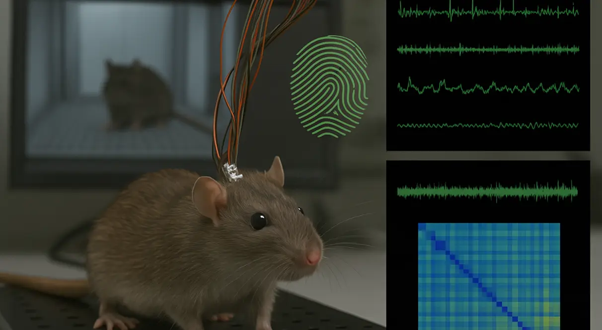
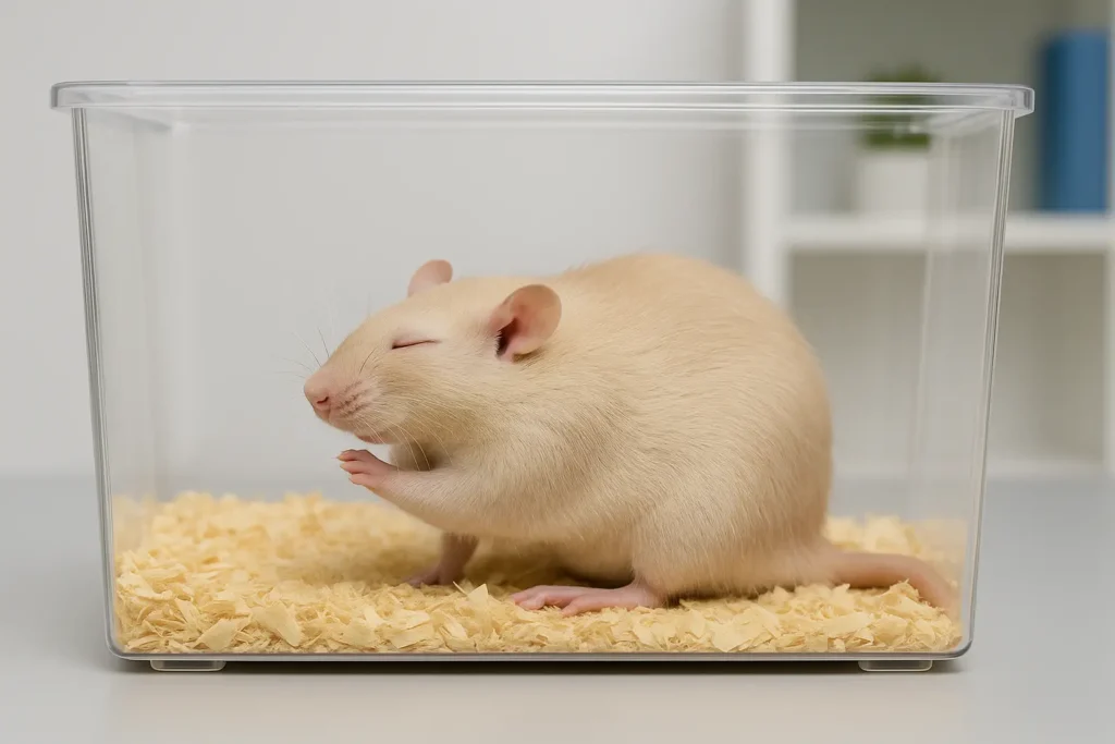
Twitching is a fundamental and well-recognized behavior in rodents, characterized by brief, involuntary muscle contractions that result in visible movements of the limbs, face, or body. This behavior can occur in various contexts, including during sleep, active exploration, stress responses, and in reaction to sensory stimuli. For researchers, twitching behavior serves as a window into the neural, genetic, and environmental factors that influence motor control and sensory processing in rodents.
This article provides a comprehensive and detailed overview of twitching behavior in rodents, exploring its types, underlying neural mechanisms, genetic influences, and environmental factors. It also highlights the significance of studying twitching for advancing our understanding of motor disorders, sleep physiology, neurodevelopmental conditions, and the effects of pharmacological agents.
Twitching behavior in rodents is a critical and versatile indicator of neural activity, motor control, sensory processing, and physiological health. Researchers study twitching behavior for multiple reasons, each providing unique insights into the functioning of the nervous system, the development of motor skills, and the impact of genetic, environmental, and pharmacological factors.
Twitching is a direct reflection of the nervous system’s ability to generate, transmit, and regulate muscle contractions. By studying twitching, researchers can explore how motor commands are initiated in the brain, transmitted through the spinal cord, and executed by muscles. Twitching provides a model for understanding the role of the motor cortex, brainstem, and spinal reflex pathways in controlling movement.
Twitching is a hallmark feature of REM (rapid eye movement) sleep, where it occurs as brief, rapid muscle contractions. Sleep-related twitching is believed to support motor learning, synaptic plasticity, and the maturation of neural circuits. Researchers use twitching as a marker for studying sleep architecture, sleep disorders, and the impact of sleep deprivation on neural function. It provides insights into how the brain remains active during sleep and how it consolidates motor memories.
Abnormal twitching is a characteristic symptom of various neurological conditions, including epilepsy, Parkinson’s disease, Huntington’s disease, amyotrophic lateral sclerosis (ALS), and multiple sclerosis. By observing and analyzing twitching behavior in rodent models of these disorders, researchers can identify neural dysfunctions, study disease progression, and test potential treatments. For example, seizure-related twitching is a key indicator of epilepsy in rodent models.
Twitching behavior is a critical endpoint in pharmacological research. Researchers assess how different drugs affect motor function, neural excitability, or muscle control by measuring changes in twitching frequency, intensity, and duration. Antiepileptic drugs are tested for their ability to reduce seizure-related twitching, while stimulants may be evaluated for their impact on spontaneous twitching. The effectiveness and side effects of drugs targeting the central nervous system can be assessed using twitching as an objective marker.
Twitching is a natural and essential part of motor development in young rodents. During early postnatal development, rodents exhibit spontaneous twitching that helps refine motor circuits, strengthen neuromuscular connections, and promote the maturation of the nervous system. Researchers use twitching as an indicator of neural development, studying how sensory inputs and motor commands are integrated. Neurodevelopmental disorders, such as autism spectrum disorder (ASD) or cerebral palsy, can be modeled using abnormal twitching patterns.
Twitching is a sensitive indicator of stress and neural excitability. Rodents exposed to stressors—such as restraint, handling, social isolation, or predator odors—often exhibit increased twitching due to heightened neural activity. Stress-induced twitching is influenced by stress hormones like corticosterone, making it a valuable model for studying the neural and hormonal responses to stress. Researchers assess stress resilience or vulnerability using twitching as a behavioral marker.
Twitching can be triggered by sensory stimuli, making it a valuable model for studying sensory processing and reflex pathways. For example, startle-induced twitching is a reflexive response to sudden sounds, bright lights, or unexpected touch. Researchers use twitching to assess sensory sensitivity, reflex strength, and sensory processing disorders, including hyperacusis or photophobia.
Genetic factors significantly influence twitching behavior in rodents. Different rodent strains exhibit varying twitching patterns due to genetic differences in neurotransmitter receptors, ion channels, muscle proteins, and neural circuitry. Transgenic rodent models allow researchers to study how specific genes, such as those associated with dopamine signaling, GABAergic transmission, or muscle contraction, impact twitching. Genetic mutations linked to neurological disorders, such as Huntington’s disease (HTT gene) or epilepsy (SCN1A gene), lead to characteristic twitching patterns.
Twitching behavior reveals the plasticity of motor circuits — the brain’s ability to reorganize neural connections in response to experience, injury, or developmental changes. Researchers studying spinal cord injury, motor rehabilitation, or neural plasticity use twitching to assess the reorganization of neural pathways and the recovery of motor function. Twitching provides insights into how the nervous system adapts to damage or training.
Twitching behavior bridges multiple areas of neuroscience, including motor control, sensory processing, sleep physiology, neurodevelopment, neurogenetics, and pharmacology. By studying twitching in rodents, researchers can explore how the brain and spinal cord generate movement, how neural circuits adapt, how sensory inputs are integrated, and how neurological disorders disrupt motor control. It serves as a versatile model for understanding how the nervous system generates, coordinates, and modulates movement.
Twitching behavior in rodents is regulated by complex neural circuits within the brain, brainstem, and spinal cord. These circuits work together to initiate, modulate, and terminate muscle contractions that manifest as visible twitching. Understanding these neural mechanisms is essential for exploring how the nervous system controls motor activity, responds to sensory inputs, and adapts to experience.
The motor cortex is the primary region of the brain responsible for initiating voluntary muscle movements. However, it also plays a role in generating involuntary twitching during REM sleep. In this context, the motor cortex generates spontaneous bursts of neural activity that result in brief, rapid muscle contractions, known as sleep-related twitching. These spontaneous motor commands are transmitted to the brainstem and spinal cord, where they trigger muscle responses.
The brainstem is a critical relay center that coordinates rhythmic motor patterns, including twitching. During REM sleep, the brainstem is highly active, generating neural signals that trigger twitching in various muscle groups. The pontine reticular formation, located in the brainstem, is particularly important for generating REM sleep-related twitching. This region communicates with the motor cortex and spinal cord to produce rhythmic muscle contractions.
The spinal cord plays a key role in mediating reflexive twitching. Sensory inputs from the skin, muscles, or joints activate sensory neurons, which transmit signals to the spinal cord. These signals can trigger reflexive muscle contractions, known as spinal reflexes, without the need for direct input from the brain. Startle-induced twitching is a prime example of spinal reflex-mediated twitching, where a sudden stimulus triggers an immediate muscle response.
Neural oscillators and central pattern generators (CPGs) in the spinal cord and brainstem are specialized neural circuits that produce rhythmic motor patterns, including twitching. These circuits generate repetitive, alternating bursts of neural activity that result in coordinated muscle contractions. CPGs are essential for producing rhythmic movements, such as walking, but they can also generate rhythmic twitching under certain conditions.
Twitching behavior is influenced by the balance of excitatory and inhibitory neurotransmitters in the central nervous system.
Sensory input is a critical factor in triggering twitching behavior. Sensory neurons detect external stimuli (touch, sound, light) and transmit signals to the spinal cord or brainstem, where they activate reflex pathways. For example:
Sleep state plays a significant role in regulating twitching behavior, especially during REM sleep. During REM sleep, the inhibitory influence of GABAergic neurons on muscle tone is reduced, allowing for brief, rapid muscle twitches to occur. In contrast, during non-REM (NREM) sleep, muscle activity is more suppressed.
The neural circuits that regulate twitching are highly plastic, meaning they can adapt in response to experience, injury, or learning. For example, repetitive exposure to sensory stimuli can enhance the sensitivity of reflex pathways, leading to more pronounced twitching. Similarly, recovery from spinal cord injury may involve the reorganization of neural circuits controlling twitching.
Twitching behavior reflects the dynamic interaction between motor and sensory systems. Motor commands from the motor cortex or brainstem can trigger muscle contractions, while sensory feedback from the muscles can modify motor output, creating a feedback loop. This interaction is essential for maintaining coordination and balance.
Hormones and neuromodulators, such as cortisol, norepinephrine, and acetylcholine, can influence twitching behavior by altering neural excitability. For example, stress-induced increases in cortisol can enhance twitching due to heightened neural activity.
Genetic factors significantly influence twitching behavior in rodents. Different rodent strains exhibit varying twitching patterns due to genetic differences in neurotransmitter receptors, neural circuitry, and muscle function. For example, transgenic rodents with mutations in the HTT gene (Huntington’s disease) exhibit abnormal twitching due to disrupted motor control.
Genetic factors significantly influence twitching behavior in rodents. Different rodent strains exhibit varying twitching patterns due to genetic differences in neurotransmitter receptors, neural circuitry, and muscle function. For example, transgenic rodents with mutations in the HTT gene (Huntington’s disease) exhibit abnormal twitching due to disrupted motor control.
Different rodent strains exhibit varying twitching behaviors due to inherent genetic differences. These variations can impact how the nervous system regulates muscle contractions, motor coordination, and reflex responses. For example:
Genetic mutations that affect neurotransmitter receptors can significantly alter twitching behavior:
Transgenic and knockout models are essential for studying how genetic alterations affect twitching:
Certain genetic mutations directly impact muscle contraction and coordination, leading to abnormal twitching:
Genetic factors also play a critical role in sleep physiology, indirectly affecting twitching:
Some rodent strains naturally carry genetic traits that result in abnormal twitching:
Environmental factors, such as diet and stress, can induce epigenetic changes that modify gene expression related to motor control:
Advanced gene-editing techniques are used to create specific mutations that influence twitching behavior:
Some genetic backgrounds predispose rodents to enhanced twitching when exposed to pharmacological agents:
Crossbreeding experiments between strains with differing twitching behaviors help identify specific gene loci responsible for variations:
Twitching behavior in rodents is not solely determined by genetic and neural factors; it is also highly sensitive to environmental conditions. Environmental factors can influence the frequency, intensity, and duration of twitching behavior, providing researchers with valuable insights into how external conditions impact neural excitability and motor control.
Stress is a potent environmental factor that can significantly increase twitching behavior in rodents. Rodents exposed to stressors such as handling, restraint, social isolation, overcrowding, or exposure to predator odors often exhibit increased twitching due to heightened neural excitability. Stress-induced twitching is mediated by the release of stress hormones, particularly corticosterone, which enhances neural activity and increases muscle contractions. Researchers use stress-induced twitching as a model to study the neural and hormonal responses to stress.
Sleep deprivation is another critical environmental factor that affects twitching behavior. Rodents deprived of sleep, especially REM sleep, exhibit increased twitching during recovery sleep (a phenomenon known as REM rebound). This increase in twitching is associated with heightened neural activity during REM sleep, where the brain compensates for lost sleep by enhancing sleep-related neural processes. Researchers use sleep deprivation studies to explore how sleep affects neural excitability and motor control.
Lighting conditions can directly influence twitching behavior in rodents. Bright lights may increase startle-induced twitching due to heightened sensory sensitivity, while darkness promotes sleep-related twitching by enhancing REM sleep. Researchers can manipulate lighting conditions to study how sensory input affects twitching and how light exposure influences sleep quality and neural activity.
Temperature is a critical environmental factor that affects muscle activity and neural excitability. Cold environments can increase muscle twitches due to shivering, which is a thermoregulatory response aimed at maintaining body temperature. Conversely, warm environments may reduce twitching by promoting muscle relaxation. Researchers study temperature-regulated twitching to understand how thermal stress affects motor control.
The social environment can influence twitching behavior in rodents. Group-housed rodents may exhibit more socially-induced twitching during social interactions, such as grooming, play, or social conflict. Conversely, isolated rodents may exhibit increased stress-induced twitching due to the lack of social interaction. Researchers use social manipulation to study how social experiences influence neural activity and motor responses.
Rodents housed in enriched environments with access to toys, nesting materials, and exploratory space often exhibit reduced stress-induced twitching and more balanced sleep-related twitching. Environmental enrichment promotes neural plasticity, reduces anxiety, and improves overall well-being, leading to healthier twitching patterns. Researchers use environmental enrichment to explore how sensory stimulation and physical activity influence motor control.
Diet can also influence twitching behavior in rodents. Rodents on high-fat or high-sugar diets may exhibit altered twitching patterns due to changes in neural excitability, metabolic rate, and body composition. Certain nutrients, such as magnesium, have been shown to reduce muscle twitching by stabilizing neural activity. Researchers study dietary influences on twitching to understand the relationship between nutrition, neural function, and motor control.
Rodents exposed to chemical agents, such as environmental toxins, pesticides, or pharmaceutical compounds, may exhibit increased or reduced twitching, depending on the neurotoxic effects of the substance. For example, exposure to neurotoxins may increase twitching due to heightened neural excitability, while sedatives may suppress twitching by reducing muscle activity.
Rodents exposed to vibration or mechanical stimulation (such as a vibrating platform) may exhibit reflexive twitching as a response to tactile stimulation. This type of twitching is used to study the impact of mechanical stress on motor control and neural excitability.
Twitching is a complex and versatile behavior in rodents that provides valuable insights into motor control, sleep physiology, sensory processing, and neurological disorders. By studying twitching behavior in rodents, researchers can explore the neural circuits that regulate motor activity, the impact of genetic mutations on muscle function, and the effects of pharmacological agents on neural excitability. Rodent models of twitching behavior are essential for advancing our understanding of motor disorders, sleep physiology, and neurodevelopmental conditions.
Written by researchers, for researchers — powered by Conduct Science.


