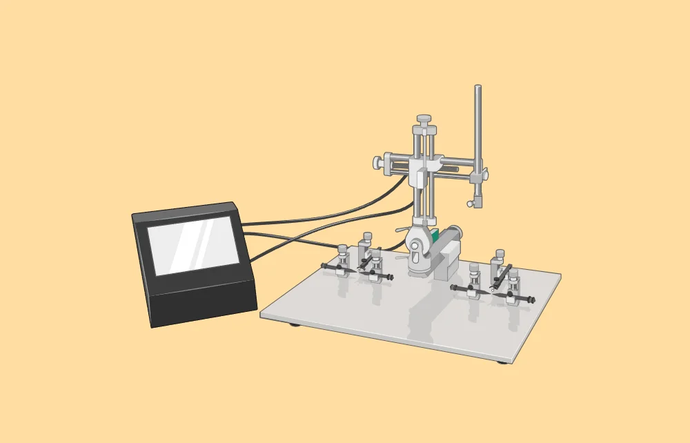

The foundation of stereotaxic surgery lies in its coordinate system. This system allows for the precise location of brain structures using three-dimensional coordinates. The two primary reference points used in this system are the bregma and lambda, which are easily identifiable on the skull. By referencing these points, researchers can navigate the brain’s complex anatomy with exceptional accuracy [1].
Misplacing probes in stereotaxic surgery often ruins experiments. These errors can happen due to experimenter mistakes or inaccuracies in the atlas. However, they are often believed to result from differences in the sex, strain, or weight of the experimental animals compared to those used to create the atlas [2].
Stereotaxic atlases are typically developed using a specific group of rats characterized by particular sex, weight, and strain parameters. Therefore, when experimental subjects differ in sex, strain, or weight from those used in the atlas, researchers must empirically determine the stereotaxic positions of each target structure [2].
Stereotaxic surgery involves multiple steps, each of which can introduce errors. Improving targeting accuracy requires minimizing errors at every stage, though the contribution of each step to overall accuracy is still unclear [3]. Here are several proposals to enhance targeting accuracy across the five main phases of the procedure:
By focusing on these proposals, researchers can improve the targeting accuracy in stereotaxic surgery, reducing both systematic and non-systematic errors and enhancing the reliability of experimental outcomes.
Targeting Specific Brain Regions
One of the primary applications of stereotaxic surgery is targeting specific brain regions. This can be achieved through various techniques, such as:
Electrode Placement for Neural Recording
Electrophysiology benefits immensely from stereotaxic surgery. By placing electrodes in specific brain regions, researchers can record neuronal activity, providing insights into how different parts of the brain communicate and process information. Accurate electrode placement relies on detailed anatomical knowledge and the use of stereotaxic coordinates [6].
Viral Vector Delivery
Stereotaxic surgery is also crucial for delivering viral vectors, which can carry genetic material to specific brain cells. This allows for the study of gene function and the development of gene therapies. Precise injection of these vectors ensures they reach the intended target without affecting surrounding tissues [7].
Optogenetics and Chemogenetics
Advanced techniques like optogenetics and chemogenetics rely on stereotaxic surgery. These methods involve genetically modifying neurons to respond to light (optogenetics) or specific chemicals (chemogenetics). Accurate placement of optical fibers or chemical injectors into the brain is necessary to control and monitor neuronal activity effectively [8].
Anatomical Navigation
Navigating the brain’s intricate anatomy requires a thorough understanding of its structure. Stereotaxic atlases, comprehensive maps of the brain, provide the necessary information for accurate targeting. These atlases include coordinates for various brain regions, allowing researchers to plan their surgeries with precision [9].
Avoiding Critical Structures
A significant challenge in stereotaxic surgery is avoiding critical brain structures, such as major blood vessels and ventricles. For example, when targeting midline brain structures, it is essential to avoid the superior sagittal sinus and third ventricle to prevent complications. This requires meticulous planning and a deep understanding of brain anatomy [9].
Stereotaxic surgery is a powerful technique that has revolutionized neuroscience research. By employing precise anatomical approaches, researchers can target specific brain regions with unparalleled accuracy. Whether it involves lesioning, implantation, neural recording, or advanced genetic techniques, stereotaxic surgery provides the tools needed to explore the brain’s complexities.
By understanding and applying these anatomical approaches, researchers can ensure that their stereotaxic surgeries are both accurate and effective, paving the way for groundbreaking advancements in neuroscience.


Dr Louise Corscadden acts as Conduct Science’s Director of Science and Development and Academic Technology Transfer. Her background is in genetics, microbiology, neuroscience, and climate chemistry.
