Introduction
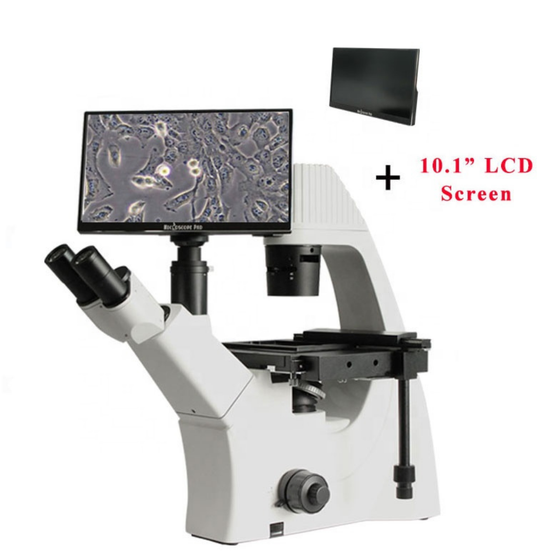
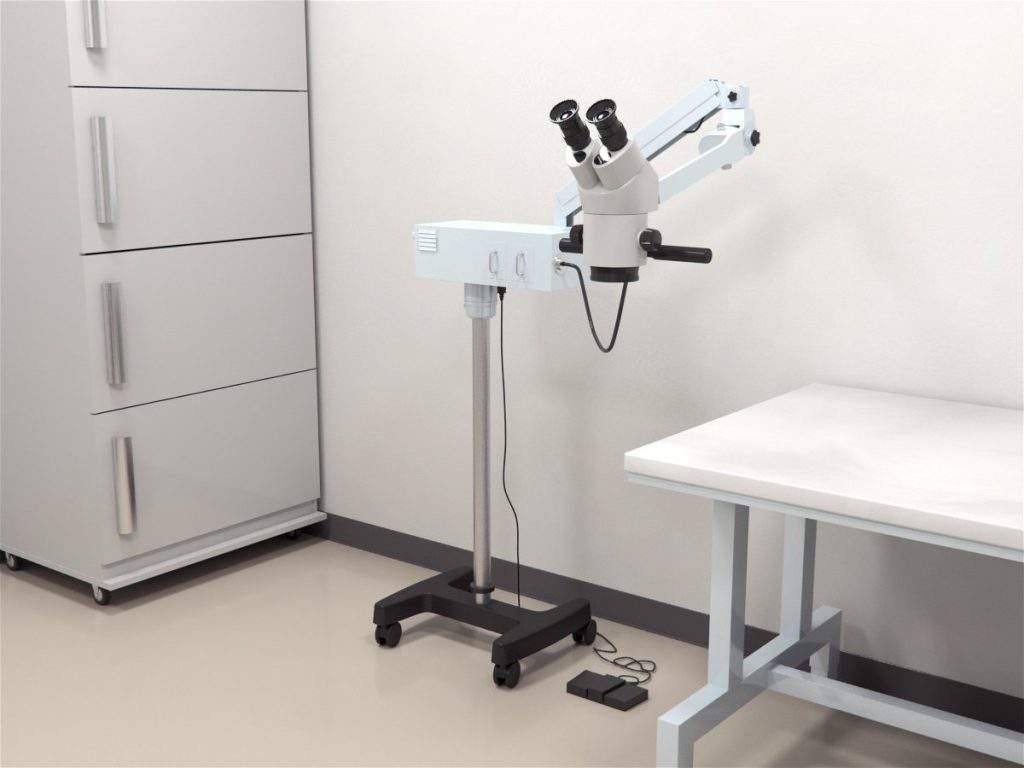
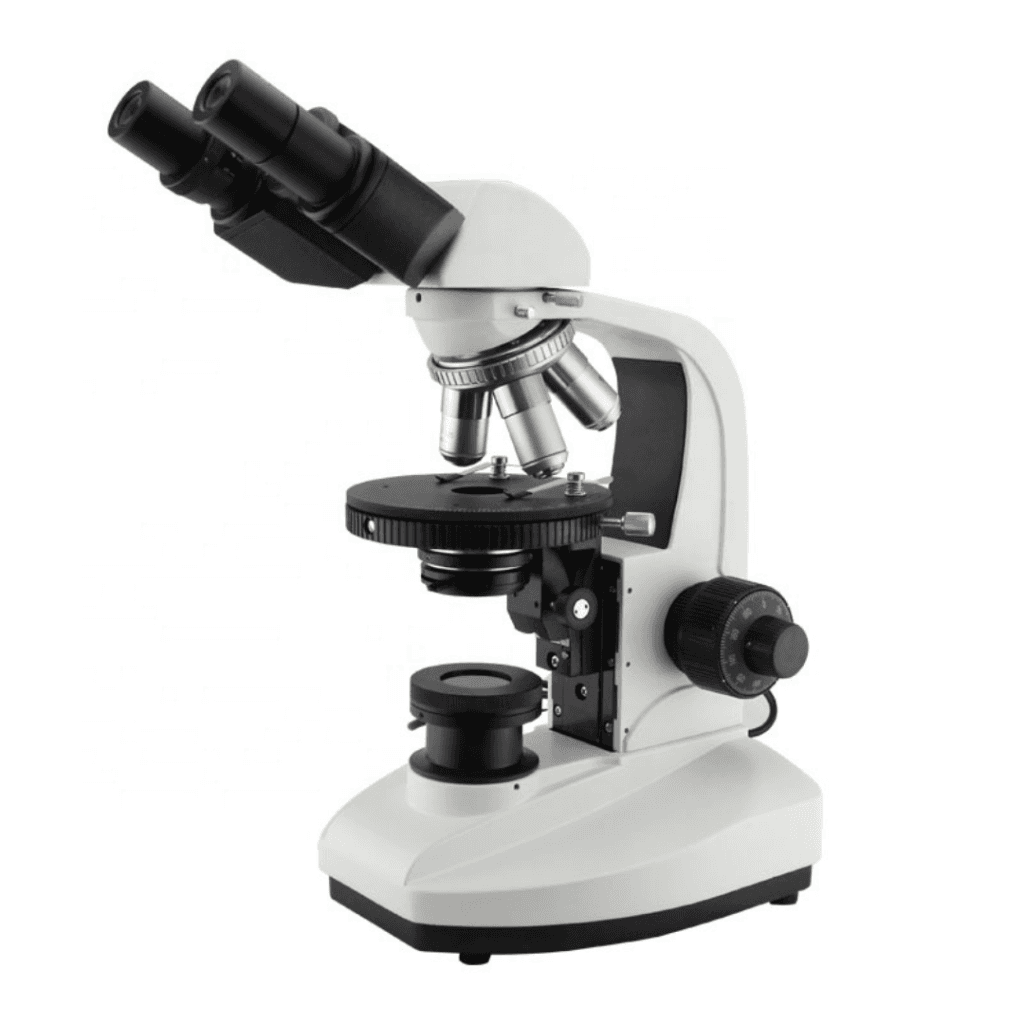
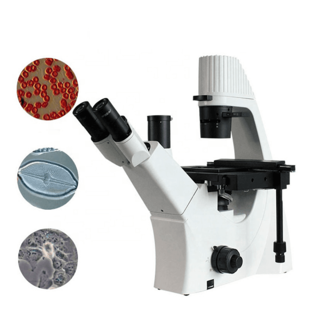
The Trinocular Phase Contrast Digital Inverted Microscope allows the study of unstained cells in medical and biological research. Phase-contrast microscopy is a technique that produces high-contrast images of translucent specimens that otherwise require staining for observation. The microscope converts refractive index differences of the sample into brightness differences, so the translucent specimen appears brighter with a darker background for proper observation.
The Trinocular Phase Contrast Digital Inverted Microscope is utilized in research laboratories, clinics, hospitals, and educational institutions. The inverted design of the microscope allows the observation of live biological tissue cultures in dishes and other specimens directly from various glass container bottoms. Therefore, the ability to observe specimens without staining them or fixing them on a slide allows them to be viewed in their natural state without needing to be killed.
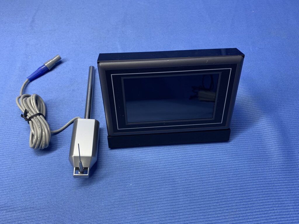
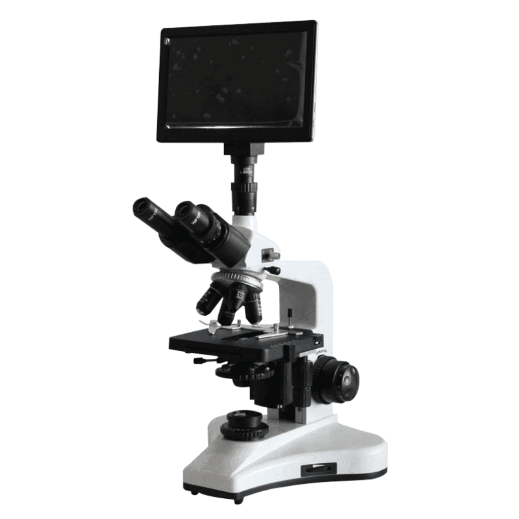
The microscope includes several advanced features. It is available and connected to a display screen or microscope camera. The digital camera produces high-quality images and captures still images and videos that you can live stream on your PC/laptop. It also comes with in-built software for Mac, Linux, and Windows to provide a multi-platform imaging solution. Additionally, you can control the image and video monitoring process easily through wireless mouse control. The microscope is also compatible with USB and HDMI output, allowing it to be connected to an external laptop or computer.
Apparatus
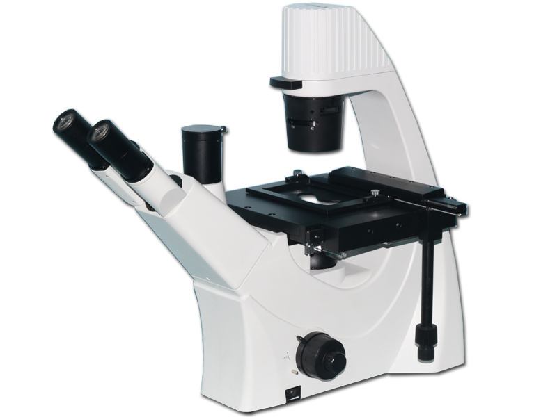
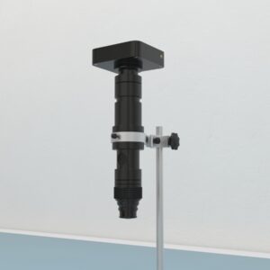
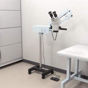
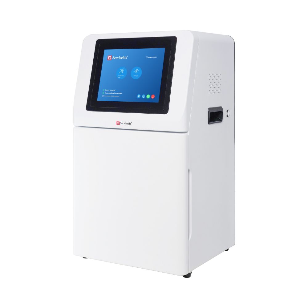
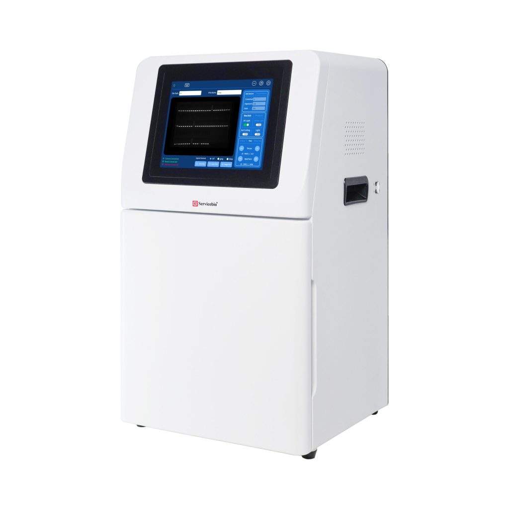
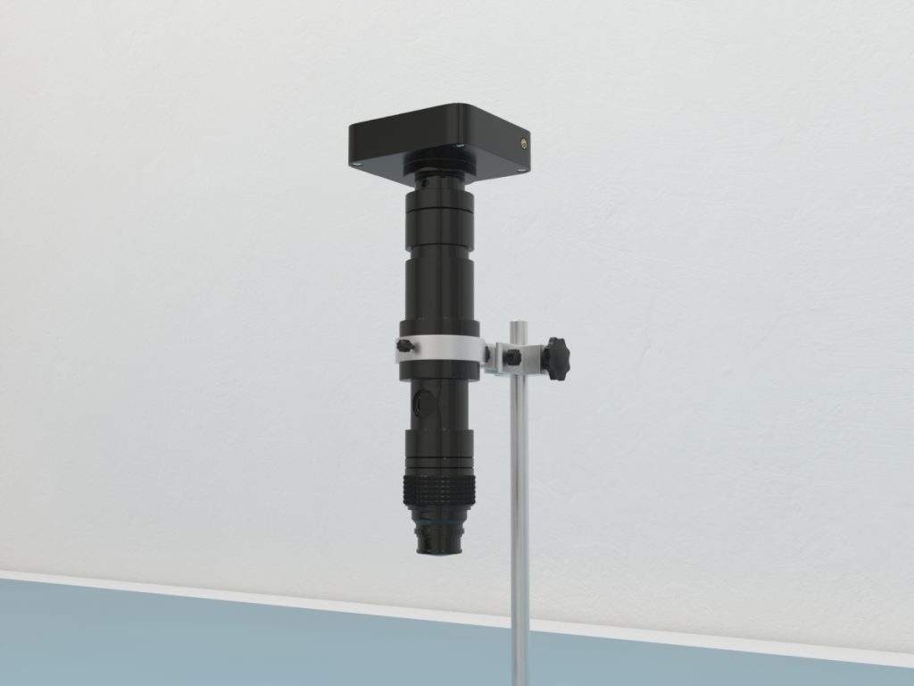
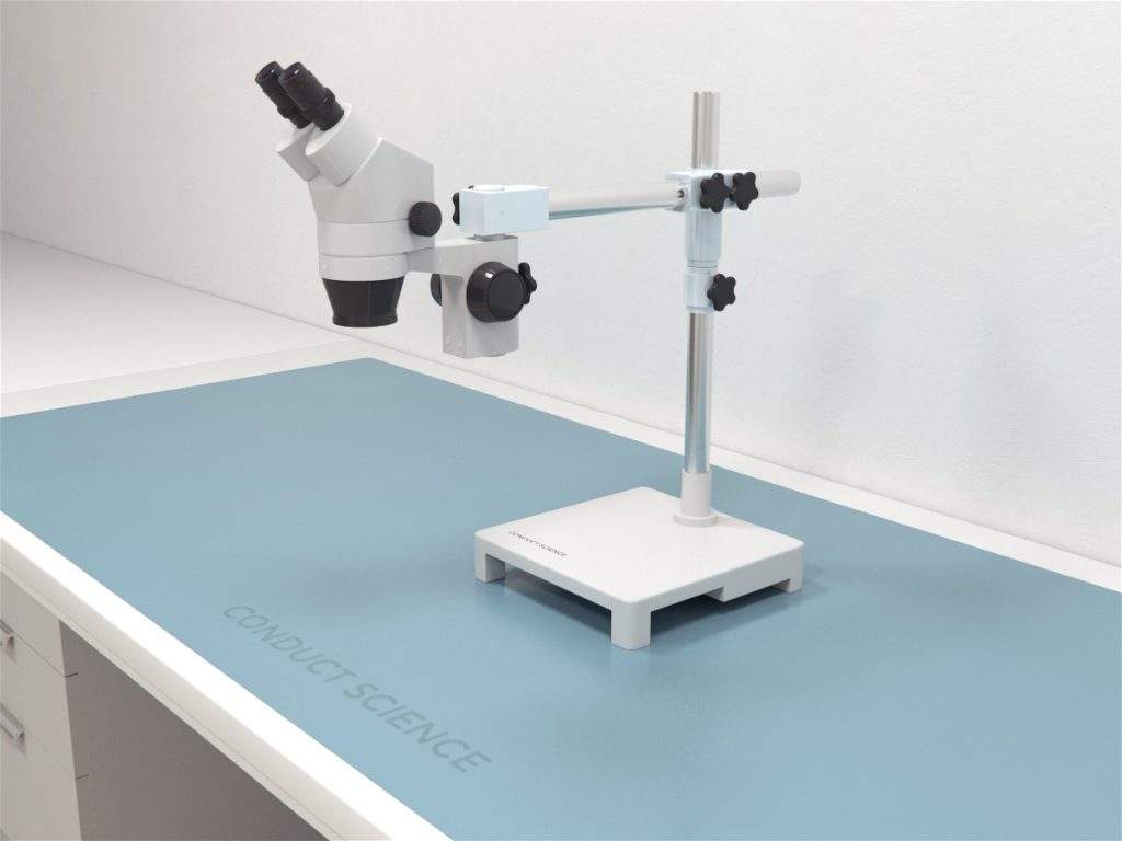
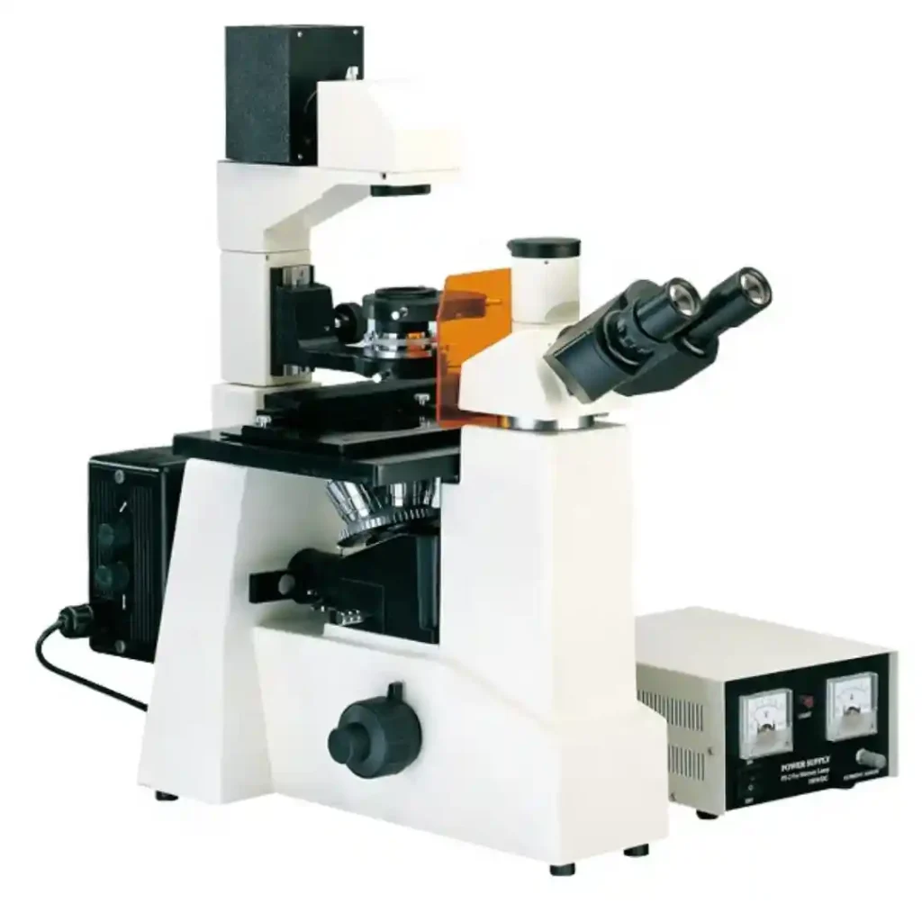
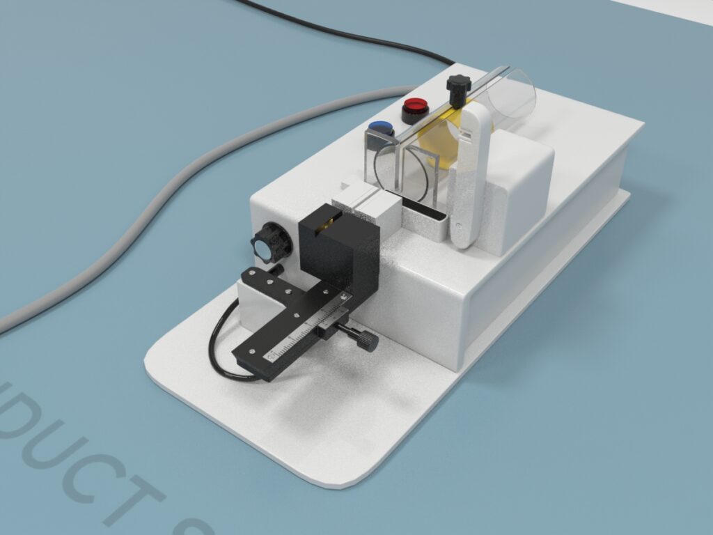
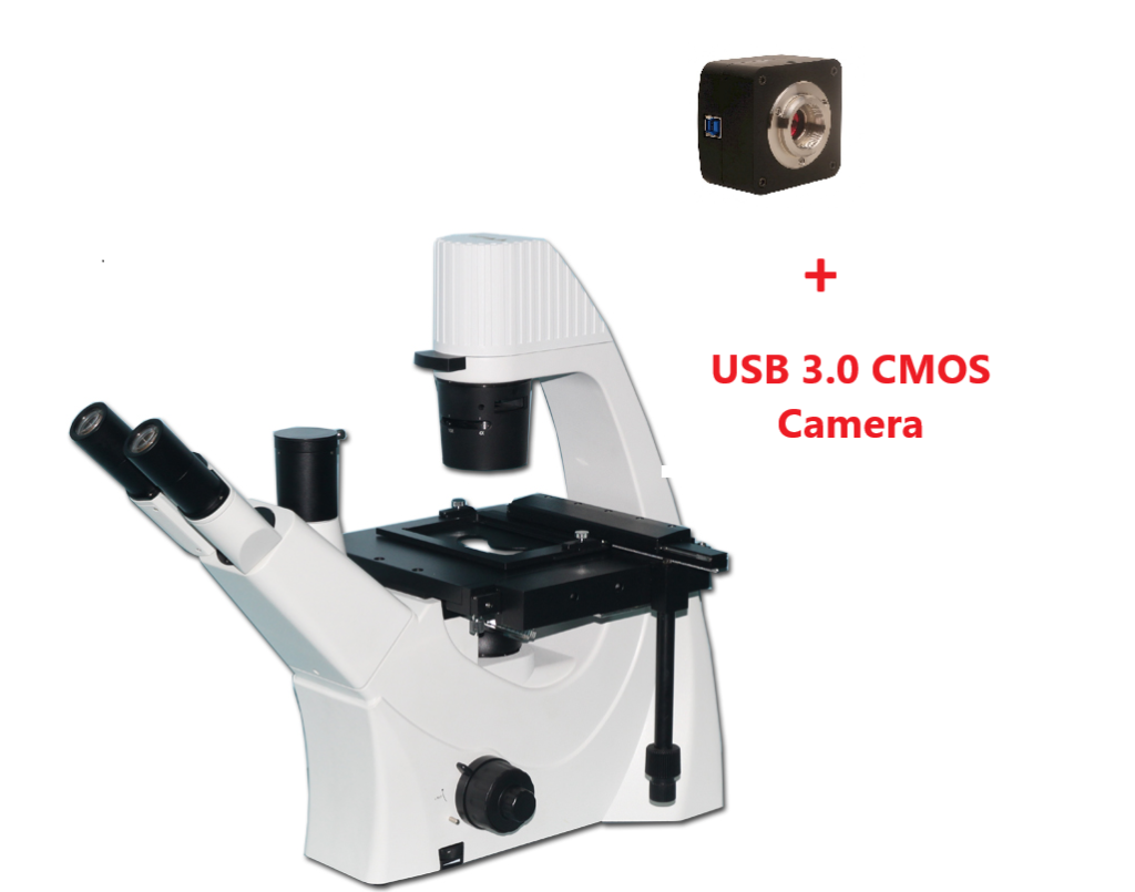
The Trinocular Phase Contrast Digital Inverted Microscope is similar in construction to the Trinocular Inverted Microscope with its components in inverted positions. However, it is available connected to a 10.1″ LCD screen that lets you view the image/video clearly and have a detailed inspection of the results. A USB & HDMI-compatible port is also present to connect it to a laptop or PC. It also includes a wireless mouse, thus making the operation even simpler.
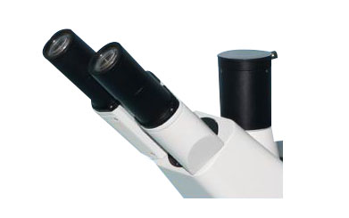
The microscope’s total magnification is between 100X-400X. It is equipped with three BF objectives (10X, 20X, and 40X) and one phase-contrast objective. The eyepiece is inclined 45°. A double-layer mechanical stage, quadruple nosepiece, coaxial coarse and fine focusing knobs, LWD condenser, 9W LED light source, and frosted blue filter are also present.
Training Protocol
Careful alignment of the microscope is needed to produce the maximum contrast effects and prevent the artifacts that degrade specimen appearance and lead to a miscalculated and erroneous interpretation. The best way is to view the image through the eyepiece while shifting the condenser annulus into and out of alignment with the objectives’ phase plate.
Before starting to align the microscope for phase contrast observation of the specimen, ensure that all objectives have the phase plates and are firmly placed in the nosepiece. Arrange the objectives on the nosepiece in sequential order, from lower to higher magnification. This sequential order will minimize the frequency changeover among different annuli on the microscope.
Applications
The phase-contrast technique combined with a trinocular inverted digital microscope is ideal for visualizing transparent specimens, microorganisms, thin tissue slices, subcellular parts like organelles, living cells, fibers, etc.
Live Intestinal Epithelial Cell Function Examination Through Time-Lapse Video and Phase-Contrast Microscopy
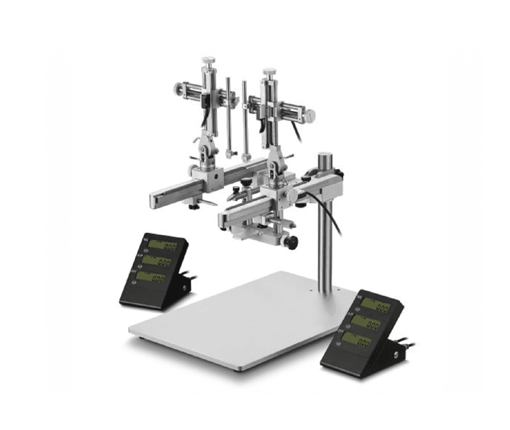
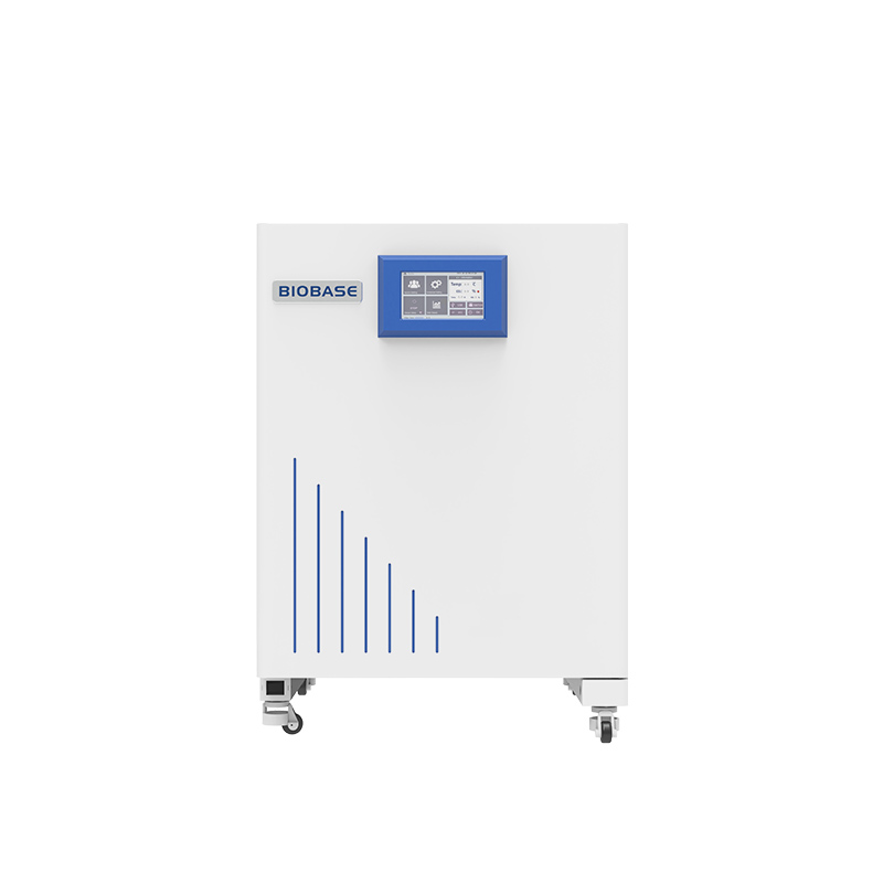
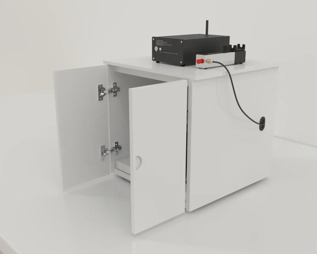
Video microscopy is the best way to understand cell physiology like cell division, migration, and membrane function. Papetti & Kozlowski (2018) used a custom time-lapse video microscopy apparatus and a trinocular phase-contrast digital inverted microscope to probe these phenomena in intestinal epithelial cells. The epithelial cells were placed in a system with controlled humidity, pH, temperature, and proliferative capacity similar to the standard tissue culture incubator for four days. The trinocular inverted microscope with phase-contrast optics and the time-lapse video microscopy apparatus helped the experimenters to observe various events in colon epithelial cells that are not visible by static imaging. The kinetics of abnormal and normal mitoses, cell migration, intracellular vesicle movements, and dynamic membrane structures were observed and identified.
Study of Microbial Cells Through the Use of Polarization and Positive Phase Contrast Microscopy
Žižka and Gabriel (2015) studied the internal structures of microbial cells through the combination of positive phase contrast and polarization. Microbial algae from different orders were collected from ponds. These were cultured in vitro under normal lighting conditions near the window at room temperature. The specimen was then observed using the Trinocular Phase Contrast Digital Inverted Microscope. The polarization microscopy showed the birefringence of the cell structures. At the same time, the positive phase contrast helped to view the fine cell structures along with a refractive index approaching the cytoplasm (where the small granules were present) and transferred the invisible phase image to the visible amplitude image.
Strengths and Limitations
Strengths
The Trinocular Phase Contrast Digital Inverted Microscope allows the transparent specimen to be observed with good image quality and contrast. It doesn’t require cells to be stained, killed, or fixed, allowing them to be viewed in their natural state. The inverted microscope setup provides the advantage of having more working distance, doesn’t require sample preparation, and allows more specimens to be viewed in a shorter period. When combined with the phase-contrast technique, it is ideal for observing thinner samples compared to upright microscopes that are good for observing thicker samples.
Limitations
The Trinocular phase-contrast digital inverted microscope often presents halos surrounding the outlines of the details with a high phase shift. So, it becomes difficult to examine the details in the boundaries. The microscope doesn’t work well for thicker specimens, as they appear distorted. Lastly, the phase annuli reduce the resolution and limit the system’s numerical aperture.
Summary
- The Trinocular Phase Contrast Digital Inverted Microscope is ideal for studying unstained cells in biological and medical research.
- It has a high-quality digital camera and 10.1″ LCD screen to view the image/video of the specimen properly.
- With an HDMI/USB port, this microscope can be connected to your PC/laptop.
- The microscope can examine colon epithelial cells, microbial cells, fibers, subcellular parts, and other thin tissue slices.
- It is a useful instrument as it offers a greater working distance for observation due to its inverted setup, and it doesn’t require the staining or killing of the specimen.
- This microscope reduces the resolution and limits the numerical aperture because of the ring or annuli.
References
- Papetti, M., & Kozlowski, P. (2018). Novel aspects of live intestinal epithelial cell function revealed using a custom time-lapse video microscopy apparatus. Cytometry. Part A : the journal of the International Society for Analytical Cytology, 93(4), 464–471. https://doi.org/10.1002/cyto.a.23334
- Žižka, Z., & Gabriel, J. (2015). Concomitant use of polarization and positive phase contrast microscopy for the study of microbial cells. Folia microbiologica, 60(6), 545–550. https://doi.org/10.1007/s12223-015-0397-8
- Spring, K.R., Parry-Hill, D. Kelly, C.D., & Davidson, M.W. (n.d.). Phase Contrast Microscope Alignment. MicrosopyU. Retrieved from https://www.microscopyu.com/tutorials/phase-contrast-microscope-alignment
- A guide to Phase Contrast. (n.d.). In Scientifica. Retrieved from https://www.scientifica.uk.com/learning-zone/a-guide-to-phase-contrast


Reviews
There are no reviews yet.