Microdialysis is an invasive surgical method to access the cranial tissues’ extracellular space. Microdialysis probes are inserted in anesthetized animals’ brains for their behavioral analysis. However, the experiments can also be performed in awake and freely moving animals. It is a sampling technique, not a measuring technique. Thus, the substance under study must be quantified from dialysate using the available high-sensitivity analytical methods (Pierce et al., 2020).
The microdialysis procedure mimics the passive function of a capillary blood vessel in which molecules show passive diffusion across the concentration gradient from an area of higher concentration to lower concentration. This technique can be used to collect or deliver the sample to the extracellular space in a process called “retro dialysis.” The dialysate can contain neurotransmitters, metabolites, metabolic precursors, or waste products. Additionally, the length of the dialysis membrane varies from 0.5-2mm. The animal is anesthetized before stereotaxic surgery, and inhalable anesthetics are preferred for this purpose as the animal can rapidly recover from their effects. These anesthetics can alter physiological parameters. For example, isoflurane administration can increase the lactate levels in the mouse brain several-fold. Injectable anesthesia is used for implanting guide cannulas, and surgery starts many days later to minimize the effect of these anesthetics. The flow rate through the probe ranges from 0.5 to 3µl/min.
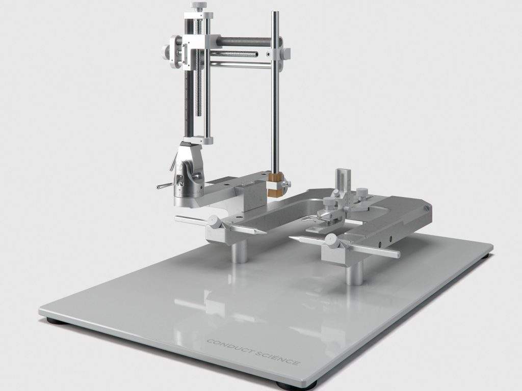
The microdialysis method is stereotaxically used to measure the level of neurotransmitters and energy metabolites. For instance, to study the effect of dopamine-releasing drugs on the subjects’ midbrain’s dopamine projections. These drugs are introduced via microdialysis probes to work precisely on specific sites. Stereotaxic surgery to implant probes or cannulas is required before the microdialysis procedure.
Microdialysis Protocol (Koeing et al., 2018)
- Once the stereotaxic surgery is complete, perfuse artificial cerebrospinal fluid (aCSF) through the porous probe. The analytes from the brain’s extracellular space will diffuse into the aCSF.
- Use two small pieces of plastic tubing to push the artificial cerebrospinal fluid through the probe and collect it post-dialysis.
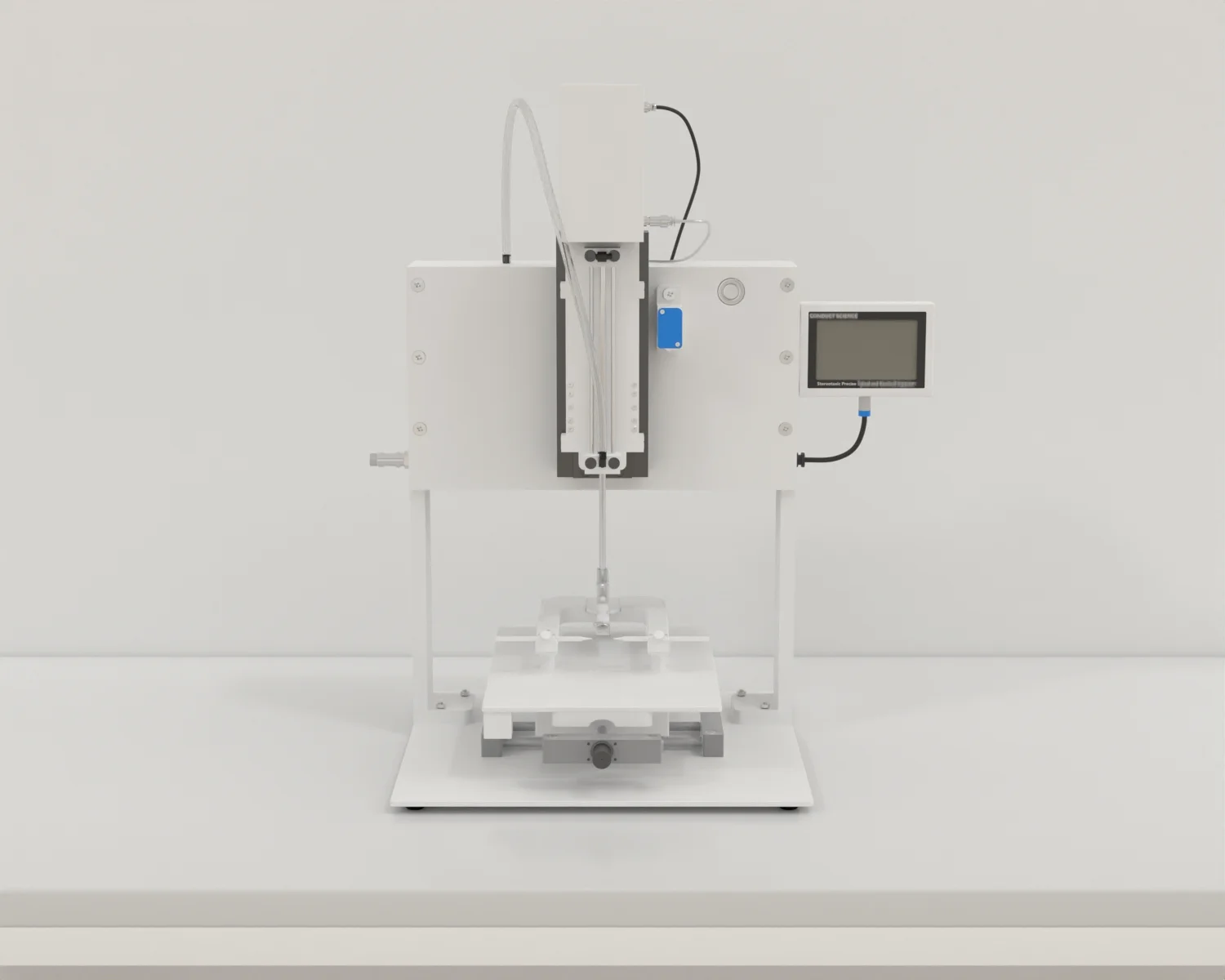
- Attach an inflow tubing to a syringe pump to push aCSF through the probe at the rate of <2.5µl/min.
- Connect the outflow tubing to a microfuge tube for collecting dialyzed aCSF.
- Attach both inflowing and outflowing tubing to a 12 inches long lightweight metal wire.
- Attach the metal wire to the tether screw at one end and a liquid swivel at the other.
- Hang the swivel on the arm in the center of the cage forming a swivel-wire-arm assembly. This assembly facilitates animal’s free movement across the cage without getting tangled in the probe.
- Habituate and equilibrate the animals for 8-10hours before sample collection.
- Collect the samples after the 6-8hours interval. The animals spend a total of 14-18hours in the cage equipped with inflow and outflow tubing without any damage.
- Provide the animals with food and water ad libitum.
 $220.00 – $384.00Select options This product has multiple variants. The options may be chosen on the product page
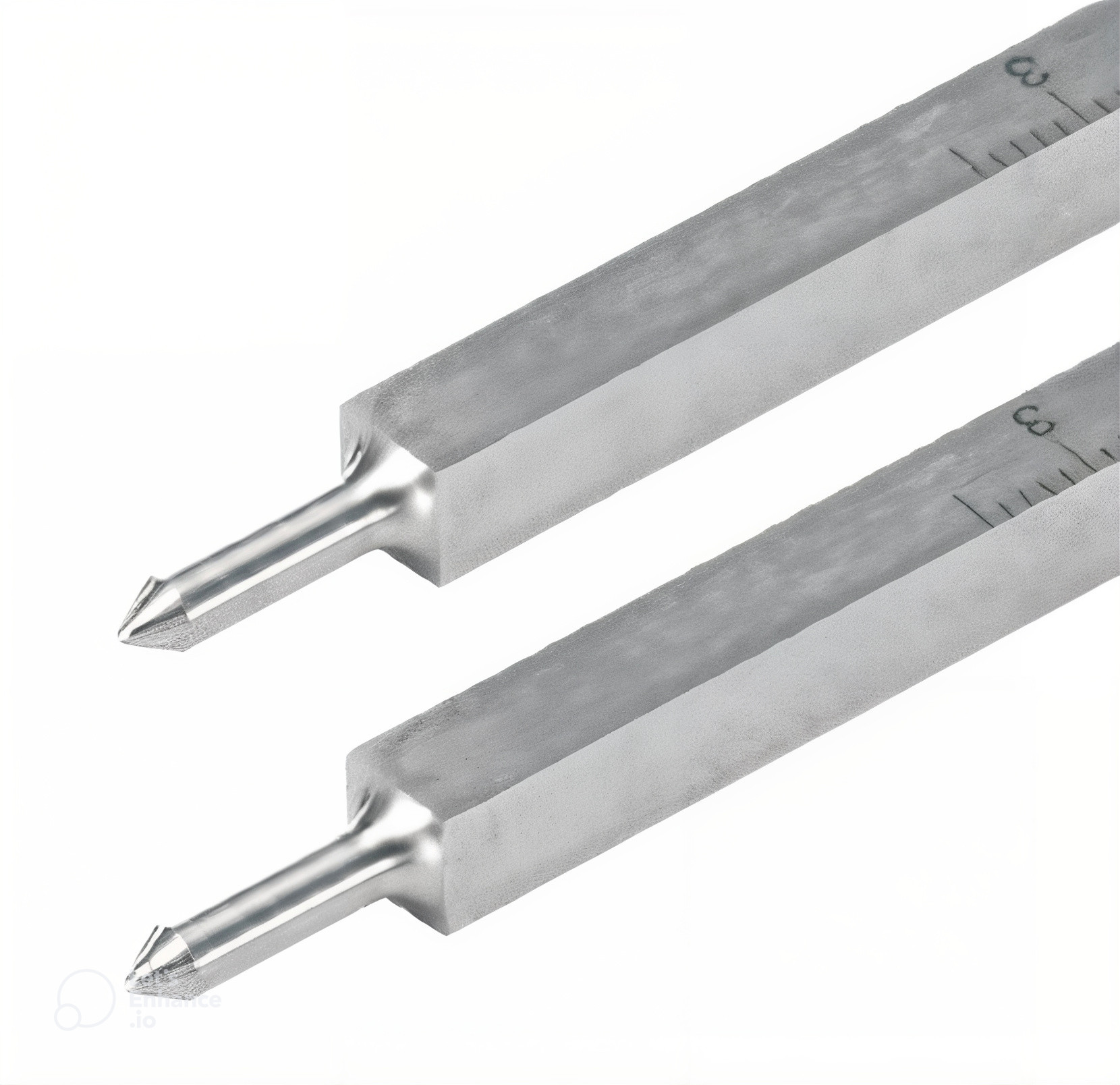 $180.00 – $495.00Select options This product has multiple variants. The options may be chosen on the product page
 $360.00 – $461.00Select options This product has multiple variants. The options may be chosen on the product page
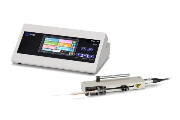
 $13,790.00 – $13,990.00Select options This product has multiple variants. The options may be chosen on the product page
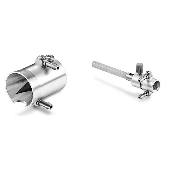
 $1,034.00Select options This product has multiple variants. The options may be chosen on the product page
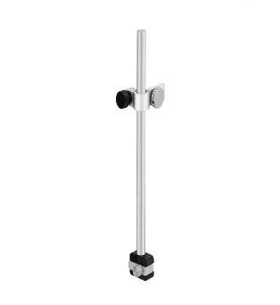 $136.00Select options This product has multiple variants. The options may be chosen on the product page
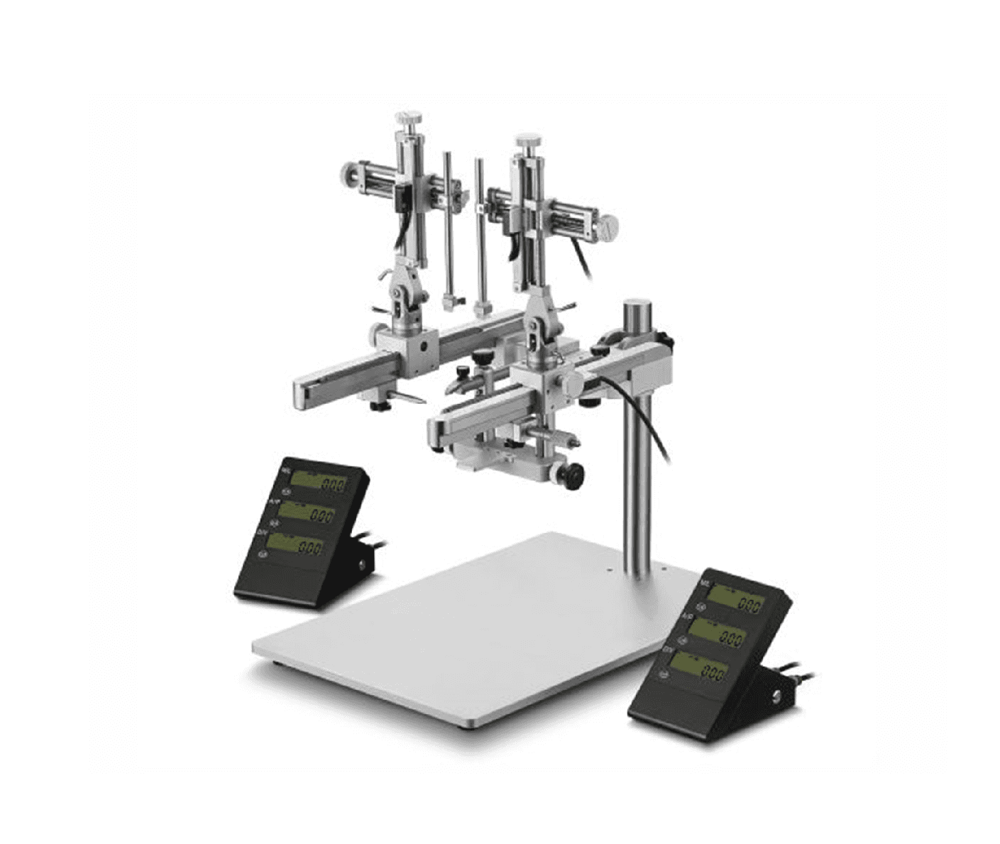 $0.00 – $8,579.00Select options This product has multiple variants. The options may be chosen on the product page


