

The biological membrane, also known as cell membrane or biomembrane, is a selectively permeable membrane that defines the boundary of the cell and separates it from the external environment.[1]
The existence of cell membranes wasn’t known until the invention of the microscope in the seventeenth century.[2] With microscopic observations, scientists came to know that organisms are composed of cells but the existence of a biological membrane was still out of the equation.
It took around 200 years (in the 19th century) to learn that there’s some form of a semi-permeable barrier separating the cell from its surrounding environment.[2] But, the composition of the biological membrane was not known until the discovery of electron microscope in the 1950s.[2]
The meticulous work and findings of several scientists like Overton, Langmuir, Gorter, and Grendel, and Davson and Danielli deduced the presence of lipids and proteins in the biological membrane.[3] The modern concept of lipid bilayer was proposed late in 1972, after a tough debate on the arrangements of these molecules in the cell membrane.[2]
This article brings you all about biological membranes, from their different models proposed by scientists, structural organization and composition, to their functions in different organisms.
Understanding all proposed membrane models in the past led to the current concept and characteristics of the fluid mosaic model. Some of the popular membrane models proposed by scientists and their significance are given below.
Evert Gorter and François Grendel hypothesized that if the plasma membrane is a bi-layer, then the surface area of the monolayer of lipids measured would be double the surface area of the plasma membrane.[3]
They supported their assumption by using the blood cells of a few mammals and measuring the total surface area of the plasma membrane of red blood cells and the area of the monolayer of lipids using Langmuir’s method.[3]
They proposed the plasma membrane structure as a lipid bilayer with the hydrophilic head of the lipid on the outside of the membrane and hydrophobic tail facing away from the aqueous environment.[3]
Apart from their proposed structure, the scientists couldn’t completely support their hypothesis. The model assumed a higher population of lipids in the membrane with no supporting ground and couldn’t describe the properties and functions of the membrane.[3]
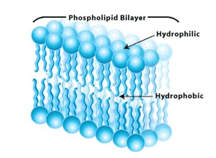
Image: Gorter and Grendel’s cell membrane model.[4]
Source: Biology4isc
Davson and Danielli proposed that the cell membrane is composed of a lipid bilayer with a thin layer of protein coating on both sides of the membrane.[3] They explained that the cell membrane contains a lipoid center covered by protein monolayers.
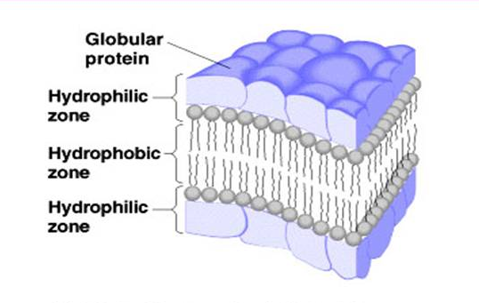
Image: An illustration of the sandwich model of the cell membrane as proposed by Davson and Danielli.[3]
Source: Biology4isc
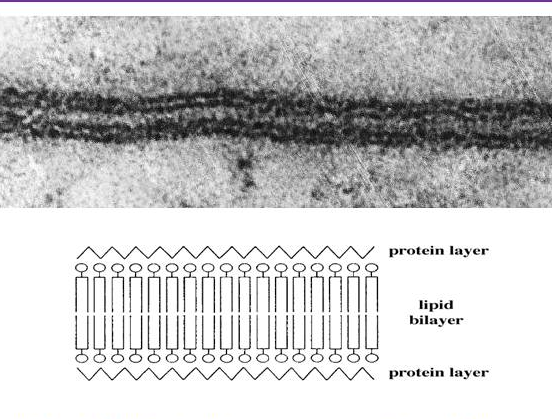
Robertson’s model supported Davson and Danielli’s sandwich model. Using high-resolution electron microscopy, he observed a trilaminar pattern of the cell membrane.[3]
From this observation, he deduced that all cellular membranes share a similar underlying structure, the unit membrane, and the membranes consist of a lipid bi-layer covered on both surfaces with thin sheets of proteins.[3]
This is the most accepted model among all the proposed models. It says that the cell membrane is composed of a lipid bilayer with proteins embedded in the membrane.
It proposes three types of proteins in the membrane in a mosaic pattern: integral protein, peripheral proteins, and lipid anchored proteins.
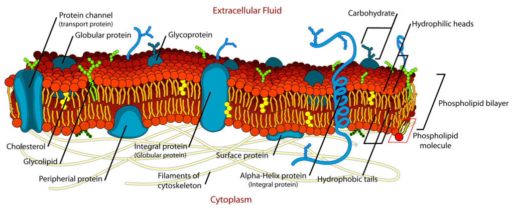
Image: An illustration of the fluid mosaic model proposed by Singer and Nicolson.[3]
Source: Wikipedia
As the fluid mosaic model proposes, the cellular membrane is composed of both lipids and proteins. All membrane structures share a common organization: a phospholipid bilayer with embedded proteins.[5] The membrane proteins are responsible for carrying out several essential functions in organisms.
Also, other than proteins and lipids, carbohydrates are a vital component of the structure. They are only present on the outer side of the cell membrane, attached by covalent bonds to some lipids and proteins. Moreover, one must know that the primary physical force that organizes the lipid bilayer is “hydrophobic force.”[5]
All membrane lipids are amphipathic – have both hydrophilic and hydrophobic ends. They constitute around 50% of the mass of most cell membranes. The proportion of lipids differs depending on the type of cell membrane. For example, the plasma membrane is composed of approximately 50% lipid and 50% proteins. Whereas the inner membrane of the mitochondria is 25% lipid and 75% protein.[5]
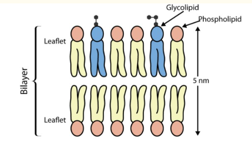
Image: An illustration of lipid bilayer showing the hydrophobic head (pink) facing an aqueous environment and the hydrophilic tail facing inwards, away from the water.
Source: Watson H. (2015). Biological membranes.[6]
Similarly, the composition of lipids in the biological membrane also differs depending on the organisms. For example, the plasma membrane of E. coli consists predominantly of phosphatidylethanolamine (constituting 80% of total membrane lipid).
In contrast, the mammalian plasma membrane comprises four different phospholipids (making up 50-60% of total membrane lipids), glycolipids, and cholesterol (which both make up the other 40%).
Below is a brief on all the three classes of lipids: Phospholipids, glycolipids, and sterols.[6]
It consists of four components: alcohol (glycerol and sphingosine), fatty acids, phosphate, and an alcohol attached phosphate.[6]
The simplest phospholipid is phosphatidic acid, in which two fatty acid residues are esterified to the OH groups at carbon atoms 1 and 2 of glycerol-3-phosphate.
Two main types of phospholipids include glycerophospholipids and sphingophospholipids.
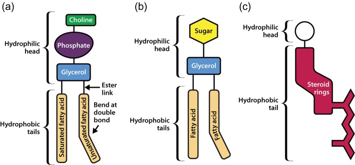
Image: An schematic representation of the structures of (a) Phosphatidylcholine, a glycerophospholipid; (b) Glycolipid; and (c) Sterol.
Source: Watson H. (2015). Biological membranes.[6]
Membrane proteins are an integral and dynamic part of the cell membrane. They account for at least half of the mass of most membranes. Their presence is essential in the membrane for signaling, communication, and several other life processes of organisms.
There are two categories of membrane proteins based on their association with the plasma membrane:[8]
These are proteins that are completely or partially embedded within the plasma membrane. They are also known as integral membrane proteins. Most of these proteins contain hydrophobic side chains that interact with the fatty acyl group of membrane phospholipids and facilitate anchoring the proteins to the membrane.[8]
One classic example of the integral membrane protein is single pass or multipass transmembrane proteins. These proteins contain one or more membrane-spanning regions (α-helices or multiple β-strands) and regions extending in the aqueous medium on each side of the bilayer.[8]
An example of single-pass transmembrane protein is RBC glycophorins, and that of multipass transmembrane proteins is band-3 proteins or chloride-bicarbonate exchange proteins of RBCs.[8]
The integrity of the transmembrane proteins with the phospholipid bilayer makes it experimentally challenging to isolate them by simple extraction procedures.
Extrinsic membrane proteins don’t span the hydrophobic core of the membrane.[8] These proteins are either indirectly bound to the membrane through interaction with integral membrane protein or directly bound to the lipid polar head group. They are bound to membranes mainly by electrostatic and hydrogen bond interactions. These proteins are also known as peripheral membrane proteins.[8]
The extrinsic membrane proteins are localized to the cytosolic face of the membrane and play an essential role in signal transduction. These proteins include cytoskeletal proteins spectrin and actin of RBCs, and protein kinase C enzyme.[8]
The extrinsic membrane proteins can be easily isolated either by using solutions of very high or low ionic strengths, solutions of extreme pH or by gentle extraction.[8]
Carbohydrates are the third major component of plasma membranes. They are attached to the outer side of the membrane while being linked to proteins (forming glycoprotein) or lipids (forming glycolipid).[9]
They also occur as polysaccharide chains of an integral membrane, called proteoglycans. These carbohydrates are composed of 2-60 monosaccharide units (that can be either straight or branched) and coat the surface of all eukaryotic cells.[9]
The lipid bilayer structure can be observed by using electron microscopy, x-ray diffraction, and freeze-fracture electron microscopy techniques. The techniques helped reveal the details of the membrane’s organization and its properties attributed to the lipid molecules.
Three such properties of the cell membrane include asymmetry, fluidity, formation of lipid rafts.
The asymmetry of the plasma membrane denotes the difference in the composition of lipids in the two monolayers of a lipid bilayer. For example, in red blood cells, almost all lipid molecules with choline—(CH3)3N+CH2CH2OH—in their head group (phosphatidylcholine and sphingomyelin) are in the outer monolayer.
While almost all phospholipid molecules containing a terminal primary amino group (phosphatidylethanolamine and phosphatidylserine) are in the inner monolayer.[10]
The asymmetric lipid distribution between the two lipid monolayers also causes a charge difference between them. At neutral pH, phosphatidylserine is negatively charged, making the cytosol side of the layer more negative than the outer side.[10] The other lipids, phosphatidylethanolamine, phosphatidylcholine, and sphingomyelin, are neutral, thus, carry no charges.
The term “fluid” in the fluid mosaic model denotes the motion of the lipids in the membrane or its fluid behavior. The membrane lipid molecules possess both rotational and lateral motion. The movement depends on three factors: temperature, lipid composition, and cholesterol.[10]
Here’s more on the factors affecting membrane permeability and fluidity.
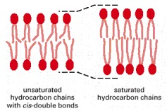
Image: An illustration of the effect of unsaturated fatty acids on membrane fluidity. The saturated fatty acids tightly pack the lipids in the membrane, thus, decreasing their fluidity.[10]
The lipid molecules in the membrane are randomly organized and connected to the neighboring fatty acids through Van der Waals attractive forces.[10] For some lipid molecules like sphingophospholipids having long and saturated fatty hydrocarbon chains, the attractive force is so strong that it links the adjacent molecules transiently in small microdomains. These microdomains are called lipid rafts.[10]
Lipid rafts are approx 70 nm in diameter and rich in sphingolipids and cholesterol. They are of two types: non-caveolar and caveolae (contain caveolin proteins). These lipids rafts are involved in signal transduction, endocytosis, and cholesterol trafficking in cells.[10]
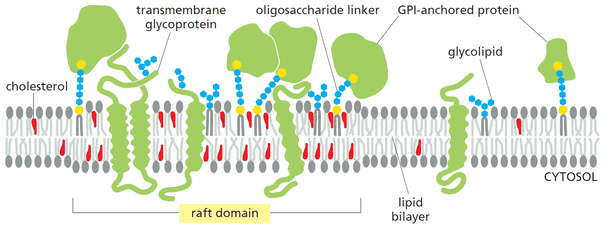
Figure: A diagrammatic representation of the lipid rafts.[7]
Source: Alberts et al. (2015) Pg. 537

Image: An illustration of types of co-transport and their examples.[6]
Source: Watson H. (2015). Biological membranes.
Biological membranes are the boundary of cells that protect them from the external environment and perform essential functions required for living organisms. The properties of the cell membranes are similarly/equally shared in both prokaryotic and eukaryotic organisms.
The bilayer of the membrane is composed of three major biomolecules: lipids, proteins, and carbohydrates. Further, the advancement in biophysical techniques and availability of substantial computational power is expanding our understanding of the lipid bilayers and providing critical insights into their structures and functions.[6]
Scientists are currently working towards understanding the properties, functions, and mechanisms of the membrane proteins, which they think might be the key to fighting deadly diseases.[6]
