Brain tissue slices taken from related brain areas are used for grasping the nature of the synaptic function in the brain.
Brain slice chambers enable examination and evaluation of the tissues by keeping them convenient for the experimental process. The tissues are exposed to oxygen and/or glucose deprivation in these chambers.
This process allows for investigating the cellular response mechanisms of the tissues. Thus, brain slice chambers offer a favorable environment for electrophysiological, ionic, and metabolic measurements. Various types of slice chambers are used in the studies based on specific applications for keeping the slice and the features. Two of them are commonly used in the studies (interface and submerged chamber types).
The main difference between these chamber types is the process of oxygen supply to the chamber. In interface chambers, oxygen is provided by the warmed, humidified gas passing over the slice.
In submerged chambers, on the other hand, oxygen is supplied by a superfusing fluid as slices are fully immersed in the chamber. However, all brain slice chambers typically aim at the same goal: to use brain tissues to examine the biological basis of brain structure and function.
Here, we will introduce semi-automated slice labs suitable for interface or submerged (sometimes even both) chamber types to allow the studies to make inferences about the related brain tissues.
What Is the Semi-Automated Slice Workstation/Lab?
Semi-automated slice workstations efficiently evaluate and observe the brain tissue slices. They enable the researchers to examine multiple slices simultaneously. Semi-automated slice workstations/labs even might allow evaluating up to 4 or 8 slices simultaneously depending on the model. They may be suitable for either interface or submerged chamber types or sometimes even both.
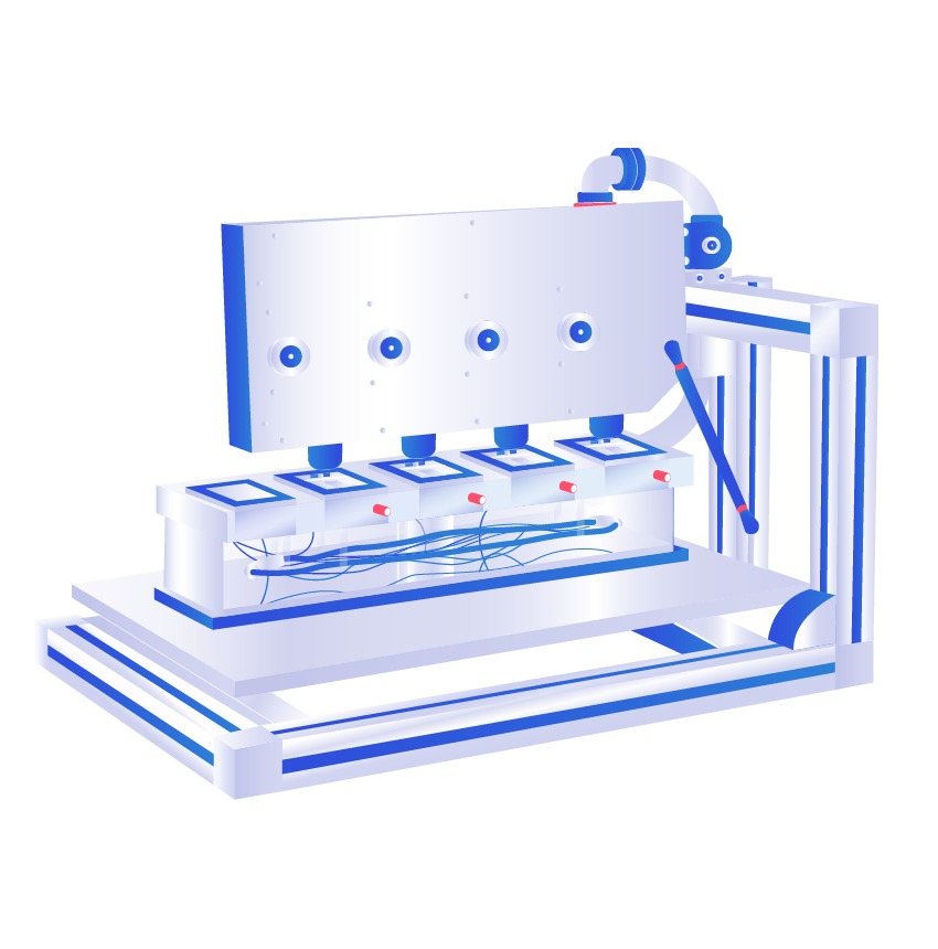
Semi-automated slices workstations generally include a support frame, anti-vibration table, camera assembly (one for 4-slices, two for 8-slices), camera power supply, quad processor, video monitor, video signal converter, and video capture software for interface to a computer.
The support frame is mounted upon an anti-vibration table and camera assembly. The camera assembly has adjustable 4 CCD color cameras for focus and can be swung upwards away from the chamber. Thus, it allows access to the chamber easily. The camera assembly is also supported by a gas spring strut and locking clamp for the raised and lowered positions.
The cameras in the camera assembly are connected to a camera power supply and quad processor. The video monitor gets its output from the quad processor. With these features, semi-automated slice workstations/labs offer favorable conditions for observing the tissues in scientific studies.
Researchers widely use semi-automated labs due to these labs’ various advantages. They help save time and make researchers work efficiently in experimental conditions. Semi-automated labs allow observation of the brain tissues with magnification up to 50x or even 100x from the chamber.
Thus, focusing on the tissues becomes more feasible. Besides, which magnification is used for the examination might be changed arbitrarily. They enable observation of the tissues simultaneously and individually, permitting more accurate results. With these superiorities, semi-automated slice workstations/labs are practical tools to investigate brain tissues in (slice) chambers.
References
- Eguchi, K., Velicky, P., Hollergschwandtner, E., Itakura, M., Fukazawa, Y., Danzl, J. G., & Shigemoto, R. (2020). Advantages of acute brain slices prepared at physiological temperature in the characterization of synaptic functions. Frontiers in cellular neuroscience, 14, 63.
- Huang, Y., Williams, J. C., & Johnson, S. M. (2012). Brain slice on a chip: opportunities and challenges of applying microfluidic technology to intact tissues. Lab on a Chip, 12(12), 2103-2117.
- Croning, M. D. R., & Haddad, G. G. (1998). Comparison of brain slice chamber designs for investigations of oxygen deprivation in vitro. Journal of neuroscience methods, 81(1-2), 103-111.


