x
[quotes_form]

Need stains for your experiments?

Introduction

Safranin is a cationic dye used in histology and cytology to distinguish and identify different tissues and cells.  It is popular in medical research for staining acidic proteoglycan that is found in cartilage tissues, enabling researchers to analyze cell chondrogenesis. The safranin is employed as a counter-stain in endospore staining and Gram’s staining.  It is mostly utilized for the identification of cartilage, mucin, and mast cell granules.

The safranin stain works by binding to acidic proteoglycans in cartilage tissues with a high affinity forming a reddish orange complex. The binding made cartilage tissues appear red when observed under the microscope. The safranin staining helps the researchers detect not only cartilage tissues but also all the body tissues and organs. The safranin stain is one of the most promising safe-lab stains used for histopathological and cytological research.


Properties

Safranin is a basic biological dye commonly used as a counter-stain in some of the staining protocols like gram staining. Following are the chemical and physical properties of safranin.

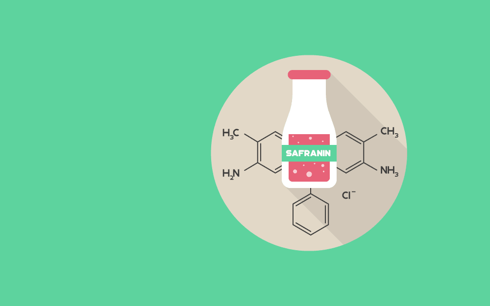
  • Chemical name: 3,7-Diamino-2,8-dimethyl-5-phenylphenazinium chloride
  • Molecular weight: 350.84
  • Molecular formula: C20H19CIN4
  • Solubility: Soluble in ethanol
  • Color: Brownish red
  • Odor: No specific odor
  • Physical structure: Powder


Staining Procedures

The following procedure should be followed for safranin staining.


To prepare the staining solution;

  1. Add 20mg safranin powder to a 100ml beaker.
  2. Pour 20ml distilled water into the beaker and make 0.1% safranin staining solution by constant stirring.
  3. Transfer 20mg of fast green dye in another 100ml beaker. Moreover, make it a 0.1% staining solution by adding 20ml distilled water in it.
  4. Filter both the staining solutions to avoid particles.


To prepare slides for observation;

  1. Hydrate the slides after deparaffinization.
  2. After washing the slides, stain them with the fast green solution for 5-10 minutes.
  3. Rinse the slides with 0.1% acetic acid for 10-15 seconds.
  4. Stain the slides with 0.1% safranin solution for 20-30 minutes.
  5. Clear and dehydrate the slides (Dehydrate in 95% and 100% ethanol)
  6. Observe the slides under the microscope for qualitative and quantitative analysis.


Gram’s staining

The Gram’s staining is performed to distinguish between two large groups of bacteria depending on their cell wall constituents. These bacterial groups are named gram-positive and gram-negative bacteria. The presence of a thick peptidoglycan layer in the gram-positive bacteria causes it to appear purple while the thinner peptidoglycan layer in gram-negative bacteria causes it to appear pink after Gram’s staining. The safranin serves as a counterstain in gram staining. The procedure followed for gram’s staining is described below:

  1. Place the specimen on the slide and heat-fix it.
  2. Add the primary stain (crystal violet) to the sample and incubate it for 1 minute. Rinse the slide with water to remove the unbound dye.
  3. Add Gram’s iodine for 1 minute (Iodine serves as mordant)
  4. Using acetone or alcohol rinse the slide for 3 seconds.
  5. Incubate the slide for 1 minute after adding the safranin stain (secondary stain).
  6. Wash the slide with water for 5 seconds.
  7. Mount the slide and observe.
 
Johansen’s method

Johansen’s method is different from the safranin staining technique. The protocol is widely used for plant tissue analysis. When plant tissues are stained with Johansen’s method the chromosomes, nuclei, lignified, or cutinized cell walls appear bright red. The staining procedure is described below:

  1. Deparaffinize the slides and bring the slides to 70% using a graded EtOH series.
  2. Stain the slides in Safranin O solution for 2-24 hours.
  3. Wash out the excess stain with distilled water.
  4. Dehydrate the slides in 95% ethyl alcohol plus 0.5% picric acid for 10s.
  5. Wash the slides for 10 s to 1 min (no longer) in 95% EtOH + 4 drops ammonium hydroxide per 100 ml to stop picric acid action.
  6. Dip the slides in 100% EtOH for 10 seconds to finish dehydration.
  7. Counterstain with fast green staining solution for 10-15 seconds. Add additional solvent to retain fast green stain as the dye evaporates with use.
  8. Rinse excess fast green solution.
  9. Wash the slides with a clearing solution by dipping for 5-10 seconds.
  10. Dip in xylene plus 2–3 drops 100% EtOH to remove the cleaning solution.
  11. Clear the slides in xylene. Repeat the step twice.
  12. Keep the slides in the final xylene solution while you mount the coverslip.
  13. Observe the tissue sections under the microscope.


Applications

The safranin staining is the most widely used staining technique for cell differentiation, cell-based assays, and stem cell culture. The safranin stain is commonly used to quantify and identify the acidic proteoglycan and glycosaminoglycan in the cartilage tissues. The safranin staining helps the researchers detect not only cartilage tissues but also all the body tissues and organs. Following are some research applications of the safranin stain.


Use of safranin for staining of frozen sections

Safranin is a cheap and safe dye that has been predominantly used to stain plant tissues for histological analysis. It was then employed to diagnose frozen sections of basal and squamous cell carcinomas accurately.  In the experiment, 1.0% lithium carbonate and a 0.5% safranin staining solution were used, which dyed the keratin a deep red, while staining the other structures like muscle cells, hair follicles, mast cell granules, and sebaceous glands pinkish red. As a result, the safranin staining provided the experimenters with high contrasting slides that are easier to analyze under the microscope for better diagnosis of frozen sections of basal and squamous cell carcinomas. The safranin staining is the most promising staining technique used for histological analysis and diagnosis of tumor cells with reasonable accuracy.

 
Safranin staining for the assessment of the selectivity of wood delignification by White Rot Fungi

In another research, the safranin staining was used to examine the Cryostat microtome sections of birch wood degraded by white-rot fungi. In the experiment, safranin was used in combination with Astra-blue. The safranin stained the lignin irrespective of the presence of cellulose, while the Astra-blue dyed the cellulose blue only in the absence of lignin. Then, the slides were observed under light microscopy. The method provided the researchers with a comfortable and reliable screening procedure to distinguish between fungi degrading cellulose and lignin, and fungi degrading cellulose only. The technique offered the understudies a detailed morphological and histological analysis of the plant cells.


Micro-spectrophotometric quantitation of glycosaminoglycan in articular cartilage sections stained with Safranin O

The safranin staining has helped the researchers to develop a new micro-spectrophotometric method for quantitation of glycosaminoglycan in cartilage tissues. The dye content of the tissues was proportional to the amount of glycosaminoglycan in the cartilage matrix. With the help of gas chromatography and thin-layer chromatography, the fixed negative charge contained in the analyte was determined. Safranin staining is the most commonly used method to detect and analyze cell chondrogenesis.


Gram’s staining

The safranin is also used as a counter-stain in Gram’s staining. In Gram’s staining, the safranin directly stains the bacteria that has been decolorized. With safranin staining, gram-negative bacteria can be easily distinguished from gram-positive bacteria.

The above-mentioned research applications of the safranin stain show that the scope of safranin staining is not only limited to plant histology, but also microbiology, oncology, stem cell differentiation, and arthrology.


Strengths and limitations
  • The safranin stain is widely used in in-vitro diagnostics. Following are the advantages and limitations of using safranin staining in biomedical research.
    • The safranin staining is the most popular counter-stain used in medical laboratories.
    • The safranin stain is a cheaper and safer-lab stain.
    • It is a certified stain for chromosomes.
    • It can be used to stain animal as well as plant cells for better cytological and histological analysis.
    • It allows easy and rapid detection of the plant’s vascular tissue.
    • It offers improved accuracy in the diagnosis of frozen sections of tumor cells.
    • It helps the microbiologists stain the decolorized gram-negative bacteria during Gram’s staining.
    • It enables the researchers to perform qualitative as well as quantitative analysis of proteoglycan in the cartilage matrix in the diagnosis of articular diseases.
    • In diseases where glycosaminoglycan loss is severe, safranin is not a sensitive indicator.


Precautions
  • Following precautionary measures should be taken while using the safranin stain.
    • Keep the safranin powder away from heat and hot surfaces as safranin is flammable.
    • Wear gloves and a face mask while handling the safranin solution as it can cause severe eye damage and skin irritation.
    • Store the powder in a cold or well-ventilated place.
    • Wash hands and the activity area before and after the staining procedure.
    • Safely dispose of the waste following the lab regulations.
References
  1. C. Shabman(2009) Tannic Acid and Iron Alum with Safranin and Orange G in Studies of the Shoot Apex, Stain Technology, 18:3, 105-111, DOI: 10.3109/10520294309105799
  2. KL, C., & SA., A. (1988). Limitations of safranin ‘O’ staining in proteoglycan-depleted cartilage demonstrated with monoclonal antibodies. Histochemistry, 89(2), 185-8.
  3. Srebotnik, E., & Messner, K. (1994). A simple method that uses differential staining and light microscopy to assess the selectivity of wood delignification by white rot fungi. American society for microbiology.
  4. Ruzin, S. E. (1999). Plant Microtechnique and Microscopy. OXFORD UNIVERSITY PRESS.
  5. Tran, D., Golick, M., Rabinovitz, H., Rivlin, D., Elgart, G., & Nordlow, R. (2000). Hemotoxylin and Safranin O Staining of Frozen Sections. Dermatol Surg, 26, 197-199.