Pig Mazes
Overview
The porcine animal model is one of the popular non-rodent models used in research. As opposed to the traditional animal models such as mice and rats, pigs as an animal model provide the advantage of representing the complexity of physiology associated with human diseases. Pigs share a lot of similarities with the human anatomy and genetics, making them a suitable model for research. Further, the similarities allow a greater translatability potential of research results into human clinical applications. Additionally, their large size makes surgical and other invasive procedures significantly easier in comparison to rodents and fishes.
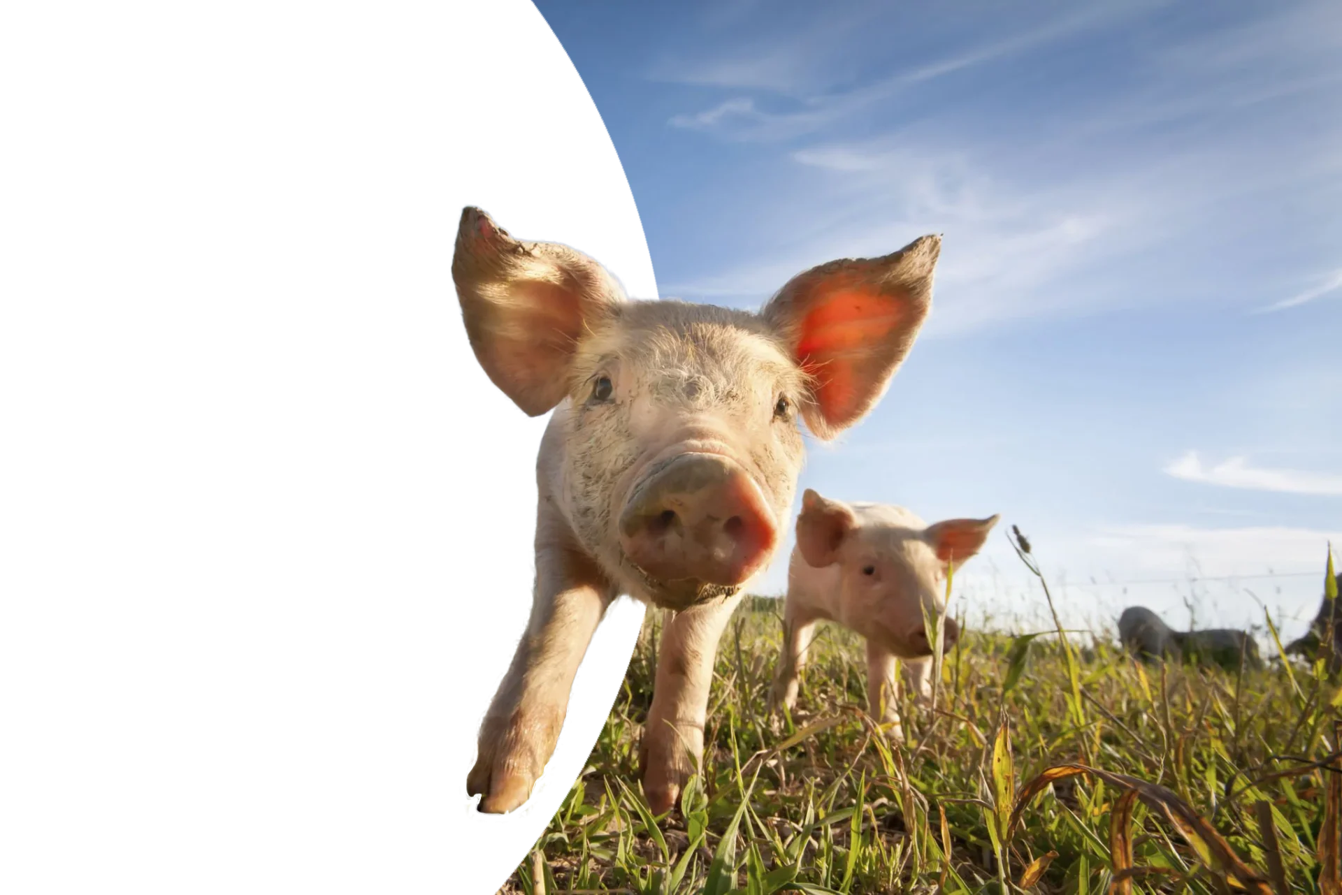
Pigs Behaviors and Characteristics
Scientific Name: | Sus Scrofa Scrofa |
Family Name: | Suidae |
Habitat: | Can survive in any productive habitat (most cases) |
Weight: | Anywhere from 10 kg to 300 kg or more |
Diet: | Omnivores |
Sexual Maturity: | 3 to 12 months (females) |
Gestation Period: | 112 to 120 days |
Litter size: | 6 to 14 piglets |
Nesting: | During the last 24 hours before the onset of farrowing; the sows begin nest building by rooting the ground to create a depression and building the nest using twigs; grasses; and leaves. |
Herding: | Pigs are very social animals and are often seen in sounders; which typically consist of two or more females and their young ones. Males can often be seen living solitary or in bachelor groups |
Hierarchy: | Dominance hierarchy can form as early as 1 week of age (confined pigs). Piglets develop teat order which once formed remains stable for the same piglet group. |
Communication: | Pigs communicate using vocalization. Rooting is also considered a form of communication. Piglets begin socialization approximately at 5 weeks of age with conspecifics and by 14 weeks of age with other species |
· Pigs have relatively poor vision. They have a panoramic vision of approximately 310° and binocular vision of 35° to 50°.
· Pigs have a highly developed olfactory sense. Sows can detect lower concentrations of smell and pheromones better than boars.
· Pigs have a well-developed hearing sense.
· Pigs lack in sweat glands. Thus, they indulge in wallowing to thermoregulate their body temperatures.
Other behaviors & characteristics
· Pigs are cognitively complex animals. They can indulge in destructive behaviors if bored.
· Pigs have a strong fear of being picked up. However, with regular interactions, they are known to be amenable.
History
Pigs were primarily used for food, though, pig-derived products such as their hides, bones, and bristles were also used for building tools and other products. The domestication of pigs is believed to have taken place around 8500 BC in the Near East. The pigs were introduced into Europe by agriculturalist migrating into northwestern Europe between 5500 and 4200 cal BC. (Caliebe, Nebel, Makarewicz, Krawczak, & Krause-Kyora, 2017; Giuffra et al., 2000; Krause-Kyora et al., 2013)
Interest in pig selection and breeding was mainly for improving their economic value as a food source. By the mid-20th century, pigs began to be considered as an alternative animal model in biomedical research. Initially, domestic farm breeds of pigs were used in research. However, their large size meant extensive husbandry and maintenance requirements leading to more expenses as opposed to the traditional animal models such as rodents. By the late 1940s, the development of minipig breeds was underway. These breeds were developed to be smaller sized, docile and easy to manage. Further, the crossbreeding of minipigs also involved the development of breeds with specific characteristics for biomedical research (Larzul, 2013).
By the early 1980s, pigs gained momentum as a popular animal model, especially as a preclinical model. Their physiologic, anatomic, and pathologic similarities to humans make them an excellent model for human diseases, disorders, and conditions. Additionally, the porcine genome project also facilitated its use as an animal model (Abbott, 2012).
Breeds
Pigs are natives of the Eurasian and African continents. Pigs belong to the Suidae family and fall under the Sus genus. The origins of the domestic pig can be traced to the Eurasian wild boar (Sus scrofa). In the United States, popular domestic farm breeds for research include Duroc, Yorkshire, Landrace, and crossbreeds while miniature breeds include Yucatan mini and micro, Hanford, Sinclair, and Göttingen.
Domestic Farm Breeds
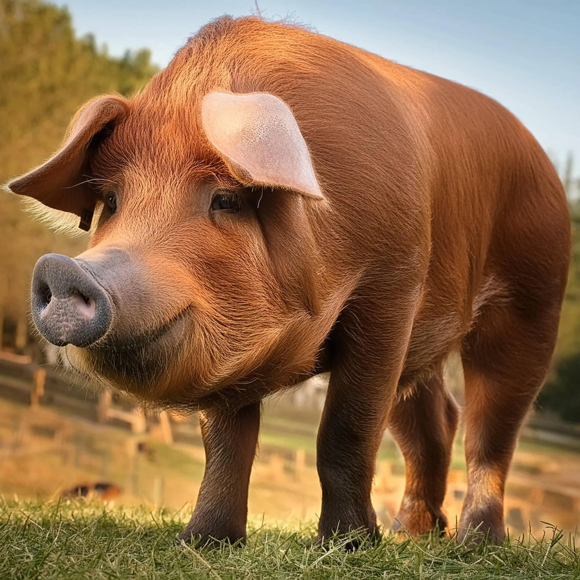
Duroc Pig
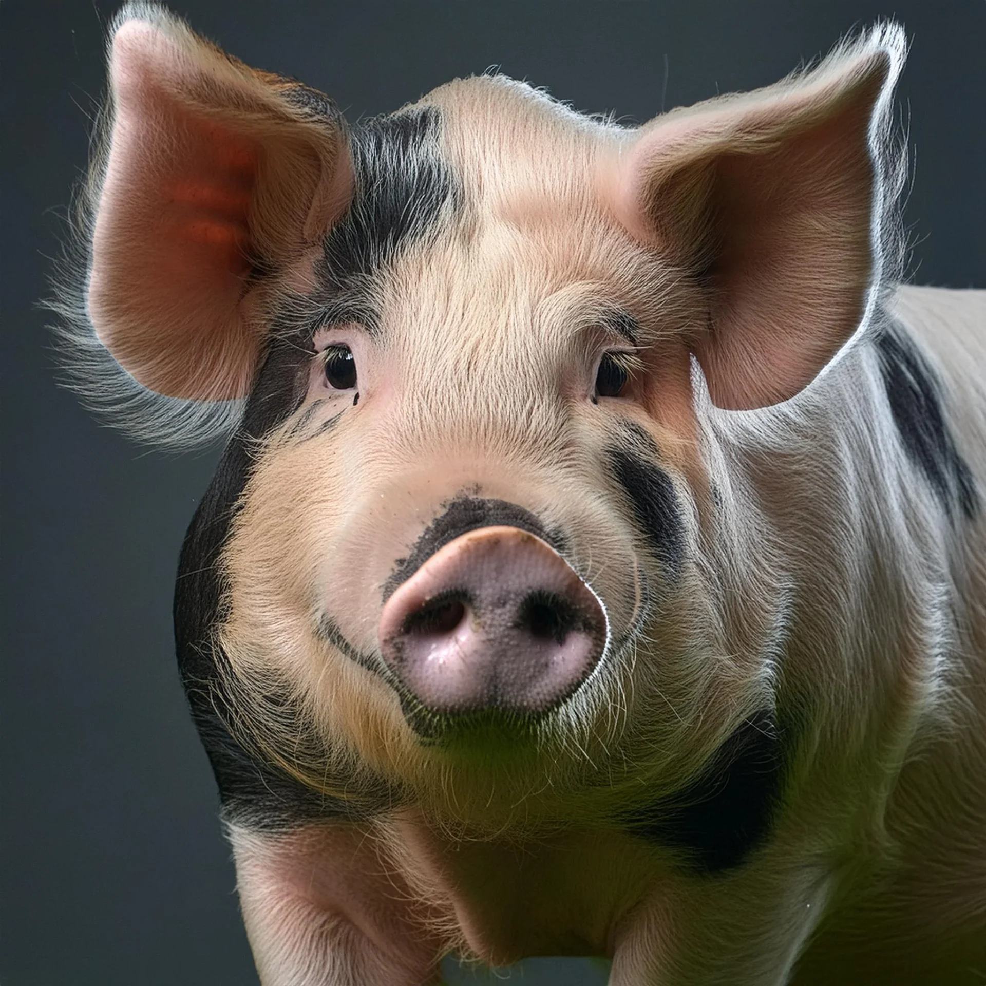
Yorkshire Pig
The Yorkshire pig was developed in Yorkshire, Northern England in 1761 as the Large White pig. The breed is a cross between large indigenous white pig of North England and the smaller, fatter, white Chinese pig. Around 1830, the Yorkshire breed was introduced to the United States in the Ohio state. The American Yorkshire pigs are white in color (sometimes with dark areas) and have erect ears, and short pugged noses. The pigs are rough and strong with good adaptability to climate and other ecological factors. Sows are known to have an average litter of 13 piglets.
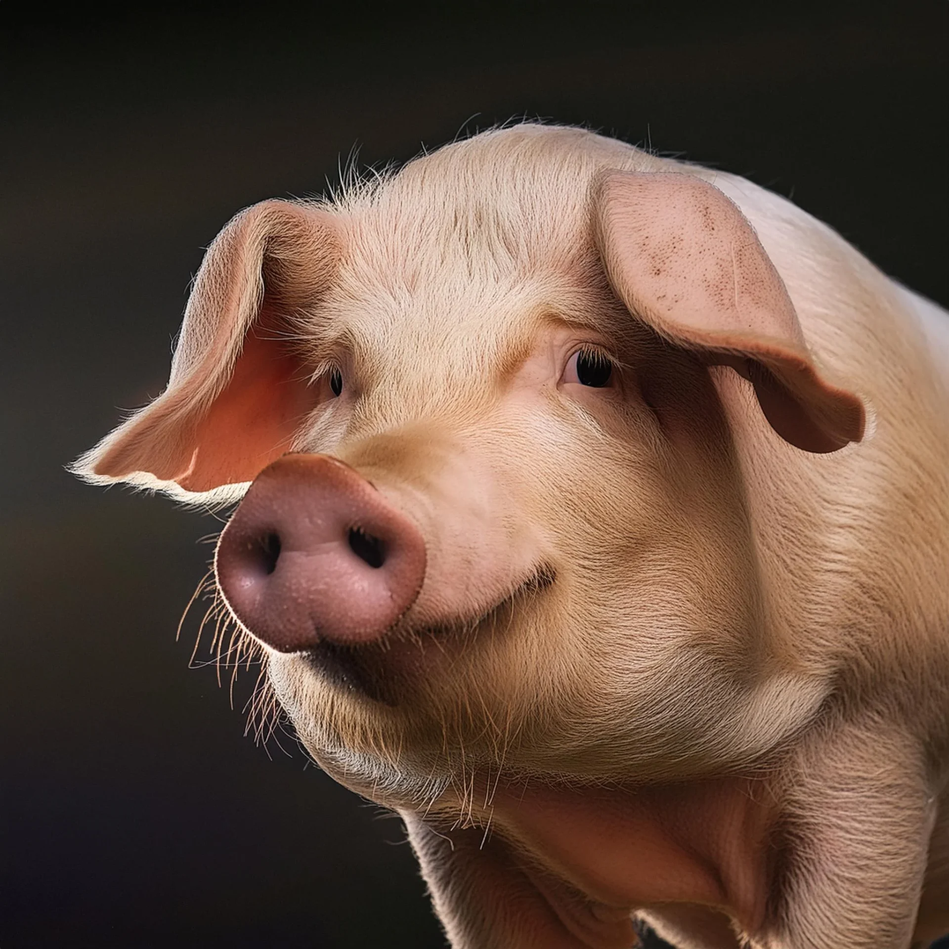
Landrace Pig
Landrace strain was first developed in Denmark around 1895 as a cross between the native Danish pigs and the Large White from England. The breed was introduced into the United States in the early 1830s. The Landrace pigs have white coloration often with small black spots. The American Landrace pigs are long and lean with a long and narrow head. The ears are large and heavy, drooping forward with the top edges almost parallel to the bridge of a straight nose. Sows of the breed are known for producing plenty of milk and raising large litters.
Miniature Breeds
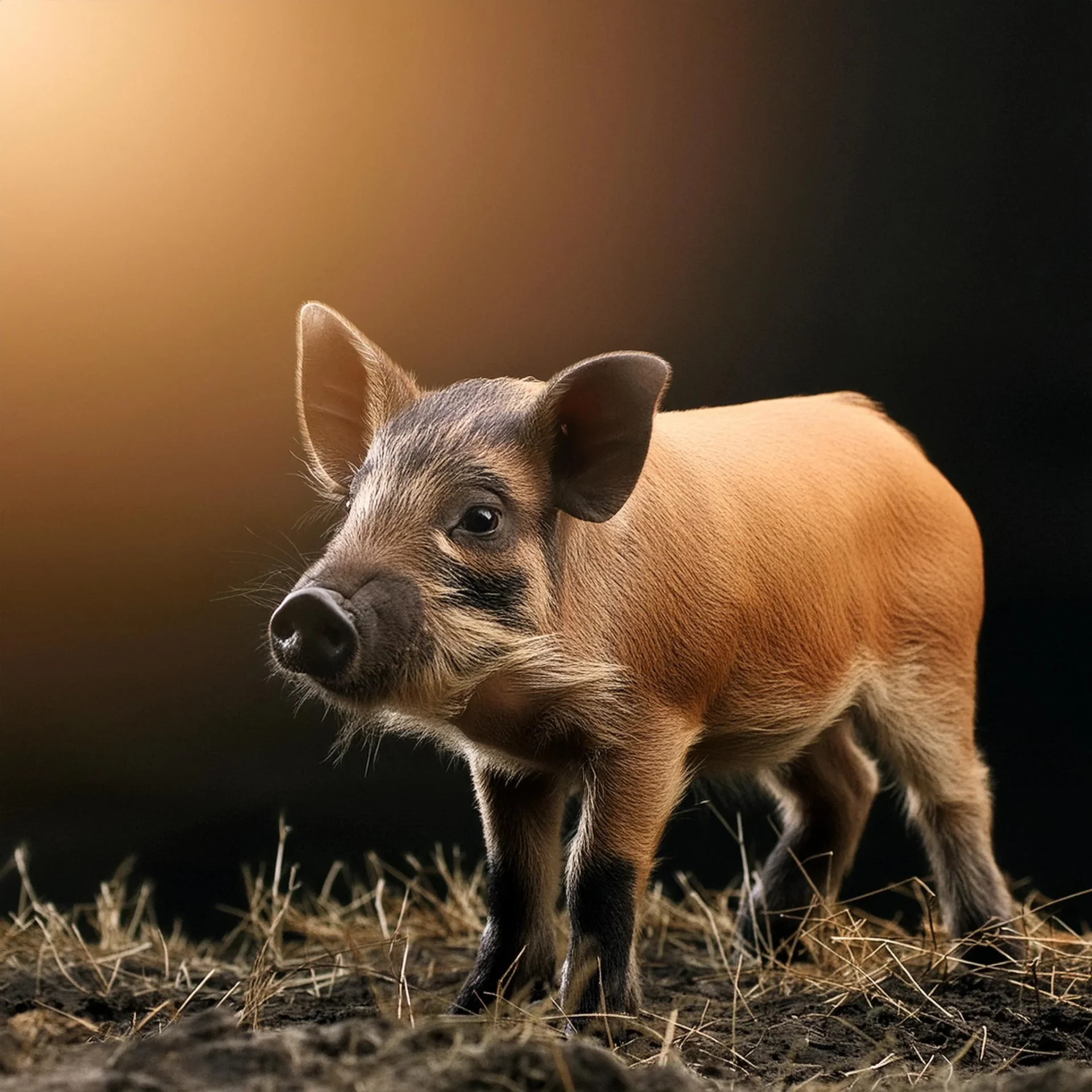
Yucatan Miniature Pig
The parent stock of the Yucatan miniature pigs was obtained from the Yucatan Peninsula of Mexico. The breed was introduced to the United States in 1960. The first micro-sized Yucatan was developed using a small Yucatan boar in the Colorado State University. The breed has a docile nature and adapts well with human interactions. Mature non-obese boars on average can weigh about 83 kg while sows can be about 70 kg, with average height and length of 57 cm and 76 cm, respectively. The breed has highly pigmented color (Slate-gray) and often are hairless or have very little hair. Since the Yucatan miniature pigs are native to the tropical region, intensive management is required for cold weather conditions.
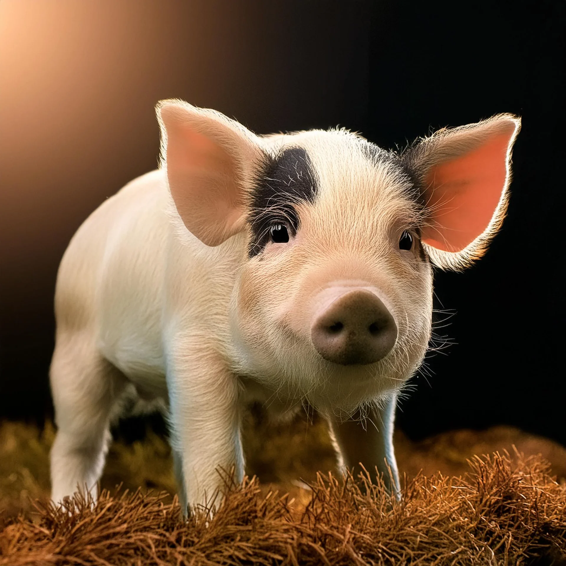
Hanford Miniature Pig
The Hanford miniature breed was developed at the Hanford Labs, Richland, Washington in 1958. The breed was a result of crossing Palouse gilts with a Pittman-Moore boar. Additional reduction in size was obtained by introducing Swamp hog from Louisiana and adding more Pittman-Moore to the mix. Mature pigs can weigh around 75 kg and tend to have less subcutaneous fat in comparison to other breeds. The breed looks much like traditional farm pigs with its white hair coat. Their calm nature makes them well-adapted for laboratory environments.
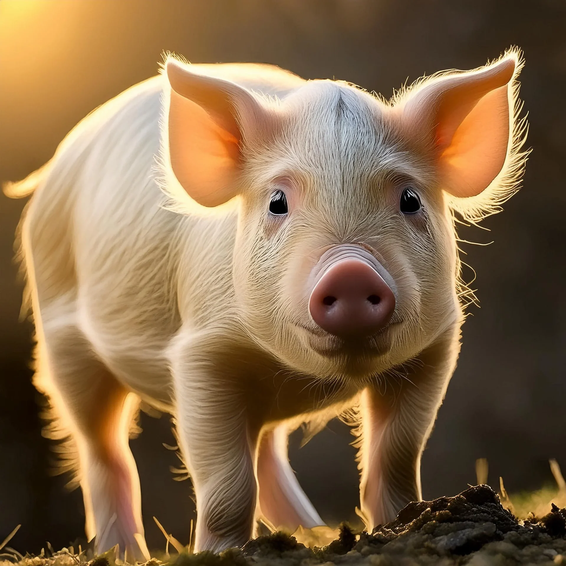
Sinclair Miniature Pig
The Sinclair miniature breed was developed by crossing four feral breeds at the Hormel Institute at the University of Minnesota in the year 1949. Addition of a Yorkshire boar to the stock resulted in white hair colored pigs. The breed was the first miniature stock to be specifically developed for research. The Sinclair breed has a slow growth rate. The miniatures are available in variations of haircoat colors.
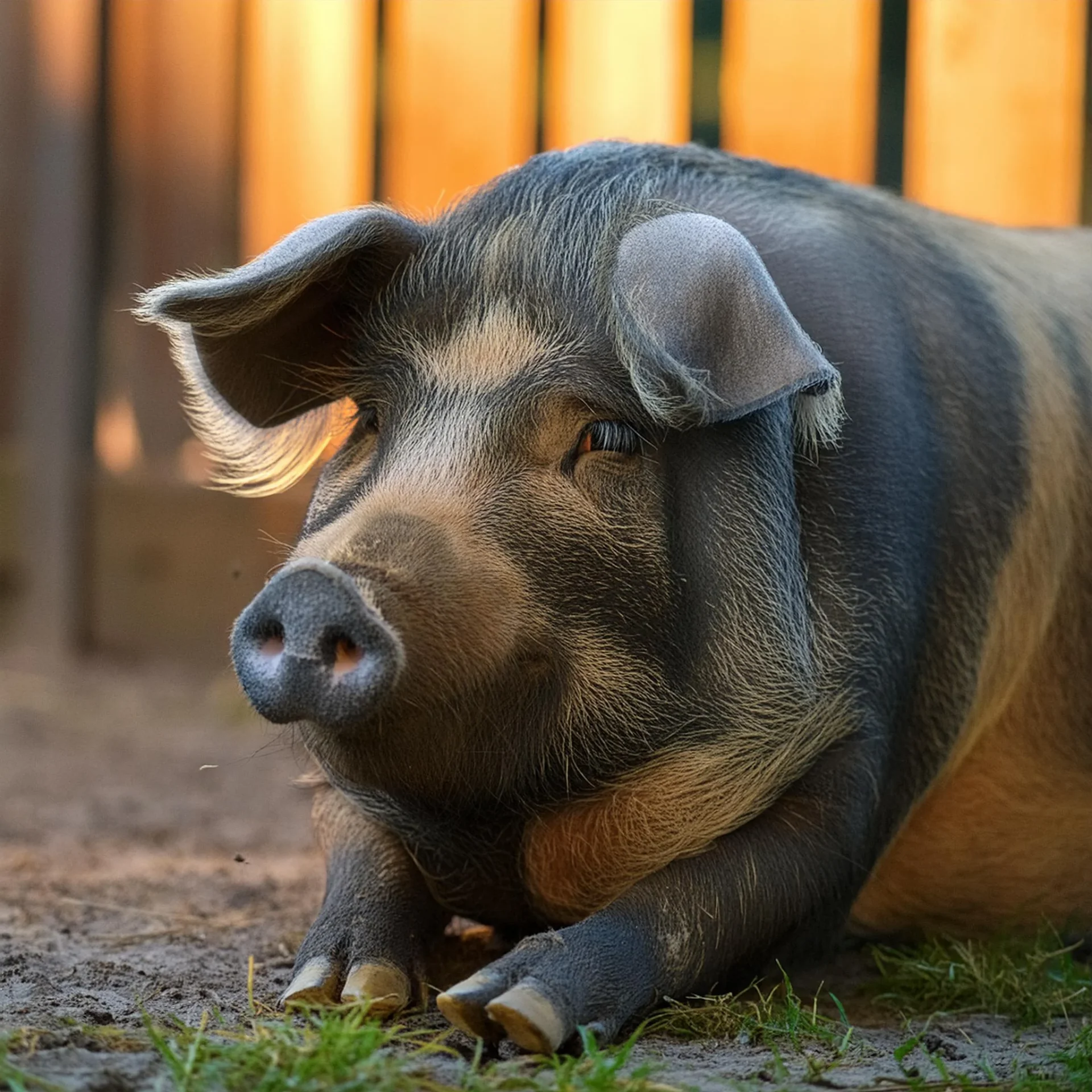
Göttingen Miniature Pig
The Institute of Animal Breeding and Genetics at the University of Göttingen, Germany, began developing of the Göttingen miniatures. The breed was developed by crossbreeding the Minnesota minipig, the Vietnamese pot-bellied pig, and the German Landrace pig. The breed is the smallest of the domestic pig breeds with adult weight around 30 to 40 kgs. The Göttingen pigs are known for their docile nature and well-characterized health status. The breed has white non-pigmented skin.
Training Considerations
Pigs fall in the category of prey animals, and thus are sensitive animals that do require a certain amount of attention to ensure their overall well-being in a laboratory set-up. The following are some considerations to take into account to ensure the animal’s welfare.
- Allow the animals to acclimate to the research environment at least one week prior to any testing. Regular, positive interactions with the pigs make them easy to handle and reduce Care must be taken to prevent any negative interactions with the animals since it will result in increased fear.
- Pigs should be maintained in an enriched environment with materials that are safe and have either of the following qualities: edible (preferably with some nutritional benefits), chewable, investigable and manipulatable.
- Enrichments used in the holding chamber should be clean since pigs will avoid interacting with soiled items.
- Pigs should preferably be maintained in groups given their social nature. However, if they are to be maintained in isolation, the holding chamber should be such that it permits visual, auditory and olfactory interactions with the other animals. Complete social isolation is not recommended since it can significantly affect the animal’s behaviors.
- Appropriate bedding and flooring must be used that either provide a thermoneutral zone or the opportunity to thermoregulate by wallowing or huddling. The right choice of flooring is also essential to ensure that animals do not suffer any discomfort or injuries. Long natural straw is an excellent choice for bedding material.
- Pigs are strong and forceful animals. Holding chambers should be made of sturdy materials and any loose objects such as feeders and waterers should be securely fastened to cages.
- Pigs should not be tethered since it will result in chronic stress.
- Physical restraining of the animals during research should be only done after the animal has acclimated to the device and the handling. Further, it is advisable to keep restraining to a minimum. Restraining devices such as the sling tend to have a calming effect on the animal, and hence such devices are recommended.
- Chemical restraint that offers minimal stress should be used for invasive procedures to prevent negative handling experiences.
- Pigs should not be subjected to forced movement and handling. Allow the subjects to run up and down the aisle at least once a week to reduce stress and make their handling easier. It is also recommended that individual animals are always taken along with a familiar conspecific for research to reduce stress and anxiety.
- Dietary requirements of pigs used as laboratory animals will defer from their farm counterparts. Diets should be adjusted to maintain the size and body condition required for the research.
- Appropriate pain management techniques should be kept in place to avoid any unnecessary discomfort to the animals.
Pigs in Research
Overall, rodents are indispensable in advancing our understanding of basic biological processes, disease mechanisms, and therapeutic interventions, contributing significantly to biomedical and scientific progress.
Shortage in human organ and cell donors makes cross-species transplantation a prospective alternative. However, interest in xenotransplantation is not newfound. Cross-species transplantation can be traced back to as far as the 17th century to Jean Baptiste Denis who began xenotransfusion between animals and humans (Taniguchi, & Cooper, 1997). Though risks such as transfer of infectious agents and chronic rejection exist, with new advancements such as genetic engineering, xenotransplantation may be viable. Xenotransplantation using pigs can be considered one of the best options out there due to their physiological and anatomical similarities with humans.
Porcine red blood cell (RBC) share many similar characteristics, such as similar diameters and counts, with that of human RBCs, though they have a shorter average lifespan. Studies have been undertaken to overcome the barriers associated with xenotransfusion of porcine RBC by developing genetically engineered pigs (Cooper, Hara, & Yazer, 2010).
In 1994, Rao and colleagues were able to demonstrate that the transgenic pig system was capable of producing hemoglobin A without translational errors. Detailed protein chemical analysis of recombinant hemoglobin A in transgenic swine revealed that the system accurately translates human α and β globin mRNA. It was also observed that the co-translation processing of the globin chains was identical to those in humans and without any unnecessary introduction of post-translational protein modification reactions.
Doucet, Gao, MacLaren, and McAlister (2004) investigated strategies that would allow porcine erythrocytes to be successfully used for blood transfusion in humans without being destroyed by human antibodies. The investigators subjected low expressers of alpha-gal(1-3)beta-galGlcNac epitopes obtained from live pigs to three different treatments; enzymatic removal of membrane alpha-gal with alpha-galactosidase, covalent attachment of cyanuric acid-linked methoxy polyethylene glycol, and both processes. Following the treatments, analysis of the treatment effects suggested that removal or masking xenoantigens on pig RBCs could, in fact, allow in vitro cross-match compatibility to human sera. The combination treatment, in comparison to controls, resulted in a reduction of the binding of human immunoglobulin M by 61%, thus, proving to be the most effective treatment of the three.
Zaidi et al. (1998) investigated the potential of genetically modified pigs as a source of kidney donors. The investigation made use of human decay-accelerating factor (hDAF) transgenic pigs that were developed to evade complement-mediated hyperacute rejection. Using cynomolgus monkeys as recipients of kidneys either from hDAF transgenic or non-transgenic control pigs a comparison was made between xenograft acceptance. The transgenic group’s xenograft resulted in a median survival time of 13 days which was higher than the 6.5 days observed in controls. While all control xenografts resulted in antibody-mediated rejection, transgenic xenografts showed no hyperacute rejection in the cynomolgus monkeys.
Iwase et al. (2015) showcased a case study of a baboon surviving for 136 days with an α1,3-galactosyltransferase gene-knockout transgenic pig’s kidney graft. This survival rate was much higher than the usually seen 30 days life-supporting kidney graft transplantation in pig to nonhuman primate. In the following years, researchers were able to attain a survival rate of more than 400 days (Kim et al., 2017). Recipient macaques received anti-CD154 (5c8) either with only anti-CD4, only anti-CD8, or both, renal transplants from hDAF transgenic pigs. Evaluation of whether only CD4 or CD8 depletion was necessary or sufficient for long-term survival resulted in the conclusion that CD4 depletion contributed to and was necessary for significantly prolonging survival.
Genetically engineered pigs lacking in galactose-α1,3-galactose (Gal) or N-glycolylneuraminic acid (NeuGc) expression have advanced the prospects of xenotransplantation. Lee et al. (2016) investigation observed no Gal nor NeuGc presence in GTKO/NeuGcKO pigs similar to human corneas. However, IgM binding was higher, in comparison to human cells, in GTKO/NeuGcKO pig corneas suggesting the presence of remaining xenoantigens. Though Gal/NeuGc deficient pigs may reduce immunologic and/or inflammatory injury to the xenograft, there still exists a need for identification and deletion of xenoantigens to be able to protect the xenograft in humans fully.
Li, Zhang, Liu, and Pan (2017) investigated the possibility of pig corneas hosting the porcine endogenous retroviruses (PERVs) by transplanting them into rhesus monkeys. Regardless of cornea preparation, polymerase chain reaction (PCR) and reverse transcription PCR results were positive only for porcine endogenous retroviruses (PERVs) subtypes A and B, and not subtype C in the pig corneas. After a 24-month period, the viral signals no longer existed and no evidence of transfer of PERV transmission could be seen in the rhesus monkeys despite the PERV-positive nature of the xenograft.
Pigs have a gyrencephalic brain that is less complex than non-human primates and is large enough to be analyzed using the same equipment that is used for humans. Pig brains share greater similarities with the human brain than other commonly used animal models like rats and mice. The anatomy, growth, and development of the brain are comparable to that of the humans (Lind et al., 2007; Ryan et al., 2018).
Axonal injury in children can lead to life-long cognitive, behavioral and motor disabilities. Friess et al. (2007) evaluated neonatal piglets in a range of functional neurobehavioral assessments including the Open Field test following closed head injury. The animals were either subjected to mild (142 rad/sec) or moderate (188 rad/sec) rapid non-impact axial rotations of the head. Immunohistochemistry (β-APP) detected injured axons in the moderately injured group only. Further, in comparison to the mild injury group, the performances of the moderately injured group were affected in the assays. The moderately injured group showed poor interest in exploring and had difficulty performing visual-based tasks.
Armstead, Riley, and Vavilala (2013) investigated the effects of dopamine on autoregulation after traumatic brain injury (TBI) using a porcine model for pediatric TBI. The study made use of newborn piglets equipped with a closed cranial window and subjected them to fluid percussion brain injury. Enzyme-linked immunosorbent assay and radioimmunoassay kits were used to quantify CSF ERK mitogen-activated protein kinase and ET-1, respectively. The investigation’s findings suggested that dopamine is protective of autoregulation after fluid percussion brain injury in both sexes. Further, it was also observed that males had greater loss of pial artery dilation during hypotension and a greater release of ET-1 and ERK mitogen-activated protein kinase after fluid percussion brain injury. Taken together, the investigators recommended pursuing sex-based therapies for the treatment of hemodynamic sequelae of pediatric TBI.
The role of sialic acid as a conditional nutrient during rapid brain growth period was evaluated by Wang et al. (2007). The study involved newborn piglets who were divided into two treatment groups, one of which received pig’s milk replacer with varying quantities of sialic acid supplemented using casein glycomacropeptide. At day twenty-one, animals were evaluated in the 8-Arm Radial Arm Maze using easy and difficult visual cues to assess learning and memory abilities. Performance analysis of controls and the group that received a dietary supplement of sialic acid showed that the latter group performed better. The performance in the difficult task was observed to be enhanced in a dose-dependent manner. Further, the dose-response relation between sialic acid supplementation amount and mRNA levels of ST8SIA4 and GNE could be significantly observed. The study concluded that sialic acid in mammalian milk could, in fact, contribute to the cognitive development of the offspring.
Nielsen et al. (2009) evaluated the spatial learning capabilities of pigs using the T-Maze (see also Pig T-Maze). Göttingen minipigs were subjected to a spatial Delayed Non-Match to Sample task that involved long delays and scopolamine treatment. Pigs were observed to have a less natural affinity to alternate between choices unlike the choice behaviors observed in rats. As expected, the scopolamine treatment increased the mean time to reach the goals and decreased performance with the increased introduction of task delays. Overall performance analysis of the pigs revealed significant deterioration of T-Maze task performance with increasing delays and scopolamine treatment.
The use of rats and mice to model human neurodegenerative diseases may often result in failure or not be comprehensive due to the biological differences between species. Large animal models, especially pigs, offer a better prospect of replicating human pathology. The similarities between pigs and humans allow pig models to mimic the neurodegenerative features of neurological diseases effectively.
Yang et al. (2010) modeled Huntington’s disease in pigs using nuclear transfer to generate transgenic Tibetan miniature pigs that express N-terminal mutant huntingtin (1–208 amino acids) with an expanded polyglutamine tract (105Q). In comparison to mice models, the transgenic pigs showed typical apoptotic neurons with nuclear DNA fragmentation in the brains. The researchers also observed hyperkinesia, a phenotype similar to chorea in Huntington’s disease patients, before death occurred, in some of the transgenic piglets.
In a similar effort, Kragh et al. (2009) developed transgenic Göttingen minipigs to model Alzheimer’s disease. Blastocysts were produced by handmade cloning using stably transfected minipig fibroblasts expressing the APP695sw (amyloid precursor protein gene with the Swedish mutation) transgene. The APP695sw transgene was inserted only at the GLIS3 locus in the opposite transcriptional direction of GLIS3 corresponding to intron 5 of human GLIS3
Human and pig hearts share similarities in terms of coronary circulation and hemodynamics. Though differences in the two heart systems exist due to the biped and quadruped stance and anatomy, pig hearts offer greater advantages over rodent models. Further, the large size of the pig heart allows implementation of human healthcare methods and equipment. The identical heart weight to body weight of pigs in the weight range 20 to 30 kgs to adult humans also makes them a feasible model for cardiovascular and cardiopulmonary studies. (Hughes, 1986; Lelovas, Kostomitsopoulos, & Xanthos, 2004)
Rixen et al. (2001) study developed a porcine model of hemorrhagic shock to overcome the limitations of traditional parameters (such as hemorrhage volume and blood pressure) of hemorrhage severity. The researchers suggested the use of oxygen debt and metabolic acidemia as severity indicators. The study made use of female pigs that were subjected to hemorrhage at an initial rate of 4 mL blood/kg min for over a period of 60 minutes. Quantification of the degree of metabolic imbalance was measured using arterial base excess and plasma lactate. Analysis of the hemorrhagic parameters suggested the superiority of the aforementioned measures and oxygen debt over traditional parameters of severity indicators.
The effect of vasopressin in improving short-term survival in uncontrolled hemorrhagic shock pig model was investigated by Voelckel et al. (2003). The investigation compared the effect of the drug with epinephrine in anesthetized 12- to 16-weeks old pigs that were subjected to a penetrating liver injury using finger-fracture dissection across the right liver lobe. When blood loss associated with shock was achieved, and the heart rate declined progressively, subjects received bolus dose and continuous infusion of either 0.4 units/kg vasopressin, 45 µg/kg epinephrine, or an equal volume of saline placebo. However, epinephrine and saline placebo treatments resulted in deaths within 15 minutes of administration. Despite high hepatic and renal artery blood flow throughout the study, vasopressin treated group did not show any further bleeding. Further, the vasopressin group survived post fluid resuscitation and surgical control of bleeding without further vasopressor therapy.
Researchers have suggested the use of pregnancy-associated plasma protein-A (PAPP-A) as a diagnostic biomarker for acute coronary syndrome (ACS). Steffensen et al. (2015) made use of 50 to 60 kg female, non-heparinized, nonatherosclerotic, Danish landrace pigs to demonstrate the elevation of PAPP-A during myocardial ischemia. Pigs had their left anterior descending coronary artery ligated for 45 minutes which was followed by reperfusion for 45 minutes. PAPP-A levels were monitored for myocardial ischemia and SHAM groups using blood samples from aorta just above the aortic valve, the coronary sinus, and the left femoral vein. After 30 minutes of ischemia, PAPP-A levels were observed to have elevated (to 0.25 ng/ml) in systemic circulation though not in the coronary sinus. Based on the observations, the researchers concluded that PAPP-A concentrations increase potentially due to myocardial ischemia in the absence of atherosclerosis or heparin; Thus, suggesting the potential role of PAPP-A as a biomarker for ACS.
Feng, Mao, Dong, and Liu (2016) established porcine models of acute myocardial ischemia with different levels of stenosis. The study also investigated the protective effects of active perfusion at a site distal to the anastomosis in comparison to the traditional method of shunt perfusion. Male pigs were divided into two groups, shunt, and active perfusion, with either 55 or 75% stenosis rate. Active perfusion led to a significant decrease in all indicators of myocardial injury as opposed to the shunt group. Further, active perfusion resulted in upregulation of Bcl-2 and downregulation of the expression level of caspase-3 in comparison to shut perfusion. Overall analysis suggested active perfusion to be a more successful approach to alleviate myocardial ischemia than conventional shut perfusion.
Animals like rabbits, guinea pigs, and rats provide the advantage of easy maintainability and handling. Additionally, they are inexpensive and hence are often used in dermatology and wound healing studies. However, skins of these animals are significantly different from human skins. As opposed to the loosely attached, sparse/dense hair coat observed in these animals, the pig’s skin is firmly attached and offers similar anatomical and physiological characteristics as human skin. Further, wound healing in pigs and humans occurs via re-epithelialization rather than contraction as observed in small animal models. (Sullivan, Eaglstein, Davis, & Mertz P, 2001; Summerfield, Meurens, & Ricklin, 2015)
Histopathological evaluations of scald and contact burns were performed by Brans, Dutrieux, Hoekstra, Kreis, & du Pont (1994) using New Yorkshire pig. Four scald burns were inflicted by application of 80°C water for 10, 20, 30 and 40 seconds, respectively. Two contact burns were inflicted using a brass block heated to 170°C and applied for 10 and 20 seconds, respectively, without exerting pressure. Contact burns led to immediate coagulation while scald burns caused initial hyperemia that is followed by delayed vascular damage. Unlike the necrotic layer developed as a result of contact burn, scalding burn led to no such layer formation thereby increasing the possibility of deep vascular plexus damage.
Pigs with streptozotocin-induced diabetes were investigated for impaired wound healing by Velander et al. (2008). The subjects underwent full-thickness wounds, and the wound fluid was collected using the chamber treatment. The analysis of wound healing and associated parameters revealed a significant delay in the healing processes of diabetic pigs. In comparison to the non-diabetic controls, diabetic pigs had significantly delayed re-epithelialization, and healing impairment was accompanied by a reduction of IGF-1. Further, it was concluded that local hyperglycemia did not contribute to delayed wound healing as observed by subjecting non-diabetic pigs to a high-glucose wound concentration. In their study (Velander et al., 2009) following observation of diabetic wound healing, the researchers demonstrated an increased re-epithelialization rate by treating wounds with autologous fibroblasts and autologous keratinocytes (86.75% and 91.3%, respectively) in comparison to saline treatment.
The Sinclair swine has been a popular animal model to study aspects of melanoma and vitiligo (Misfeldt & Grimm, 1994). Millikan, Hook, and Manning (1973) observed that Sinclair pigs had more than one type of lesion and that not all lesions progressed from Types I to V. By observing ultrastructure under an electron microscope, similarities between human tumor tissues and the animal tissues could be seen. The Sinclair pigs showed the range of comparable tumors to human melanocyte disorders. Further, the superficial spreading of malignant tumors was comparable to that of human melanoma and Klug’s type-A melanoma.
Other selectively bred minipigs include the Libechov minipig (Vincent-Naulleau et al., 2004) and the Munich miniature swine Troll (Müller, Wanke, & Distl, 2001). These models allow the observation of inheritance of the disease and the factors affecting the progression and regression. Further, pig models also allow observation of the interplay between endogenous retroviruses, melanomagenesis, and adaptive immune response.
Strengths and Limitations
The porcine model offers many advantages over traditional animal models such as the rodents. Pigs tend to have a docile temperament and with regular human interactions become amenable to handling. The calm nature facilitates repeated and frequent sampling. Further, their reproductive cycle and breeding characteristics permit genetic manipulations and selections. The large litter production proves advantageous in reducing experimental variability. However, the primary advantage of the swine model is their similarity to human physiology and genetics. Pig organs are comparable in size and physiologic character to those of humans. Using a porcine model ensure readily available tissue for research studies. Pigs and humans also share similar dietary habits and metabolism. These similarities make them an excellent model for studying human conditions and diseases.
The availability of miniature pigs significantly reduces the management efforts that are usually required for the large domestic variety. Miniature pigs allow easy handling and maintenance. The variety also tends to be relatively calmer than the large variety. The development of transgenic miniature pigs for specific disease model further facilitates the use of pigs to study human diseases.
The domestic breeds of pigs tend to be very large and hence require trained personnel and extensive husbandry management. This results in an increase in expenses that could have been reduced with animal models such as rats and mice. The domestic variety also often tends to carry the gene for porcine stress syndrome (PSS). Further, pigs tend to have increased cardiac irritability and are susceptible to gastric ulcers, which require additional efforts and use of drugs that may interfere with the results of the investigation. Since pigs are prey animals, they are easily frightened by loud sounds or being lifted. Further, negative interactions with the handler can significantly increase the fear of humans and stress. Social isolation can have a detrimental effect on the animal’s behavior. Dietary requirements of the laboratory pigs must be adjusted to maintain their size and weight required for research.
Summary
- Pigs are evolutionarily closer to humans. Interest in pigs as animal models peaked in the 18th
- Porcine model is a popular non-rodent model due to its physiologic, anatomic, and pathologic similarities to humans.
- The large size of pigs is comparable to humans and allows the use of human clinical devices.
- Popular domestic breed pigs include Duroc, Yorkshire, Landrace, and crossbreeds.
- Popular miniature breeds include Yucatan mini and micro, Hanford, Sinclair, and Göttingen.
- Miniature pigs are easy to handle and maintain in comparison to the domestic variety. Further, the development of transgenic miniature varieties allows for a more cohesive investigation of human diseases.
- Pigs tend to have large litters which permit reduction of experimental variability.
- Physical or chemical restraining that offers minimal stress to the animals should be used when required.
- Pigs are very social and possess complex cognition. Social isolation, poor holding environments, and negative interactions have a significant effect on the animal’s behaviors.
- Holding environments should be constructed taking into consideration the lack of sweat glands in pigs.
- Dietary requirements of laboratory pigs will require appropriate adjustments to meet the experiment’s requirements.
References
- Abbott, A. (2012). Pig geneticists go the whole hog. Nature, 491(7424):315-6. doi: 10.1038/491315a.
- Almond, G. W. (1996). Research Applications using Pigs. Veterinary Clinics of North America: Food Animal Practice, 12(3), 707–716. doi:10.1016/s0749-0720(15)30394-7
- Armstead, W. M., Riley, J., & Vavilala, M. S. (2013). Dopamine prevents impairment of autoregulation after traumatic brain injury in the newborn pig through inhibition of Up-regulation of endothelin-1 and extracellular signal-regulated kinase mitogen-activated protein kinase. Pediatric Critical Care Medicine, 14(2):e103-11. doi: 10.1097/PCC.0b013e3182712b44.
- Bassols, A., Costa, C., Eckersall, P. D., Osada, J., Sabrià, J., & Tibau, J. (2014). The pig as an animal model for human pathologies: A proteomics perspective. PROTEOMICS – Clinical Applications, 8(9-10), 715–731. doi:10.1002/prca.201300099.
- Brans, T. A., Dutrieux, R. P., Hoekstra, M. J., Kreis, R. W., & du Pont, J. S. (1994). Histopathological evaluation of scalds and contact burns in the pig model. Burns, 20, S48–S51. doi:10.1016/0305-4179(94)90090-6
- Caliebe, A., Nebel, A., Makarewicz, C., Krawczak, M., & Krause-Kyora, B. (2017). Insights into early pig domestication provided by ancient DNA analysis. Scientific Reports,7:44550. doi: 10.1038/srep44550.
- Cooper, D. K. (2012). A brief history of cross-species organ transplantation. Proceedings (Baylor University, Medical Center), 25(1):49-57.
- Cooper, D. K. C., Hara, H., & Yazer, M. (2010). Genetically Engineered Pigs as a Source for Clinical Red Blood Cell Transfusion. Clinics in Laboratory Medicine, 30(2), 365–380. doi:10.1016/j.cll.2010.02.001.
- Doucet, J., Gao, Z. H., MacLaren, L. A., & McAlister, V. C. (2004). Modification of xenoantigens on porcine erythrocytes for xenotransfusion. Surgery, 135(2):178-86.
- Ekser, B., Li, P., & Cooper, D. K. C. (2017). Xenotransplantation: past, present, and future. Current Opinion in Organ Transplantation, 22(6):513-521. doi: 10.1097/MOT.0000000000000463.
- Feng, Z., Mao, Z., Dong, S., & Liu, B. (2016). Protective effect of active perfusion in porcine models of acute myocardial ischemia. Molecular Medicine Reports, 14(4):3581-7. doi: 10.3892/mmr.2016.5665
- Friess, S. H., Ichord, R. N., Owens, K., Ralston, J., Rizol, R., Overall, K. L., …, Margulies, S. S. (2007). Neurobehavioral functional deficits following closed head injury in the neonatal pig. Experimental Neurology, 204(1):234-43.
- Gieling, E. T., Nordquist, R. E., & van der Staay, F. J. (2011). Assessing learning and memory in pigs. Animal Cognition, 14(2):151-73. doi: 10.1007/s10071-010-0364-3. https://link.springer.com/article/10.1007/s10071-010-0364-3
- Giuffra, E., Kijas, J. M., Amarger, V., Carlborg, O., Jeon, J. T., & Andersson, L. (2000). The origin of the domestic pig: independent domestication and subsequent introgression. Genetics, 154(4):1785-91.
- Gutierrez, K., Dicks, N., Glanzner, W. G., Agellon, L. B., & Bordignon, V. (2015). Efficacy of the porcine species in biomedical research. Frontiers in Genetics, 6:293. doi: 10.3389/fgene.2015.00293.
- Hughes, H. C. (1986). Swine in cardiovascular research. Laboratory Animal Science,36(4):348-50.
- Iwase, H., Liu, H., Wijkstrom, M., Zhou, H., Singh, J., Hara, H., …, Cooper, D. K. (2015). Pig kidney graft survival in a baboon for 136 days: longest life-supporting organ graft survival to date. Xenotransplantation, 22(4):302-9. doi: 10.1111/xen.12174.
- Kim, S., Higginbotham, L., Mathews, D., Breeden, C., Stephenson, A., Larsen, C., …, Adams, A. (2017). CD4 Depletion Is Necessary and Sufficient for Long-Term Nonhuman Primate Xenotransplant Survival. [abstract]. American Journal of Transplantation, 17 (suppl 3).
- Kragh, P.M., Nielsen, A.L., Li, J., Du, Y., Lin, L., Schmidt, M., …, Jørgensen, A.L. (2009). Hemizygous minipigs produced by random gene inserti on & handmade cloning express the Alzheimer’s disease-causing dominant mutati on APPsw. Transgenic Research, 18(4):545-58.
- Krause-Kyora, B., Makarewicz, C., Evin, A., Flink, L. G., Dobney, K., Larson, G., …, Nebel, A. (2013). Use of domesticated pigs by Mesolithic hunter-gatherers in northwestern Europe. Nature Communications, 2013;4:2348. doi: 10.1038/ncomms3348.
- Larzul, C. (2013). Pig genetics: insight in minipigs. Bilateral Symposium on Miniature Pigs for Biomedical Research in Taiwan and France., Taïnan, Taiwan. pp.1-6, 2013. <hal-00958583>
- Lee, W., Miyagawa, Y., Long, C., Ekser, B., Walters, E., Ramsoondar, J., …, Hara, H. (2016). Expression of NeuGc on Pig Corneas and Its Potential Significance in Pig Corneal Xenotransplantation. Cornea, 35(1):105-13. doi: 10.1097/ICO.0000000000000635.
- Lelovas, P. P., Kostomitsopoulos, N. G., & Xanthos, T. T. (2004). A comparative anatomic and physiologic overview of the porcine heart. Journal of American Association for Laboratory Animal Science, 53(5):432-8.
- Li, A., Zhang, Y., Liu, Y., & Pan, Z. (2017). Corneal Xenotransplantation From Pig to Rhesus Monkey: No Signs of Transmission of Endogenous Porcine Retroviruses. Transplantation Proceedings, 49(9):2209-2214. doi: 10.1016/j.transproceed.2017.07.018.
- Lind, N. M., Moustgaard, A., Jelsing, J., Vajta, G., Cumming, P., & Hansen, A. K. (2007). The use of pigs in neuroscience: modeling brain disorders. Neuroscience & Biobehavioral Reviews, 31(5), 728–751. doi: 10.1016/j.neubiorev.2007.02.003
- Lunney, J. K. (2007). Advances in swine biomedical model genomics. International Journal of Biological Science, 3(3):179-84.
- Marchant-Forde, J. N., & Herskin, M. S. (2018). Pigs as laboratory animals. Advances in Pig Welfare, 445–475. doi:10.1016/b978-0-08-101012-9.00015-0.
- Millikan, L. E., Hook, R. R., & Manning, P. J. (1973). Immunobiology of melanoma. Gross and ultrastructural studies in a new melanoma model: the Sinclair swine. Yale Journal of Biology and Medicine, 46(5):631-45.
- Misfeldt, M. L., & Grimm, D. R. (1994). Sinclair miniature swine: an animal model of human melanoma. Veterinary Immunology & Immunopathology, 43(1-3):167-75.
- Müller, S., Wanke, R., & Distl, O. (2001). Inheritance of melanocytic lesions and their association with the white colour phenotype in miniature swine. Journal of Animal Breeding & Genetics, 118 275–283. 10.1046/j.1439-0388.2001.00280.x.
- Nielsen, T. R., Kornum, B. R., Moustgaard, A., Gade, A., Lind, N. M., & Knudsen, G. M. (2009). A novel spatial Delayed Non-Match to Sample (DNMS) task in the Göttingen minipig. Behavioral Brain Research, 196(1):93-8. doi: 10.1016/j.bbr.2008.07.019.
- Nunoya, T., Shibuya, K., Saitoh, T., Yazawa, H., Nakamura, K., Baba, Y., & Hirai, T. (2007). Use of Miniature Pig for Biomedical Research, with Reference to Toxicologic Studies. Journal of Toxicologic Pathology, 20(3), 125-132. doi:10.1293/tox.20.125
- Rao, M. J., Schneider, K., Chait, B. T., Chao, T. L., Keller, H., Anderson, S., …, Acharya, A. S. (1994). Recombinant hemoglobin A produced in transgenic swine: structural equivalence with human hemoglobin A. Artificial Cells, Blood Substitutes, and Immobilization Biotechnology, 22(3):695-700. DOI: 10.3109/10731199409117900
- Rixen, D., Raum, M., Holzgraefe, B., Sauerland, S., Nagelschmidt, M., & Neugebauer, E. A. M. (2001). A PIG HEMORRHAGIC SHOCK MODEL: OXYGEN DEBT AND METABOLIC ACIDEMIA AS INDICATORS OF SEVERITY. Shock, 16(3), 239–244. doi:10.1097/00024382-200116030-00012
- Ryan, M. C., Sherman, P., Rowland, L. M., Wijtenburg, S. A., Acheson, A., Fieremans, E., …, McGuire, S. A. (2018). Miniature pig model of human adolescent brain white matter development. Journal of Neuroscience Methods, 296:99-108. doi: 10.1016/j.jneumeth.2017.12.017.
- Seaton, M., Hocking, A., & Gibran, N. S. (2015). Porcine Models of Cutaneous Wound Healing. ILAR Journal, 56(1), 127–138. doi:10.1093/ilar/ilv016
- Smith, A. C., & Swindle, M. M. (2006). Preparation of Swine for the Laboratory. ILAR Journal, 47(4), 358-363. doi:10.1093/ilar.47.4.358.
- Steffensen, L. B., Poulsen, C. B., Shim, J., Bek, M., Jacobsen, K., Conover, C. A., …, Oxvig, C. (2015). Myocardial and Peripheral Ischemia Causes an Increase in Circulating Pregnancy-Associated Plasma Protein-A in Non-atherosclerotic, Non-heparinized Pigs. Journal of Cardiovascular Translation Research, 2015 Dec;8(9):528-35. doi: 10.1007/s12265-015-9656-y.
- Sullivan, T. P., Eaglstein, W. H., Davis, S. C., & Mertz, P. (2001). The pig as a model for human wound healing. Wound Repair & Regeneration, 9(2):66-76.
- Summerfield, A., Meurens, F., & Ricklin, M. E. (2015). The immunology of the porcine skin and its value as a model for human skin. Molecular Immunology, 66(1):14-21. doi: 10.1016/j.molimm.2014.10.023.
- Suzuki, Y., Yeung, A. C., & Ikeno, F. (2011). The representative porcine model for human cardiovascular disease. Journal of Biomedicine & Biotechnology, 2011;2011:195483. doi: 10.1155/2011/195483.
- Swindle, M. M., Makin, A., Herron, A.J., Clubb, F. J. Jr, & Frazier, K. S. (2012). Swine as models in biomedical research and toxicology testing. Veterinary Pathology, 49(2):344-56. doi: 10.1177/0300985811402846.
- Taniguchi, S., & Cooper, D. K. (1997). Clinical xenotransplantation: past, present and future. Annals of Royal College of Surgeons of England, 79(1):13-9.
- Velander, P., Theopold, C., Bleiziffer, O., Bergmann, J., Svensson, H., Feng, Y., & Eriksson, E. (2009). Cell Suspensions of Autologous Keratinocytes or Autologous Fibroblasts Accelerate the Healing of Full Thickness Skin Wounds in a Diabetic Porcine Wound Healing Model. Journal of Surgical Research, 157(1), 14–20. doi:10.1016/j.jss.2008.10.001.
- Velander, P., Theopold, C., Hirsch, T., Bleiziffer, O., Zuhaili, B., Fossum, M., … Eriksson, E. (2008). Impaired wound healing in an acute diabetic pig model and the effects of local hyperglycemia. Wound Repair and Regeneration, 16(2), 288–293. doi:10.1111/j.1524-475x.2008.00367.x
- Vincent-Naulleau, S., Le Chalony, C., Leplat, J.-J., Bouet, S., Bailly, C., Spatz, A., … Geffrotin, C. (2004). Clinical and Histopathological Characterization of Cutaneous Melanomas in the Melanoblastoma-Bearing Libechov Minipig Model. Pigment Cell Research, 17(1), 24–35. doi:10.1046/j.1600-0749.2003.00101.x
- Voelckel, W. G., Raedler, C., Wenzel, V., Lindner, K. H., Krismer, A. C., Schmittinger, C. A., …, Königsrainer, A. (2003). Arginine vasopressin, but not epinephrine, improves survival in uncontrolled hemorrhagic shock after liver trauma in pigs. Critical Care Medicine, 31(4):1160-5.
- Wang, B., Yu, B., Karim, M., Hu, H., Sun, Y., McGreevy, P., …, Brand-Miller, J. (2007). Dietary sialic acid supplementation improves learning and memory in piglets. American Journal of Clinical Nutrition, 85(2):561-9.
- Yang, D., Wang, C. E., Zhao, B., Li, W., Ouyang, Z., Liu, Z., …, Li, X. J. (2010). Expression of Huntington’s disease protein results in apoptotic neurons in the brains of cloned transgenic pigs. Human Molecular Genetics, 19(20):3983-94. doi: 10.1093/hmg/ddq313.
- Zaidi, A., Schmoeckel, M., Bhatti, F., Waterworth, P., Tolan, M., Cozzi, E., …, Friend, P. (1998). Life-supporting pig-to-primate renal xenotransplantation using genetically modified donors. Transplantation, 65(12):1584-90.
