

Cells are building blocks of organisms. Thus, understanding the whole metabolic processes and dynamics of organisms requires us to look clearly inside cells.
Advanced microscopy technologies combined with protein labeling approaches have enabled scientists to understand cellular processes and investigate the dynamic processes in organisms with better resolution and sensitivity.[1]
The quality optical conditions can only be obtained using high numerical aperture oil-immersion objective lenses that work with a cover slip of a very specific refractive index (η = 1.515) and thickness (0.15 mm).[2] Any deviation from these parameters can impact the resolution and quality of the obtained image.
Also, a system that can meet the physical requirements of such high-throughput experiments is required. These requirements are chemoattractant gradient generation, shear flow, electrophysiology electrodes, the introduction of stimuli, and multiple wells for multiplex or multimodal imaging.[2] All this can only be achieved by using high-quality imaging chambers.
Imaging chambers are tools used in labs working on microscopy techniques to conserve specimens and control the environment around them, such as temperature, air quality, and humidity. With them, specimens are observed with minimal invasion at high resolution.
After the development of mammalian cell culture techniques, the first live-cell imaging chamber was introduced in the early 20th century.[3]
From simple coverslip sealed on a microscope to sophisticated open and closed perfusion chambers in open-well or channel format, the technology of the imaging chambers has evolved progressively. Here are some things to know about imaging chambers:
Allows for easy changes in the culture media; this ensures that tissues grow at a healthy pace and samples are imaged at the required high numerical apertures.[4]
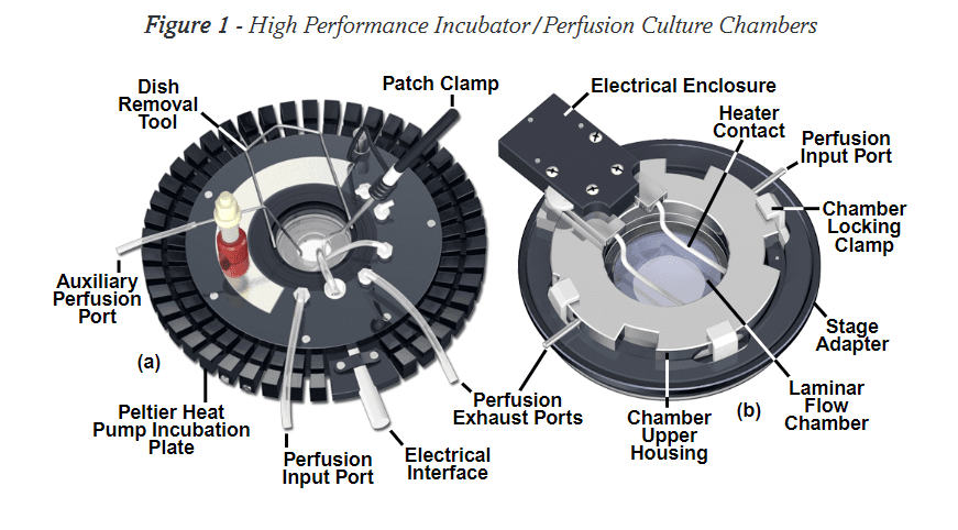
Figure: Image of a Perfusion Imaging Chamber.[4]
Simple microscope slides with a coverslip are used for short-term (20-30 minutes) experiments.[4] It’s enough to protect adherent cells from physical stress that can lead to autofluorescence.
Coverslips are sealed using molten agarose, vacuum grease, rubber cement, or a useful preparation known as VALAP (a 1:1:1 mixture of vaseline, lanolin, and paraffin).[4] This prevents the evaporation of culture media and maintains viable cells.
The prepared slide can be stored on a small heating block near the microscope or carbon dioxide incubator between image gathering sessions.[4]
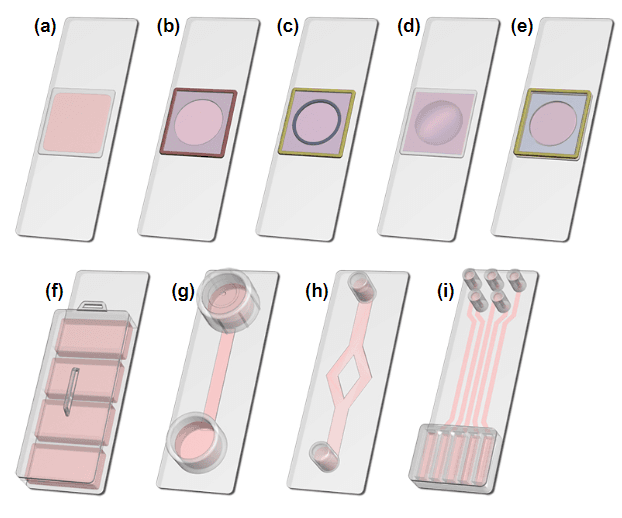
Figure: Different types of simple microscope slide imaging chambers.[4]
However, the Petri dish culture and imaging chamber is a standard 35 or 50-millimeter disposable Petri dish with a 10-, 14-, and 20-mm circular opening and covered with a borosilicate coverslip to observe living cells at high resolution.[4] They always contain large amounts of media which minimize cell damage due to sudden changes in pH and temperature.
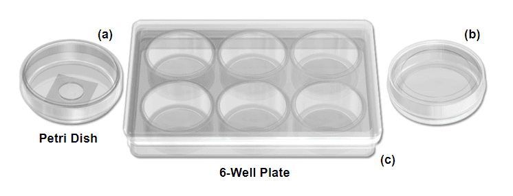
Figure: Different types of Petri-dish imaging chamber.[4]
They are available as both open and closed perfusion chambers. The open perfusion chamber is similar to custom microscopy slides and Petri dishes. Its advantages include:[4]
One limitation of the open perfusion chamber is that it does not provide sufficient control over the cells’ environment.[3] The technique is more suitable for experiments in which high-resolution transmitted light techniques are not required.
The closed perfusion chamber is sealed to protect cell cultures against environmental variables, such as temperature, osmolarity, pH, and carbon dioxide concentration.[4]
In these systems, fresh medium or other chemicals are added through a port, regulated by either a motor-driven syringe, a peristaltic pump, or through a gravity-controlled manifold.[4] They are most suited for long-term experiments.
One limitation of closed perfusion imaging chambers is that they do not allow easy access to the cells during experiments.
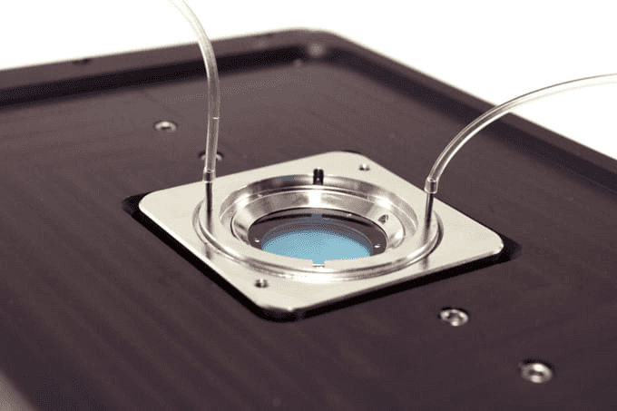
Figure: Image of an advanced perfusion imaging chamber.[3]
While designing a microscopy experiment, researchers should first determine working distance restrictions, necessary magnification range, and optimum numerical aperture of the depth field of the microscope.[4] The chamber used in the experiment must be chosen in a way that it is perfectly paired with the set physical conditions.
For example, in transmitted light applications that require a high numerical aperture condenser, the imaging chamber should accommodate the physical size, working distance, and proximity of the condenser front lens element.[4] And to ensure optimum image clearance, the dimension of the culture chamber should match the geometry of the microscope stage.
For such situations, the advanced open and closed system imaging chamber works best for the experiments.
While using an open system imaging chamber, consider characteristics like construction material, chamber geometry, volume, coverslip thickness, aperture size, and biocompatibility.[4] Factors affecting your experiment with these systems include ambient light, evaporation (and condensation), and lab conditions.
Closed system imaging chambers are used when the culture must be completely isolated from the external environment or for transmission optical microscopy applications that require advanced contrast-enhancing techniques.[4]
For closed system imaging chambers, other factors in addition to those involved in the open system are laminarity, shear stress, the separation distance between optical surfaces, fixed or variable chamber volume requirements, and flow channel geometry.[4]
It’s a combination that can either be utilized as a static chamber or a perfusion device to simulate the conditions found in a humidified carbon dioxide incubator. In the device, a circular, heated water tank is equipped with carbon dioxide injectors, fed by the remote gas-mixing unit to maintain high humidity inside the chamber.[4] Fresh medium and metabolites are added in the chamber through an access port in the cover glass.
It’s most suited for applications like polarized light imaging techniques and transmitted light differential interference contrast at a low numerical aperture.[4]
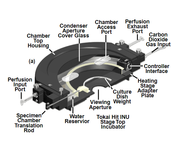
Figure: Image of a Tokai hit stage incubator.[4]
Cell-based analyses or tissue incubations are carried out in imaging chambers, followed by processing of the microscopy slides carrying the sample.[5] These imaging chambers have many applications in labs including:[5]
Imaging chambers are essential tools in biological labs for imaging live cells and tissues. They have major applications in the short-term storage of cells or tissues and help maintain a suitable optimum environment for cells.
Today, a range of designs for imaging chambers are available that are employed based on the microscopy experimental goals. Some known imaging chambers are simple microscopy slide chambers, petri dish chambers, perfusion chambers (extensively used ones), and high-stage incubators.
Live cell imaging is gaining more popularity in cell and molecular biology for its extensive applications in providing better insight into cellular processes and dynamics.[4]
Check out our Visual Patching and Imaging Chamber if you want a high-quality imaging device for your lab.
