Scientific Equipment for Every Lab
From behavioral mazes to stereotaxic instruments, lab consumables to analytical tools. Trusted by 1,200+ research institutions worldwide.

Shop by Research Area
View All Departments


Popular in Each Department

Broome Rodent Restrainers
$80.00
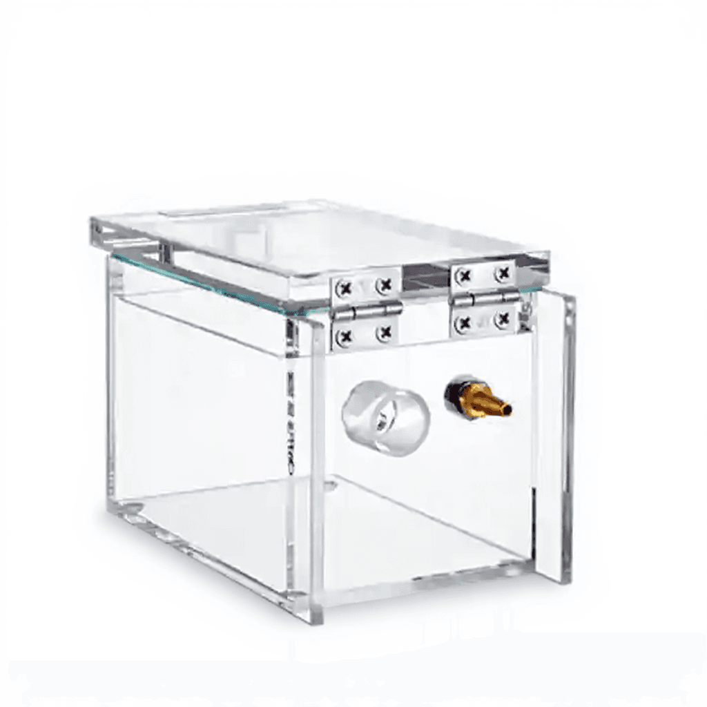
Anesthesia Induction Chamber
$190.00

Drosophila Y Maze
$150.00

Anesthesia Cone Masks for Mouse and Rat
$87.80

Rodent Heating Pad
$125.00

Euthanasia Chamber/Containment System
$345.00

Silicone Coated MCAO Monofilament Suture
$400.00

Injection Cone with Light
$690.00
ConductVision
AI-Powered Vision for Life ScienceCutting-edge behavioral analysis using deep learning and markerless tracking. Quantify posture, movement, and interaction in rodents, zebrafish, drosophila, and more.


Maze Engineers
World-class behavioral mazes and neuroscience apparatus. From open-field arenas to complex decision-making paradigms, designed for reproducible research.


Pathology & Histology
Browse All


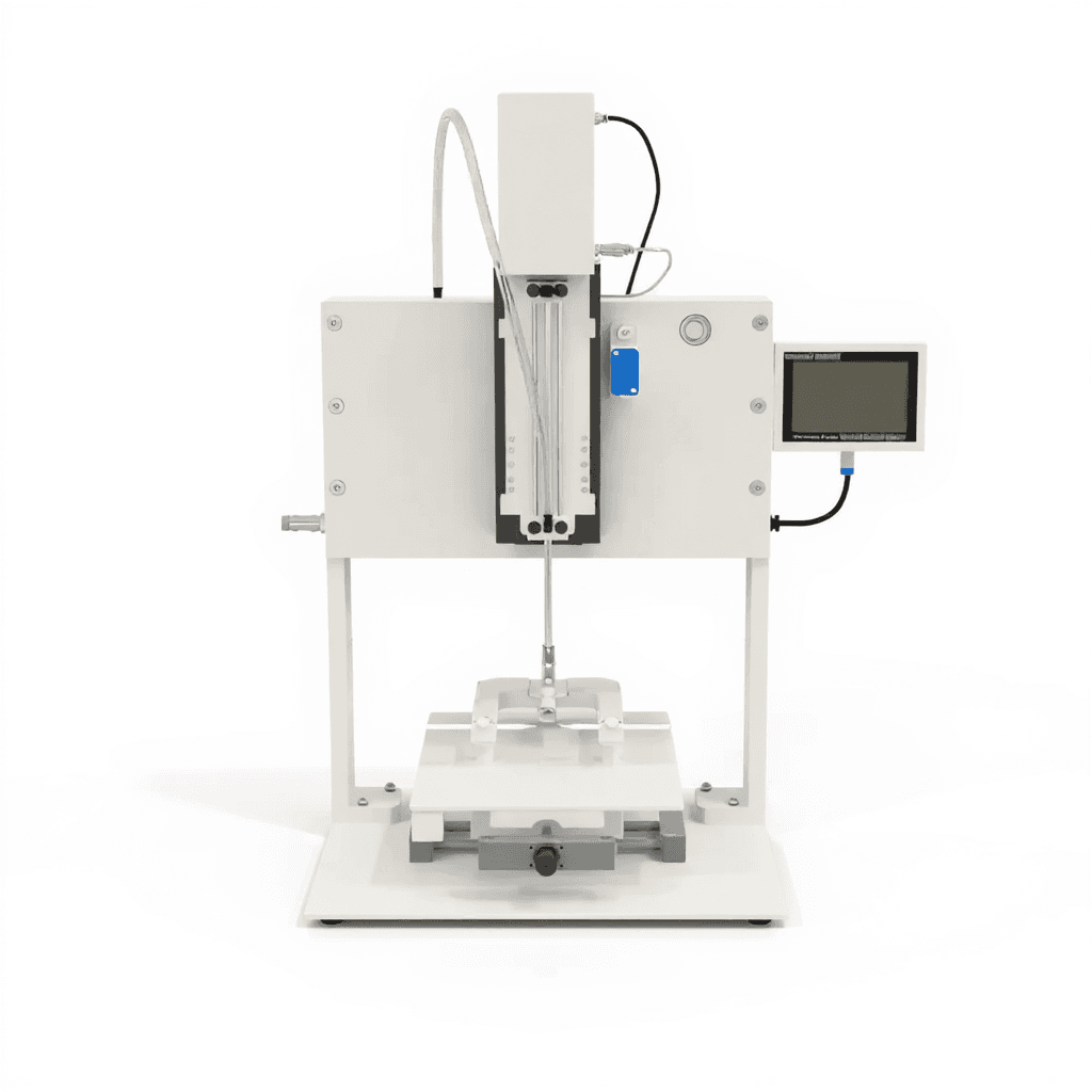
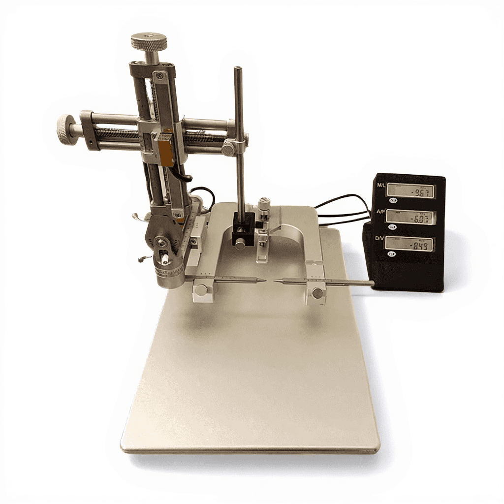
Stereotaxic Equipment
Browse All


Why ConductScience
Custom Configurations
Every lab is different. We customize equipment dimensions, materials, and specifications to match your exact research protocol.
Fast Quote Turnaround
Average 6-day quote-to-order cycle. Get pricing within 24 hours for standard equipment, 48 hours for custom builds.
Trusted by Academia
From NIH and Calico to Cornell and Emory — over 1,200 institutions trust ConductScience for their research equipment.
24/7 Technical Support
Expert support whenever you need it. Our team includes scientists who understand your research — not just the equipment.
Can’t Find What You Need?
We build custom equipment for unique research protocols. Tell us what you’re working on.
Request a Custom Quote
Trusted by World-Class Researchers