Benchtop centrifuges are the equipment utilized in laboratories to separate and purify molecular mixtures in a liquid medium based on their density gradient. Centrifugation is a significant practice employed in biochemistry labs for analyzing and isolating cells, subcellular fractions, molecular complexes, and biological macromolecules like proteins, DNA, and RNA. Centrifuges are high-speed devices that require vacuum, gravitational acceleration, and centrifugal force to separate the desired molecules from the liquid mixtures without overheating the samples. Theodor Svedberg, a Nobel Laureate, developed the first analytical centrifuge in 1924 for monitoring the sedimentation process. Later, in the 1940s, Claude and his coworkers refined the centrifugation technique, making it the heart of biomedical and biological research for the upcoming decades (Wohlleben and Wendel, 2020). Currently, small-capacity benchtop centrifuges serve as a crucial tool in routine biomedical research.
The particles are uniformly distributed in a medium before centrifugation. The denser particles in the medium move towards the bottom, and the lighter particles move upwards upon centrifugation. After centrifugation, the top liquid fraction collected is called “supernatant,” The fraction that settles at the bottom is called “pellet.” An interface separates the supernatant and the pellet. The particle recovery in the pellet is the fraction of particles in the pellet post-centrifugation. This recovery depends on the particle density and diameter (Rikkert et al., 2018).
Analytical centrifugation is a separation technique that separates the samples based on their density by exposing them to centrifugal force. It is a method of studying a molecule’s hydrodynamic properties as it flows through the liquid medium. Analytical centrifuges are high-speed ultracentrifuges with optical systems for recording the sedimentation process. They characterize biological macromolecules based on their properties like molecular weight, density, diffusion, sedimentation coefficients, etc. Naturally, biological macromolecules possess random thermal motion unaffected by Earth’s gravitational field in an aqueous environment. However, isolated biomolecules show distinct sedimentation under high accelerations. A typical analytical centrifuge creates a centrifugal force of up to 200000xg. Analytical centrifuges work on the principle that molecules with higher molecular weight and density move more quickly and settle down faster than the smaller molecules with low density.
There are two main types of analytical centrifugation experiments: (1) sedimentation velocity (2) sedimentation equilibrium experiments. In sedimentation velocity experiments, a change in concentration distribution in the centrifuge cell at a high-speed rotor is recorded. In contrast, the steady-state concentration distribution is achieved in sedimentation equilibrium experiments. The hydrodynamic properties of the system are defined by their sedimentation coefficients. Three types of optical systems for analytical centrifuges are available, i.e., absorbance, interference, and fluorescence, that facilitate the selective and precise recording of sedimentation in real-time (Ohlendieck and Harding, 2018).
Preparative centrifugation is a separation technique that separates submicroscopic particles. Unlike analytical centrifuges, preparative centrifuges cannot be used in analytical procedures. Preparative centrifuges can process large sample quantities. There are two types of preparative centrifugation.
Differential centrifugation separates the biological particles of different sizes and densities based on the difference in their sedimentation rates. It involves a gradual application in the centrifugal field to divide crude tissue homogenates containing membrane vesicles, organelles, and structural fragments into different fractions. Differential centrifugation allows the initial sedimentation of the homogenate based on their density and relative size. The larger particles are sedimented in the initial centrifugation steps, leaving only smaller particles in the supernatant (Livshits et al., 2015). The cellular debris pellets can be homogenized repeatedly to increase the yield of membrane structures and protein aggregates. The largest particles sediment at the bottom of the centrifuge tube forming a pellet, whereas the smaller particles remain suspended in the supernatant.
Density gradient centrifugation is a technique in which the macromolecules move through a density gradient until they find a density equal to their own. This method is used for separating molecules of similar sizes yet different densities. For instance, cesium chloride centrifugation is employed for the plasmid and DNA isolation, and sodium bromide or sodium iodide are used for lipoprotein fractionation, and DNA/DNA banding. The sample and the gradient-forming solution are loaded in the centrifuge tube and rotated in a centrifuge. The centrifugal force distributes the cesium salts forming a density gradient from top to bottom. The sample molecule moves to the region that equals its own density. The two types of density gradient centrifugation include rate-zonal and isopycnic centrifugation.
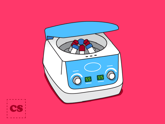
Different types of benchtop centrifuges are listed below.
Microcentrifuges are the benchtop equipment suitable for processing low-volume sample tubes, with a small footprint and capacity to process up to 48 microtubes. They can provide a speed of approximately 6000rpm and process sample volumes of up to 2ml.
Mini centrifuges occupy even less space as compared to other benchtop centrifuges. They can process a maximum of eight tubes and have a maximum speed of 6000rpm. While such centrifuges are well-suited for research labs with less space, they might not be the best choice for high output laboratories.
Plate centrifuges are mainly used in PCR labs. These centrifuges ensure that all reagents are at the bottom of the wells for accurate concentrations and precise results. Plate centrifuges allow a horizontal spin at a maximum of 400xg speed. These benchtop centrifuges have a peculiar “wing-out rotor design” to prevent spillage.
The researchers use refrigerated centrifuges for the temperature-sensitive samples as even a mild change in temperature can ruin the samples. These are almost similar in design to their non-refrigerated benchtop counterparts. However, they allow temperature control within -10oC to 40oC.
Depending on the use in low-speed, high-speed, or ultra-centrifuge, different types of centrifugal forces are experienced by rotors. According to their use, the rotors can be made of different materials. For instance, low-speed rotors are made of brass or steel, whereas high-speed rotors are made of titanium, aluminum, or fiber-reinforced composites. The types of rotors mainly used in benchtop centrifuges include fixed-angle rotors, swinging bucket rotors, and vertical tube rotors.
Fixed angle rotors are used to separate biomolecules where the sedimentation rates significantly vary, such as nuclear, mitochondrial, and microsomal separation. For isopycnic separation, centrifugation is continued until the sample particles reach their lowest isopycnic gradient. It means that the sample has reached a position where the sedimentation rate is zero. Centrifugation tubes are held at a fixed angle ranging from 14o to 40o to the vertical, and the particles move radially outwards. Since the centrifugal field is exerted at an angle, they need to travel a short distance to reach their isopycnic position. They are used for differential centrifugation.
The tubes are held parallel to the rotational axis in vertical rotors and secured in the rotor cavities by screws, washers, and plugs. The samples are collected across the tube’s diameter instead of its length. Therefore, isopycnic separation time is significantly shorter. The tubes are held at an angle of 7° to 10°. They are usually used for density gradient centrifugation.
In-swinging bucket rotors hinge pins or crossbars are used to attach rotor buckets. The buckets are hung vertically, and when accelerated initially, the buckets swing out horizontally.
Benchtop centrifuges work on the sedimentation principle, i.e., substances separate according to their density under the influence of Earth’s gravitational force ‘g’ (g = 9.81ms-2). The sedimentation rate increases when these samples undergo acceleration in a centrifugal field (G > 9.81ms-2). The relative gravitational field is often expressed as a multiple of gravitational acceleration. Underlying points must be considered while working with benchtop centrifuges.
The frictional force experienced by a biological medium in a viscous medium is in a direction opposite to that of sedimentation. It is equal to the product of the particle’s velocity and frictional coefficient. As described earlier, the centrifugal field is related to Earth’s gravitational field. The relative centrifugal field (RCF) is the ratio of centrifugal force at a specified radius and speed to the standard gravitational acceleration (Ohlendieck and Harding, 2018). The RCF can be calculated from the formula presented below:
Insert the sample tube in one of the portals. Based on the number of samples you are centrifuging, add tubes filled with water for balancing. Secure the lid and select desired settings. Press the “start” button and wait for the centrifuge cycle to complete. Once the centrifuge stops spinning, take out the sample and the balances. Notice that the sample is differentiated into two parts, i.e., supernatant and pellet. Analyze the sample as per your sample requirement.
Conduct Science presents the best range of benchtop centrifuges with an easy-to-use ergonomic design that complements your laboratory requirements. These benchtop equipment are made of durable and high-quality material and bring efficiency and consistency in experiments. Different benchtop models possess two types of rotors: (1) out-swung rotors (2) fixed-angle rotors. These centrifuges have a maximum 250 ml instrument capacity and can provide a variable speed of up to 6000rpm. Moreover, they can give a maximum RCF of 20,000xg. Other features of these instruments include continuous hold-spin function, rubber suction feet for grip at the benchtop, and last-spin memory to record the last centrifuge experiment. The researchers can also buy microcentrifuges or ultracentrifuges with a compact design best suited for their workspace.
Benchtop centrifuges have various applications in microbiology, biochemistry, and biomedical research. Differential centrifugation and density gradient centrifugation isolate microsomal fractions from muscle homogenates, isolate highly purified sarcolemmal vesicles, and sub fractionation liver mitochondrial membrane systems. A few applications of benchtop centrifuges are given below.
Platelet Rich Fibrin (PRF) Production
Second-generation platelet-rich fibrin (PRF) is now used as a “therapeutic strategy for promoting implant healing and bone and soft tissue integration” in dental implants. Platelet-rich fibrin (PRF) or leukocyte and platelet-rich fibrin (L-PRF) are generally obtained from patients’ blood and centrifuged at 700xg RCF for 12 minutes without additives. Feng et al. (2020) studied the antibacterial properties of PRF prepared by horizontal centrifugation against E. coli and S. aureus. The researchers reviewed that PRF’s horizontal centrifugation yields better layer separation by minimizing cell accumulation at distal ends of centrifugal tubes. The swung-out buckets used in horizontal centrifugation produce a completely horizontal tube. It causes substantial differences between RCF-min and RCF-max due to the difference in minimum and maximum tube radii. They reviewed that horizontal centrifugation could increase the yield fourfold compared to fixed-angle centrifugation. Therefore, they aimed to compare the antimicrobial effects of horizontal centrifugation (H-PRF) and L-PRF were produced on a fixed angle rotor against S. aureus and E. coli and determined whether these antibacterial effects were correlated with immune cell number. In the case of L-PRF preparation, the experimenters centrifuged the samples using fixed-angle rotors and observed an angular red blood layer formed within the centrifuge tube. On the contrary, in the case of horizontal centrifugation, the researchers observed a horizontal division between red blood cells and platelet-rich fibrin. Since cell content in each PRF layer is different, and H-PRF can harbor up to four times more leukocytes than L-PRF, the researchers divided liquid state PRF into five equal portions following centrifugation. From the CFU examination and flow-cytometric analysis of these samples, the experimenters concluded that solid PRF exhibits better antimicrobial activity than liquid PRF.
Isolation of Biopsy-based Tumor Biomarkers
Cancer patients’ blood contains numerous biomarkers, including circulating tumor cells (CTC), circulating cell-free DNA (ccfDNA), and tumor-educated platelets (TEPs). As these biomarkers differ in size and density, the lab personnel usually centrifuge blood to isolate or concentrate the biomarker of interest. Rikkert et al. (2018) devised Stokes’s law-based model to estimate the effect of centrifugation on biomarker purity and then applied this model to predict biomarker behavior during centrifugation. According to Stoke’s law, the acceleration due to gravity depends on the distance from the rotational axis in a centrifuge. This gravitational acceleration is given by:
g = Rω2
where R is the distance to the axis of rotation and ω is the angular velocity. Stoke’s equation is usually used in the case of the swing-out rotor. They diluted polystyrene beads in phosphate buffer saline (PBS) or blood plasma to validate the suggested model. They subjected the sample to centrifugation at 300g for 20 minutes, 2700g for 22 minutes, or 60 minutes at 15,800g. They also measured bead concentration by flow cytometry before and after centrifugation. The experimenters concluded that Stokes law could be used to predict biomarker behavior in blood. They also concluded that centrifugation alone could not isolate a single biomarker since other coisolated biomarkers remained present in significant amounts.
Benchtop centrifuges are a perfect fit for the research laboratories with less benchtop space due to their compact design. These centrifuges are easy-to-use and offer fast start-up and shut down. They are fully automated hence reducing the possibility of experimental failure due to human error. Moreover, their enclosed design keeps the specimens uncontaminated. However, a potential disadvantage of centrifuges is their higher energy demand (Brandt et al., 2017).
Wohlleben, W., Coleman, V. A., & Gilliland, D. (2020). Analytical centrifugation. In Characterization of Nanoparticles (pp. 225-247). Elsevier.
Livshits, M. A., Khomyakova, E., Evtushenko, E. G., Lazarev, V. N., Kulemin, N. A., Semina, S. E., … & Govorun, V. M. (2015). Isolation of exosomes by differential centrifugation: Theoretical analysis of a commonly used protocol. Scientific reports, 5(1), 1-14.
Brandt, M. J., Johnson, K. M., Elphinston, A. J., & Ratnayaka, D. D. (2017). Chapter 13—Energy Use, Sustainability and Waste Treatment. Twort’s Water Supply, 7th Ed.; Brandt, MJ, Johnson, KM, Elphinston, AJ, Ratnayaka, DD, Eds, 553-580.
Rikkert, L. G., Van Der Pol, E., Van Leeuwen, T. G., Nieuwland, R., & Coumans, F. A. (2018). Centrifugation affects the purity of liquid biopsy‐based tumor biomarkers. Cytometry Part A, 93(12), 1207-1212.
Feng, M., Wang, Y., Zhang, P., Zhao, Q., Yu, S., Shen, K., … & Zhang, Y. (2020). Antibacterial effects of platelet-rich fibrin produced by horizontal centrifugation. International journal of oral science, 12(1), 1-8.
Ohlendieck, K., & Harding, S. E. (2018). Centrifugation and Ultracentrifugation. Wilson and Walker’s Principles and Techniques of Biochemistry and Molecular Biology.
Get the entire package for up to 50% discount with our Replication program.
DISCLAIMER: ConductScience and affiliate products are NOT designed for human consumption, testing, or clinical utilization. They are designed for pre-clinical utilization only. Customers purchasing apparatus for the purposes of scientific research or veterinary care affirm adherence to applicable regulatory bodies for the country in which their research or care is conducted.