Magnetic Resonance Imaging (MRI)

Introduction
Magnetic resonance imaging is an advanced, non-invasive diagnostic medical imaging technique that provides three-dimensional images of organs. The discovery of MRI involves a series of successful events that started in 1946 when Felix Bolch proposed the new magnetic properties of the atomic nucleus[1]. He explained the behavior of atoms at tiny magnets, and based on these, NMR tools were developed[2].
Paul Lauterberg, the pioneer of MRI, introduced the concept of “tissue characterization” after the discovery that the affected and normal tissue shows different NMR (nuclear magnetic resonance) parameters by Raymond Damadian[1]. Then, the phenomenon of tissue characterization or tissue/organ imaging using the magnetic field was termed Magnetic resonance imaging.
Techniques other than MRI, like X-ray, produce gray and flat images without contrast compared to MRI which produces images with excellent contrast resolution in any dimension. This property helps in capturing even small lesions in tissues and to identify multiple sclerosis, tumors, tendonitis, strokes, and many other abnormalities[1].
This article poses the workings of magnetic resonance imaging, its principle, application, advantages, and disadvantages[1]. You will also learn what safety measures you should take while using the machine. So, let’s get started.
The Concept of MRI and Its Physics
The atomic nucleus is made of protons that contain positive charge, neutrons having neutral charge, and electrons that carry a negative charge. The similarity between these particles is to spin about their axis and possess an intrinsic spin of ½ which can be positive or negative. The spin is also called the angular momentum of the particles[2].
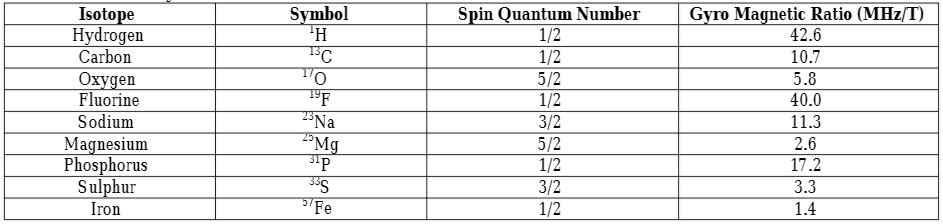
Each nucleus has its spin characteristic depending on the number of protons and neutrons present in it. For example, if a nucleus has an even number of electrons and protons, then, they can be arranged in such a way that their spin gets canceled. However, when the particles are in odd numbers, some of the spins will not be canceled, which creates a net spin-charge and produces magnetic properties known as a magnetic moment[2]. So, when the spin is placed in a magnetic field, it rotates around its axis by absorbing photons at certain frequencies which depends on the gyromagnetic ratio of the particle[2]. The equation explains the relation:
V = 𝝲B, for hydrogen, 𝝲 = 42.58 MHz/T
The magnetic property of the atomic nuclei is utilized in MRI to produce images of organs and the elements having unpaired protons are preferred (see table for MRI friendly elements). However, the hydrogen element is mostly preferred as an MRI imaging source because of the following three[2] reasons:
- Our body is rich in hydrogen atoms
- Hydrogen has the largest gyromagnetic ratio.
- Hydrogen has a large magnetic moment.
Elements preferred for MRI[1]
Spin, Precession, and Larmor frequency
The protons present in the atom are randomly oriented in the absence of an external magnetic field, however, when the magnetic field is applied to the sample, all the protons are arranged according to the applied field. This occurs in two[1] ways:
- The protons are aligned in the opposite direction of the applied magnetic field (Bo) i.e higher energy state.
- The arrangement of protons in the direction of the applied magnetic field (Bo) i.e lower energy state.
The north and south poles of protons do not exactly align in the direction of the applied magnetic field and somewhat tilt from the direction parallel to the applied magnetic field. This wobbling/oscillation of protons is called precession and the rate or frequency of precession is called Larmor frequency[1]. The equation expressing Larmor frequency is:
F = r B
Here, F = Larmor or resonant frequency (MHz)
r = gyromagnetic ratio of protons (MHz/T)
B = strength of applied magnetic field (T)
Larmor frequency determines the frequency of the MRI. The Larmor frequency of hydrogen in the 1T applied magnetic field is 42.58 MHz and the MRI requires the magnetic field strength ranging from 0.1T to 4.0T. The net magnetization of elements in the applied magnetic field strength is denoted by MZ[1].
Radio Frequency Pulse and RF flip
The radio-frequency is sent through the patient’s body to manipulate the net magnetization vector. The radio-frequency when matches the center frequency, creates a resonance, and only protons that spin at the radio frequency, respond to the applied pulse.
The radio-frequency interacts with every component attached to the patient’s body (tissue, any foreign element, or metallic implants). During imaging, it is transformed into heat and the absorption and generation of heat take place in tissues[1]. This thermal characteristic is different for different organ systems. For example, limbs dissipate thermal energy more rapidly than the abdomen whereas the eyes take much more time to dissipate thermal energy[1].
The radio pulse should be provided for sufficient duration to generate an intensity that rotates the net magnetization vector and produce signals which help to transform and create the image. The bound state and presence and absence of hydrogen in tissue are some of the factors that play a role in the generation of the signal. For example, hydrogens in the bone present in a tightly bound state and do not produce any usable signal.
T1 and T2 relaxation
T-1 relaxation is defined as the time tissues take for the longitudinal magnetization to reach 63% of the original magnetization. When the pulse stops, the protons in the higher energy state want to go back to the lower energy state[1]. The bound state of hydrogen also determines the differences in the rate of relaxation of protons in tissues. For example, in fat tissues, hydrogens are in a bound state so the rate of relaxation is low as compared to water. This specific property is involved in creating a contrast image in MRI.
When the radio-frequency is applied at 90o, all magnetization flipped in the XY plane, called transverse magnetization[1]. At the same time, the protons start to rotate “in-phase” i.e all vectors align in one direction. However, this alignment is disturbed by the other surrounding vectors. So, the rate of the flip will be different in different vectors. This will disturb the alignment of vectors in the same direction and start “de-phasing”. This phenomenon of vectors getting from in-phase to out-of-phase or de-phase is called T2 relaxation[1].
It should be noted that the T1 and T2 relaxation processes are independent of each other but occur simultaneously. This whole physics of protons during MRI help to create a contrast image[1].
Summary of MRI workings
The magnets in MRIs produce a strong magnetic field that forces the protons in the body to align with the field in the same direction, an equilibrium is maintained. This equilibrium is disturbed when an external radiofrequency pulse is applied through the patient[5]. In this condition, the protons reach a higher energy state and are opposite to the applied magnetic field.
However, when the external radiofrequency pulse is stopped, the protons return to the lower energy state to realign with the magnetic field which is detected by the MRI sensors.
The time protons take to align with the magnetic field and the amount of energy released depends on the environment and chemical nature of the molecule. These differences in protons of different tissues based on the magnetic field help physicians to tell the difference between two tissues[5].
Contrast agents are given to patients intravenously to fasten the process of alignment of protons. The faster the realignment of protons, the brighter the images are produced.
Applications of MRI
MR imaging has the potential to produce a clear picture of the anatomy and detect anomalies in the body. Here, clinical applications of MR imaging based on the region of the body is explained[1] in brief.
- Head: MR imaging has the potential to detect brain tumors, aneurysms, bleeding in the brain, damages due to stroke, nerve injury, and problems associated with eyes and optic nerves, ears, and auditory nerves[1].
- Chest: By using MR imaging, one can identify any damages in the heart and lungs. It can be used to picture heart valves, coronary blood vessels, breast, and lung cancer[1].
- Magnetic Resonance Angiography: It is a type of MRI scan which is used to look at blood vessels and the flow of blood through them. It can detect any defects in arteries and veins such as aneurysms, blocked blood vessels, the torn lining of the blood vessels[1].
- Abdomen and Pelvis: MRI scans help to detect problems associated with the stomach, liver, pancreas, gallbladder, and kidneys. The diseases that can be studied using the technique include tumors in these areas, bleeding, infection, and blockage. It can also picture any abnormalities in the uterus and ovary in females and prostate in males[1].
- Bone and Joint: The diseases studied by MRI related to bones and joints include arthritis, problems with the temporomandibular joint, bone marrow problems, bone tumors, cartilage problems, torn ligaments or tendons, and infection. It produces a much clearer image compared to X-ray and commonly used for the bone associated diseases[1].
- Spine: It can be used to check discs and nerves of the spine for anomalies like stenosis (narrowing of duct, passage, or vessels of the body), disc bulges, and spinal tumors[1].
- Neck: The diseases that can be observed using MRI in the neck area include thyroid nodules, cysts, parathyroid adenomas, abnormal lymph nodes, and tumors in the laryngeal tracts[4].
Advantages of MRI
The advantages[1] of MR imaging include:
- It doesn’t involve the use of ionizing radiation so it doesn’t cause any harmful effects on the individual.
- MR imaging is non-invasive which means its procedure doesn’t involve any insertion or break in the skin[1].
- It produces contrast images which is the principal advantage over other available techniques. This property allows the adjacent soft tissues to be differentiated by altering the radio-frequency pulses.
- It is possible to take direct, sagittal, coronal, and oblique (multiplanar) images using MRI[1].
- The artifact (abnormal appearances in the image produced by MRI) produced due to dental filling is not significant in MR imaging.
- MR imaging helps to differentiate between acute and chronic transit parallelly with the microscopic examination of tissues to study diseases[1].
- It is efficient to determine and examine spreads within the spinal cord or bone marrow.
- MRI can determine if cancer has spread to other locations in the body and that helps to determine the best treatment[6].
- No significant adverse effects have been observed till now.
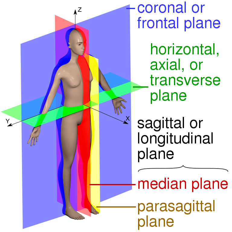
Figure: The image showing different anatomical planes that MRI can capture[7].
Source: https://commons.wikimedia.org/wiki/File:Human_anatomy_planes,_labeled.svg
Disadvantages of MRI
- Any tiny metal attached to a patient’s body can create a distorted MRI image. So, patients with surgical clips or stents, and any metallic artifacts are excluded from this process.
- In an MRI scanner, patients are kept in long cylinders and if the regions being imaged are in the head, the patient may experience a sense of isolation and become claustrophobic and anxious after a long period of confinement. Generally, patients refuse to go inside the cylinder looking at deep space[3].
- It’s difficult to scan ill patients using MR imaging techniques because they experience vomiting, respiratory distress, and unnoticed movements with other emotional reactions[3].
- The patients have to remain immobile for 5-15 minutes of the scanning period. The long scan time is a big disadvantage of magnetic imaging[3].
- When the current is passed through a conducting metal (coil) immersed in a magnetic field, the coils are displaced slightly and abruptly producing a sharp sound. During the scanning process, the gradient is turned on and off multiple times which produces a machine gun-like noise. These noises disturb the patients during MR imaging and some patients even can not tolerate it[3].
- The patients having pacemakers are avoided because of the machine’s magnetic field tampers with this vital device. So, it’s unwise to place this kind of patient in MR imaging[3].
- MR imaging is the most complex technology yet used for medical purposes[3].
- Magnetic and electrical shielding is required for the optimal performance of MR imaging. Also, the setup is needed to set up at a particular distance from all the other machines which adds up the site cost in addition to the shielding cost. So, MR imaging is a very expensive technology[3].
- MRI can’t detect all types of tumors and it generates false-positive results because of its limited performance in distinguishing the malignant and benign tumors[6].
- A contrasting agent is applied to the patient’s body for a clearer image that can cause allergy in some patients or cause infection at the injection site.
Recent Advancements in MRI
- 3-D Imaging: The improvements in MRI have now made it possible to capture the image in volume rather than tomographic slice[1].
- Echo imaging: It’s a multi-echo spin that records different parts by using different echo spins and completely records the image in 1/4th of the time[1].
- Chemical Shift Imaging: This advancement allows us to produce the whole image from just one chemical shift in the sample components.
- Echo Planar Imaging or Functional MRI: It helps to visualize the function in the brain and the change of blood flow to its different parts. In this, the tomographic image is taken at a video rate[1].
- Magnetization transfer contrast: This helps in increasing the contrast between the tissue and brings more clarity to the image. However, it involves physical aspects rather than chemical means.
- MR Elastography: This utilizes the elastic modulus property of the tissues. To capture the image, it sends ultraviolet waves and locates any pathogenesis in soft tissues[1].
Conclusion
Despite certain limitations, magnetic resonance imaging is an efficient tool serving several clinical applications. The best thing is the absence of harmful radiation during the whole procedure. It has made it possible to produce a high quality and contrast image of affected parts. In terms of safety and sensitivity, it can replace many other invasive and risky techniques. It helps to detect any pathological changes during the early phase.
However, making the procedure more comfortable for the patients by the reduction in coil noise and tube length may help many other individuals in their diagnostic procedures. Further, advances in the process of imaging and dynamic scanning will enhance and broaden its application in the future.
References
- Idrees, Muhammad. (2014). AN OVERVIEW ON MRI PHYSICS AND ITS CLINICAL APPLICATIONS. International Journal of Current Pharmaceutical & Clinical Research. 4. 185-193.
- Katti, Girish & Ara, Syeda & Shireen, Dr. (2011). Magnetic resonance imaging (MRI) – A review. Intl J Dental Clin. 3.
- Oldendorf, W., & Oldendorf, W. (1988). Advantages and Disadvantages of MRI. Basics of Magnetic Resonance Imaging, 125–138. doi:10.1007/978-1-4613-2081-4_9
- William R. Hendee and Kathleen A. (1985). Clinical applications of magnetic resonance imaging-Current status. West J Med; 143:793-803.
- https://www.nibib.nih.gov/science-education/science-topics/magnetic-resonance-imaging-mri
- https://www.cancerquest.org/patients/detection-and-diagnosis/magnetic-resonance-imaging-mri#
- https://commons.wikimedia.org/wiki/File:Human_anatomy_planes,_labeled.svg