


As an Amazon Associate Conductscience Inc earns revenue from qualifying purchases
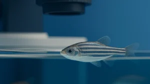
Total internal reflection fluorescence microscopy (TIRFM) is a cutting-edge optical technique that excites the fluorophores in a fragile axial region to visualize the cellular events occurring close to the cell surface. The evanescent field, a near-field wave which decays in intensity over a sub-wavelength distance, is used as the basis for total internal reflection fluorescence microscopy (TIRFM). The evanescent field occurs when the incident light is totally reflected at the interface of two transparent media having different refractive indices. The TIRFM has a wide range of applications in visualizing the biological events and quantifying their kinetic rates. The total internal reflection fluorescence microscopy is used to study the protein-protein and protein-nucleic acid biochemical interactions. The total internal reflection fluorescence microscopy has made it easier to understand the mechanism and function of cellular components including signaling cascades, membrane proteins, and molecular motors. The relative ease of use of TIRF and the high sensitivity in single-molecule detection makes it an indispensable technique in biomedical science to tackle a wide array of cellular questions.
The total internal reflection fluorescence microscopy is based on the evanescent field, which exclusively illuminates a thin plane just below the glass coverslip. The evanescent field occurs as a result of the total internal reflection of the light rays at the interface of the imaging surface and an aqueous medium. The refractive index of the optical medium tells about the propagation of the electromagnetic waves through it relative to the propagation of the wave through the vacuum. When light rays traveling through one medium strike at the interface of another medium with a different refractive index, the subsequent direction of the light rays is changed depending on the angle at which the light meets the interface. The energy of the evanescent field decreases as it travels to the interface, only the fluorophores close to the coverslip are excited. This creates the images with an outstanding signal-to-noise ratio, as the rest of the fluorophores in the cell are hardly excited. Therefore, the membrane-associated processes like cell adhesion, molecule transport, hormone binding, and exocytotic and endocytotic processes are observed (Yildiz. & Vale., 2015).
The total internal reflection fluorescence microscopy consists of an objective lens, an excitation beam path which passes the light through the objective lens to the sample, and a coupling element arranged in the back focal plane of the objective lens. The coupling element consists of two areas; one for relaying light to the objective lens for total internal reflection illumination and the second for separating the light emitted by the sample and passing it through the excitation beam path in reverse direction. The laser beams are joined with a dichroic mirror and expanded via Gaussian beam expander. These laser beams are focused on the back focal plane of the objective with an achromatic doublet lens. A set of multiband dichroic and emission filters reflect the laser beams on the objective and transmit the fluorescence simultaneously. The fluorescence is then separated by a Dual View instrument which is equipped with a dichroic mirror to split the fluorescence, and band-pass emission filters to reduce the cross talk between the two fluorescence channels (Fish, 2009).
Coverslip preparation
Cell culture and transfection
Microscope and sample preparation
Stimulation and imaging
Cleaning and coating of dishes
Cell transfection
Preparation of mouse β cells
Infection
Infect the cells with adenoviruses at the rate of 30–100 infectious particles per cells for 4 hours, and then change the culture medium.
Imaging of exocytosis
Insulin-responsive GLUT4 storage vesicles (GSV) helps in the glucose uptake by translocating to the cell surface. In the study, the total internal reflection fluorescence microscopy was used to understand the events involved in glucose uptake and insulin-regulated GLUT4 translocation in both cultured 3T3-L1 adipocytes and primary adipocytes isolated from the rodents and humans. The cells were prepared, transfected, and imaged under the TIRF microscope. It was found that the GSV traffic is decreased as the vesicles lead to the plasma membrane followed by fusion. It was observed that the insulin-induced GSV fusion is followed by the release of GLUT4 monomers into the plasma membrane. The total internal reflection fluorescence microscopy has been found as a powerful tool to observe the vesicle translocation across the plasma membrane.
The total internal reflection fluorescence (TIRF) microscopy has been widely used to visualize single molecules in the cells to unveil fundamental aspects of cell biology as it selectively excites a very thin fluorescent volume close to the substrate on which the cells are grown. TIRFM has been used to track single receptors having a SNAP-tag, and to compare their arrangement, mobility, and supramolecular organization. The studies presented that the G-protein coupled receptors (GPCRs) possess varying degrees of di-/oligomerization. Whereas β1- or β2-Andregenic receptors are freely diffusive on the cell surface. These results suggest that GPCRs are located on the cell surface in a dynamic equilibrium, with constant formation and dissociation of new receptor complexes that can be stimulated or targeted, in a ligand-regulated manner, to different cell-surface micro-domains. The total internal reflection fluorescence microscopy has become a promising technique in profiling the receptor pharmacology in clinical applications.
Cell-substrate contact regions demonstration is one of the applications of total internal reflection fluorescence microscopy. The evanescent excitation has been used to image the arrangement of fluorescent probes for different membrane components in cell-substrate contact regions. The fluorescent reporters attached to cytoskeletal elements are visualized in rat myotube membranes adjacent to glass substrates. TIR-FPPR was used to probe the lateral mobility of fluorescent antibodies which linked the rat basophil leukemia cells to supported planar membranes. It was found that the fluorescence is intense if the cell-to-substrate distance is large and is weak if the distance is small. The TIRFM has also been used to measure the spatial distribution of fluorescence intensities that provides a two-dimensional map of cell-to-substrate contact distances and the binding kinetics of the cell-substrate contact.
The total internal reflection fluorescence microscopy has been used to visualize the trafficking of plasma membrane-localized intracellular estrogen receptors following estradiol stimulation in living cells. To visualize estrogen receptor trafficking N-38 neurons were used as a model for membrane-initiated estradiol signaling. The TIRFM permits observation of live, intact cells while allowing visualization of the receptor activation cascade following estradiol activation. The TIRFM yielded high-contrast real-time images of fluorescently labeled E6BSA molecules on and just below the cell surface and was found a powerful tool for studying estrogen receptor trafficking in living cells.
The total internal reflection fluorescence microscopy has also been used to demonstrate different steps of intracellular signaling. The TIRF has been instrumental in delineating plasma membrane recruitment and spatial distributions of signaling molecules. The plasma-membrane-targeted biosensor enabled the imaging of temporal oscillations of cAMP (cyclic Adenosine monophosphate) signaling, instigating the research in the regulation of upstream targets. The single plasma membrane Ca2+ channels have also been imaged with spatial and temporal resolution revealing uneven molecular kinetics. The TIRF and patch-clamp methods have successfully demonstrated the localization and signaling of open calcium channels and calcium-sensing molecules, explaining the spatial dynamics of intracellular calcium signaling.


Monday – Friday
9 AM – 5 PM EST
DISCLAIMER: ConductScience and affiliate products are NOT designed for human consumption, testing, or clinical utilization. They are designed for pre-clinical utilization only. Customers purchasing apparatus for the purposes of scientific research or veterinary care affirm adherence to applicable regulatory bodies for the country in which their research or care is conducted.