Types of Nucleic Acids and Their Biological Significance

Introduction and History
Nucleic acids are an essential class of macromolecules present in all cells of organisms including viruses.[1] They were first discovered by Friedrich Mischer in 1869. He wanted to study the composition of leukocytes from the pus cells of discarded surgical bandages. During his study, he observed nucleic acids in the form of a precipitate when the cells were treated with acid. He called this component nuclein. Later in 1889, Altmann named the constituent nucleic acid.[2]
A few other major historical events in the discovery of nucleic acids are mentioned below:[2]
- Kossel showed that the nucleic acids are composed of purine and pyrimidine bases, sugar, and phosphate.
- Around 1930, many scientists characterized nucleic acids and identified the four bases and deoxyribose groups in DNA (this is when it was named deoxyribonucleic acid).
- In 1939, the role of RNA in protein synthesis was discovered.[3]
- Astbury and Bell published the first x-ray diffraction pattern of DNA.
- Erwin Chargaff discovered that DNA from a particular species contains the same amount of cytosine (C) and guanine (G), and the same amount of adenosine (A) and thymine (T).[2]
- Avery, MacLeod, and McCarty proved through an experiment that DNA is the genetic material — a carrier of genetic information.
- In 1955, Watson and Crick designed and presented the structure of DNA.[4]
- In 1959, Severo Ochoa won a Nobel prize for the discovery of the mechanism of RNA synthesis.[3]
Nucleic acids act as a blueprint of all the information to build and develop organisms. They are the chemical basis for the transmission of genetic information or traits from parents to offspring.
This article describes the structure, biochemical properties, and functions of nucleic acids in organisms.
Types of Nucleic Acids and their Basic Structures
Nucleic acids are biopolymers built from several monomer units of nucleotides that are composed of three components: sugar, phosphate, and a nitrogenous base.
Nitrogenous Bases
Nitrogenous bases are planar, heterocyclic, and water-soluble molecules. They are of two types: purines and pyrimidines.[5]
Purines: Purines are two carbon-nitrogen rings. It includes adenine (6-aminopurine) and guanine (6-oxy-2-aminopurine). Adenine contains an amino group at the C-6 position of the ring while guanine has an amino group at the C-2 position and a carbonyl group at the C-6 position.[5]
Pyrimidines: The primary structure of pyrimidines is composed of a single carbon-nitrogen ring. Thymine (5-methyl-2,4-dioxypyrimidine) and cytosine (2-oxo-4-aminopyrimidine) are two pyrimidines found in DNA, while uracil (2,4-dioxypyrimidine) and cytosine are found in RNA.[5]
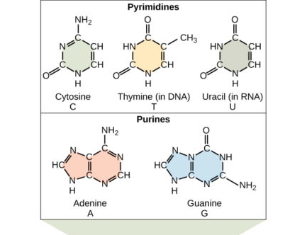
Image: The structural diagram of two purines and three pyrimidines found in DNA and RNA.[6]
Source: lumenlearning
Sugar
Two types of sugar occur in nucleic acids: Ribose and deoxyribose sugar. The difference between these two types of sugars is due to the presence of the hydroxyl group on the second carbon of the ribose, and hydrogen on the second carbon of the deoxyribose. All the sugars present in nucleic acids exist in D-stereoisomeric forms.[5]
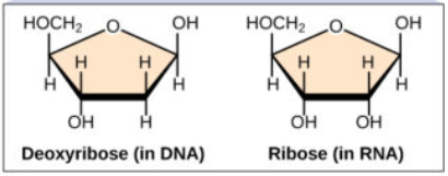
Image: The structural configuration of deoxyribose and ribose sugar.[6]
Source: Lumenlearning
The pentose sugar present in nucleic acids is planar and puckered around C2’ or C3’ carbon. Purines are in C2’- endo pucker conformation while pyrimidines prefer C3’- endo conformation.[5]
Nucleic acids are divided into two main classes, based on the sugar involved in the formation of nucleic acid structure: Deoxyribonucleic Acid (DNA) and Ribonucleic Acid (RNA).
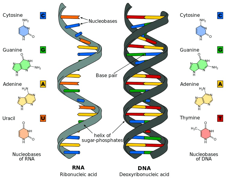
Image: The structure of DNA and RNA with the representation of their nitrogenous bases and helix formation.[7]
Source: Biologydictionary
Phosphate
The phosphate group present in the nucleotides distinguishes them from nucleosides (composed of only sugar and nitrogenous base and have entirely different functions). Nucleic acids are formed from the joining of two or more nucleotides.
The condensation reaction occurs between the alcohol of a 5’- phosphate group of one nucleotide and the 3’- hydroxyl group of a second nucleotide. The reaction leads to the formation of phosphodiester bonds between the molecules.[5]
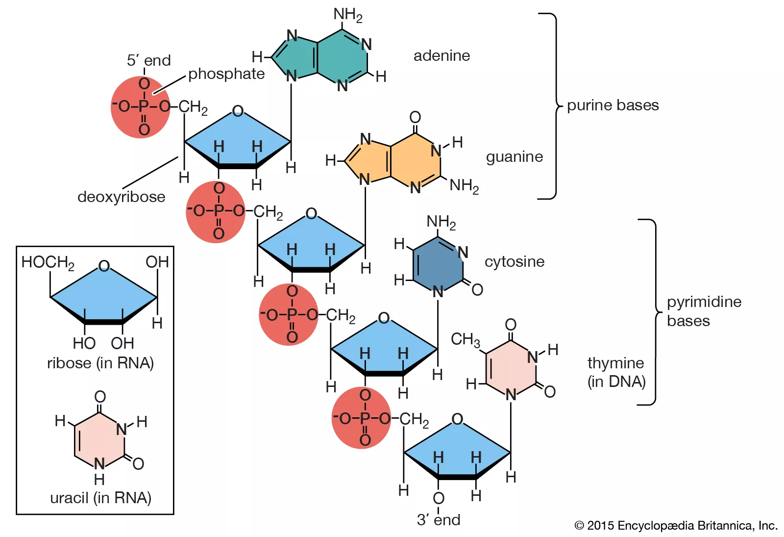
Image: A representative complete structure of nucleic acids.[8]
Source: Encyclopedia Britannica
Deoxyribonucleic Acid (DNA)
In 1962, Watson, Crick, and Wilkins were awarded the Nobel prize for discovering the double-helix molecular structure of DNA. The four bases present in DNA structure include adenine (A), guanine (G), cytosine (C), and thymine (T).
Structure of DNA
The structure proposed by Watson and Crick is the B-form of the DNA double helix. However, DNA also exists in two other forms — A-form and Z-form — that are also biologically significant structural forms. The conformation adopted by DNA, out of these three forms, depends on the hydration level, DNA sequence, chemical modification of the bases, and the concentration of metal ions in solutions.[5]
The nitrogenous bases constituting DNA always form a pair by establishing hydrogen bonds between them. For example, adenine and thymine bond together with two hydrogen bonds, while guanine and cytosine bond together with three hydrogen bonds between them.[8] The bonding has a significant biological role in the replication mechanism of DNA and the transfer of genetic information.[8]
The bases act as a bridge between two sugar-phosphate chains of the DNA. Sugar and phosphate are the backbones of the DNA strands.
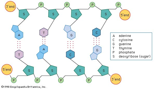
Image: A schematic representation of hydrogen bonds between the nitrogenous bases, linking together two strands of the DNA.
Source: Encyclopedia Britannica[8]
Some of the major features of B-DNA are given below:[5]
- It has a central axis around which two polynucleotide strands of the DNA are coiled together to form a helical structure.
- It has a right-handed helix.
- The two strands of the structure are present in an antiparallel fashion (one strand in 5’-3’ and the other in 3’-5’ orientation).
- The interaction between base pairs forms major and minor grooves.
- The diameter of the helix is 20 Å, the helix rise per base pair is 3.32 Å, and the helix pitch is 33.2 Å.
- The structure has 10.4 base pairs per helical turn.[5]
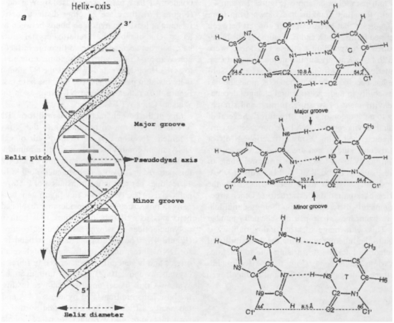
Image: The schematic diagram of Watson-Crick double-helical B-DNA.[9]
Source: Bansal, M. (2003). DNA structure: Revisiting the Watson–Crick double helix.
Given below is a chart of features and differences between B-, A-, and Z-forms of the DNA structure:[5]
| Content | 1 | Helix sense | Right-handed | Right-handed | Left-handed | Content | 2 | Repeating units | 1 bp | 1 bp | 2 bp | Content | 3 | Twist angle | 33.6° | 34.3° | 60°/2 | Content | 4 | Mean bp/turn | 10.7 | 10.4 | 12 | Content | 5 | Base pair tilt | 20° | -6° | 7° | Content | 6 | Rise/base pair | 2.3 Å | 3.32 Å | 3.8 Å | Content | 7 | Pitch/helix turn | 24.6 Å | 33.2 Å | 45.6 Å | Content | 8 | Mean propeller twist | +18° | +16° | 0° | Content | 9 | Glycosidic bond | Anti | Anti | Anti for C, syn for G | Content | 10 | Sugar pucker | C3’-endo | C2’-endo | C-C2’-endo, G-C3’ endo | Content | 11 | Diameter | 23 Å | 20 Å | 18 Å | Content | 12 | Major groove | Narrow and deep | Wide and deep | Flat | Content | 13 | Minor groove | Wide and Shallow | Narrow and deep | Narrow and deep |
Properties of DNA
1. Thermal denaturation
DNA double helix, when exposed to specific conditions of pH, ionic strength, or temperature, disrupts the hydrogen bond connecting the two strands of DNA. When the temperature is the denaturing agent, the process is known as the melting of DNA.[5]
Denaturation changes the physical properties of DNA and increases the absorbance of the DNA solution by 40% at 260 nm (termed as hyperchromic shift). The temperature at which the change in absorbance is half-maximal, or at the midpoint, is known as melting temperature (Tm).[5]
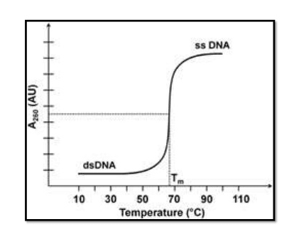
Image: The graph for thermal denaturation of dsDNA to ssDNA.[10]
Source: Slideshare
The denatured DNA strands can be re-natured by the method of slow cooling, but the graph will not be similar to denatured DNA.
The melting temperature of dsDNA depends on several factors including GC (guanine and cytosine) content of DNA, ionic strength, and change in pH.
2. Stability of DNA Helix
The DNA helix is stabilized by noncovalent interactions which include stacking interaction between adjacent bases and hydrogen bonds between adjacent strands. The stacking interaction between bases involves hydrophobic interaction and Van der Waals interaction that provides overall stability and minimizes contact of the bases with water.[5]
The hydrogen bond is present between the nitrogenous bases (connecting two strands) and in the sugar-phosphate backbone (connecting with water molecules). A sum of all these interactions provides overall strong stability to the DNA helix.
3. Chemical modification
DNA can be modified by enzymes like DNA methyltransferase, physical agents like oxidants or ionizing radiation, or chemical carcinogens.[8] It can also be cleaved and degraded by enzymes like endonucleases or exonucleases. The abnormal modifications in the DNA by any of the mentioned agents lead to severe fatal diseases in the organisms.[8]
4. Supercoiling
Supercoiling is referred to as the coiling of a DNA double helix upon itself. It represents the structural strain in the DNA structure. If a B-DNA contains 10.4 base pairs per turn, it’s referred to as being in a relaxed state. However, if the DNA structure has base pairs less or more than 10.4 per turn, it’s considered to be supercoiled — a situation created due to torsional stress in the helix.[5]
The supercoiling can be positive (overwound, having fewer base pairs per turn than relaxed DNA) or negative (under wounded, having more base pairs per turn than relaxed DNA).
Supercoiling occurs in circular DNA or plasmid due to cleavage and resealing of DNA molecules. It is measured by the linking number, which is the number of times one strand crosses over the other.[5]
The formula to calculate the linking number of the closed supercoiled DNA is: Lk = Tw + Wr, where Tw represents twist, that is the total number of helical turns in DNA and Wr represents wreath number, which refers to the supercoiling of helix in space.[5]
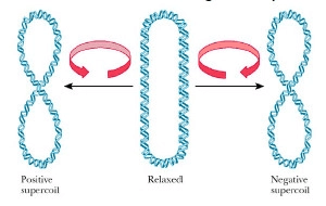
Image: A schematic diagram of supercoiling of the DNA.[11]
Source: Quizlet
Functions of DNA
- DNA is the hereditary material that stores all the information required for the functioning of life.
- Three nucleotides (called codons) are considered as a genetic code for the production of an amino acid residue. So, the polynucleotides lead to the formation of different amino acids that together constitute proteins required for the body. And, the individual proteins interact with each other to regulate the proper functioning of the body.
- DNA is also involved in processes like replication, cellular metabolism, transcription, and mutation.
Ribonucleic Acid (RNA)
RNA is a nucleotide polymer consisting of four bases — adenine (A), guanine (G), cytosine (C), and uracil (U) — sugar, and phosphate. The information contained by DNA is transferred to the whole body at specific locations through RNA. They are involved in several essential metabolic functions required for living organisms.
Structure of RNA
RNA is a single-strand helical structure. The phosphate group present in the structure makes it a charged molecule (polyanion). A distinguishing feature in the RNA structure is the presence of hydroxyl group at the 2’ position of the ribose sugar, which gives it an A-form geometrical structure.[5]
RNA structures have the capability of self-folding, that is, interchain complementary base pairing between the bases of the strand. It leads to the formation of bulges and helices in the RNA strands. In perchlorate solution, a hybrid structure of RNA/DNA form is more stable than either RNA/RNA or DNA/DNA duplexes.[5]
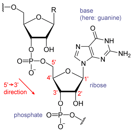
Image: A fragment of an RNA structure, showing a guanosyl subunit.[12]
Source: Wikipedia
Types and Functions of RNA
RNA molecules can be found in multiple copies and several forms inside the cells. A few major classes of RNA include mRNA, rRNA, tRNA, snRNA, snoRNA, miRNA, XIST, siRNA, tmRNA, and telomerase RNA.[5] Out of all these RNAs, the most well-known and well-studied RNA molecules are: mRNA, rRNA, and tRNA.
They play diverse roles in organisms including transfer of genetic information during protein synthesis, gene expression, enzymatic activity, and storage of genetic information in RNA viruses and viroids.[5]
1. mRNA
mRNA refers to messenger RNA. It copies and carries the information, encoded in one or more genes, from the DNA to ribosome for protein formation. Some differences between the RNA molecules of eukaryotes and prokaryotes are mentioned below:[13]
| Content | 1 | Mostly monocistronic mRNA (encodes only a single protein) with an average size of 1500-2000 nucleotides. | Polycistronic mRNA that encodes several proteins. | Content | 2 | It has a cap-like structure at the 5’-end. | It doesn’t have any cap-like structure at the 5’-end. | Content | 3 | It has a long poly-A tail at the 3’ end that helps to stabilize RNA. | It has a short poly-A tail at the 3’-end that acts as a targeting signal for RNA destruction. | Content | 4 | It doesn’t have intercistronic regions. | It has intercistronic regions. | Content | 5 | It requires extensive processing and transport. | It doesn’t require any processing. |
2. rRNA
rRNA molecules are ribosomal RNA. These are the most abundant forms of RNA in a cell, forming 80% of the RNA molecules in eukaryotes.[13] It associates with ribosomes to form a complex structure that moves in a 5’ to 3’ direction to catalyze the formation of proteins. These molecules play an active role in recognizing the conserved positions of mRNA and tRNA.[13]
In eukaryotes, the four different rRNA molecules include 18s, 5.8s, 28s, and 5s rRNA, whereas, in prokaryotes, it includes 16s, 5s, and 23s rRNA. [13]
3. tRNA
tRNA is referred to as transfer RNA or adaptor RNA. It carries individual amino acids into the ribosomes to assemble the growing polypeptide chains. tRNA has clover leaf-like structures containing 70-80 nucleotides. The structure has well-defined stems and loops that make up the acceptor arm, D-stem and loop, anticodon stem and loop, and the T-stem and loop.[13]
Each amino acid has its specific type of tRNA. Each tRNA binds to the specific amino acids and carries them to the growing polypeptide chain. The prokaryotes have 30-45 different tRNA and the eukaryotes contain 50 or more tRNA.[13]
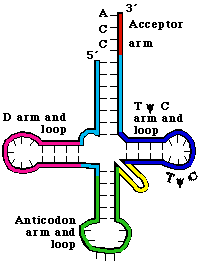
Images: The labeled clover leaf-like structure of tRNA.[14]
Source: bx.psu.edu
RNA Processing
Eukaryotes and prokaryotes have different RNA processing mechanisms. In eukaryotes, the process of mRNA and protein synthesis occurs in different compartments of the cell. However, in prokaryotes, both events simultaneously occur in the single compartment of the cell. Because of this reason, the mRNA of prokaryotes undergo little or no modification after synthesis, whereas, pre-tRNA and pre-rRNA undergo processing like cleavage, the addition of a nucleotide, and chemical modifications after synthesis.[13]
In eukaryotes, after pre-mRNA is synthesized, it undergoes different processing stages including 5’-capping, 3’cleavage/polyadenylation, splicing, and RNA editing before they are transported to the cytoplasm for protein synthesis.[13] The rRNA and tRNA of eukaryotes undergo the same modification processes as of prokaryotes.
Conclusion
Nucleic acids are one of the major biomolecules required for the proper functioning of the body. These are the molecules responsible for carrying the genetic information from parents to offspring, gene expression, and synthesis of proteins required for metabolic functions.
After the discovery of nucleic acids in 1869, research on these molecules has come a long way. Several regions in the DNA have been identified to be responsible for particular traits or mutations leading to fatal diseases. The study of several other RNAs including snoRNA, XIST, and siRNA needs an in-depth understanding of their functioning in the organism’s body.
The nucleic acid study is an ongoing and popular research area in clinical studies because of its importance in the study of the mechanisms, cures, and treatments of several diseases, whose mysteries are hidden in the genetic codes of these molecules.
References:
- Nucleic acids. Retrieved from https://www.genome.gov/genetics-glossary/Nucleic-Acid.
- Minchin, S., & Lodge, J. (2019). Understanding biochemistry: structure and function of nucleic acids. Essays in biochemistry, 63(4), 433–456. https://doi.org/10.1042/EBC20180038.
- Mandal Ananya. RNA discovery. Retrieved from https://www.news-medical.net/life-sciences/RNA-Discovery.aspx.
- Nucleic acids. Retrieved from https://en.wikipedia.org/wiki/Nucleic_acid.
- Kumar, Pranav & Mina, Usha. (2016). Life Sciences, Fundamentals, and Practice, Part I.
- Nucleic acids. Retrieved from https://courses.lumenlearning.com/suny-wmopen-biology1/chapter/nucleic-acids/
- Gabe Buckley (2021). Nucleic acids. Retrieved from https://biologydictionary.net/nucleic-acid/.
- Nucleic acids. Retrieved from https://www.britannica.com/science/nucleic-acid/Ribosomal-RNA-rRNA.
- Bansal, M. (2003). DNA structure: Revisiting the Watson–Crick double helix. Current Science, 85(11), 1556-1563. Retrieved June 18, 2021, from http://www.jstor.org/stable/24110017.
- Factors affecting the structure of DNA. Retrieved from https://www.slideshare.net/punya08/denaturation-and-renaturation-of-dna.
- Lecture 33. Retrieved from https://quizlet.com/203493874/lecture-33-flash-cards/.
- RNA. Retrieved from https://en.wikipedia.org/wiki/RNA#/media/File:RNA_chemical_structure.GIF.
- Kumar, Pranav & Mina, Usha. (2016). Life Sciences, Fundamentals, and Practice, Part II.
- Gene Expression and Protein Synthesis. Retrieved from https://www.bx.psu.edu/~ross/workmg/TranslationCh14.htm.