Discover the versality and power of ConductVision as it revolutionizes behavorial research. From standard tests to specialized experiments, our software offers unparalleled automation, accuracy, and efficiency. Explore the diverse applications of ConductVision and unlock new possibilities in your research endeavors.
Our platform provides advanced tools for behavioral research, including:
Welcome to our support section. We’re here to assist you with any questions or issues you may have. Our dedicated team is committed to providing prompt and effective solutions.
After ServicesNeed help after your purchase? Our After Services team is here to support product setup, troubleshooting, and post-sale inquiries—ensuring you get the most from your ConductVision tools.
ConductCareEnjoy personalized assistance from our ConductCare specialists, offering expert guidance, training, and tailored solutions to help you meet your research goals with confidence.
Try our software for free and get 10 hours of FREE behavioral data analysis! Experience powerful features that deliver raw data or beautiful data visualizations—no commitment required. Sign up now and start transforming your results effortlessly!
Our platform provides cutting-edge tools for behavioral research, enabling precise tracking and analysis of movement and interactions.
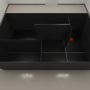
Our platform is designed to support a wide range of model organisms. From rodents to aquatic and flying species, we provide tools tailored to the specific needs of each, enabling scalable and accurate behavioral analysis.🐭 Rodents are a cornerstone in neuroscience and behavioral studies. Our tools provide detailed, high-resolution tracking for both individual and social behaviors.🐟 Zebrafish provide a transparent and scalable system ideal for high-throughput behavioral research in pharmacology and genetics.🪰 Drosophila melanogaster is a key model in genetic and neurobehavioral research. Our system captures detailed movement and interactions in both solo and group setups.
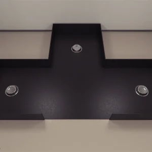
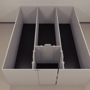
Browse our lab hardware collection for reliable tools in neuroscience, surgery, and behavioral research. Built for precision and performance.
Explore our complete selection of animal lab equipment and research tools built to support preclinical studies, surgical procedures, anesthesia, and behavioral monitoring.
 Skip to content