$7,945.00
An automatic gel imaging system works on the principle that when ultraviolet light (of wavelength 254nm-302nm) is directed towards a gel stained with ethidium bromide, the dye intercalates with DNA’s groove, gets excited, and emits fluorescent light. This phenomenon can be observed and recorded in the visible range, i.e., 590nm using a digital camera.
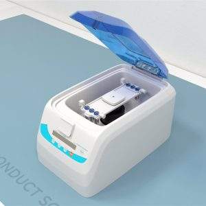
The Gel-Doc system comprises four main parts: a darkroom hood, a UV illuminator, a camera, and computer software for recording and analyzing gel images. The automatic gel imaging system offered by Conduct Science is a completely automated and highly integrated gel imaging system having 340x475x725mm dimensions. It can operate at different wavelengths such as 254nm, 302nm, 365nm, and 590nm and has an exposure time of 1ms-1000ms.
ConductScience offers the Automatic Gel Imaging System.

Servicebio is a company that specializes in providing high-quality products and services for biomedical research and diagnostics.


The automatic gel imaging system is also known as Gel Documentation System, Gel-Doc, or Gel Imager. These systems are used to visualize nucleic acid, and protein samples run on polyacrylamide or agarose gels containing ethidium bromide, coomassie blue stain, silver nitrate, etc. Gel documentation systems help record, identify, quantify, optimize, and store data regarding DNA, RNA, and protein samples.
An automatic gel imaging system comprises:
The gel imager is designed to capture gel images after running gel electrophoresis. Nucleic acid bands that appear on the gel, as a result, are captured. The digital camera is connected to a computer display for adjusting the image before capturing it. These gel images are then utilized for data storage, research purposes, and detailed analysis (Bhardwaj et al., 2011).
Camera |
|---|
Product
| Quantity |
|---|---|
PixelResolution | 6.3 million pixels |
Resolution | 3072×2048 |
Pixel Size | 2.4×2.4μm |
Target Size | 1/1.8”(7.37×4.92mm) |
Full Well Capacity | 10.4ke- |
Sensitivity | 760mv |
Readout Noise | 2.14e- |
Dark Current | 0.15mV |
Signal-to-Noise Ratio | 40.2dB |
Exposure Time | 17us-15s |
Binning Mode | 1×1,2×2,3×3,4×4 |
Grayscale | 8-bit(256levels) |
Camera Type | Black and White Camera |
Lens |
|---|
Product | Quantity |
|---|---|
Aperture | F1.0-F16 |
Focal Length | 8-48mm |
Type | Motorized zoom lens |
close-up lens | 2 times |
Filter | 590/60nm |
Light Source |
|---|
Product | Quantity |
|---|---|
Aperture | F1.0-F16 |
Focal Length | 8-48mm |
Type | Motorized zoom lens |
close-up lens | 2 times |
Filter | 590/60nm |
Light Isolation | Fully light-sealed, isolates environmental light |
Dark Box |
|---|
Product | Quantity |
|---|---|
Door Control | Door control sensor can control the on/off of the bright field light source |
Field of View | Effective field of view is140×140 mm |
Gel Cutting | After opening the door,the UV light source can be pulled out cutting,in conjunction with a UV protection shield |
Software Functions |
|---|
Product | Quantity |
|---|---|
Camera Settings | Adjust contrast, exposure time, and gain |
Lens adjustment | Choose adjustment range: coarse adjustment, fine adjustment, super-fine adjustment Lens: Zoom, focus, aperture adjustment |
Image cropping | Image cropping: Crop the necessary parts of the bands |
An automatic gel imaging system works on the principle that when ultraviolet light (of wavelength 254nm-302nm) is directed towards a gel stained with ethidium bromide, the dye intercalates with DNA’s groove, gets excited, and emits fluorescent light. This phenomenon can be observed and recorded in the visible range, i.e., 590nm using a digital camera. The researchers can choose from a variety of cameras, transilluminators, and imaging software according to their convenience and experimental requirements (Bhardwaj et al., 2011).
The Gel-Doc system comprises four main parts: a darkroom hood, a UV illuminator, a camera, and computer software for recording and analyzing gel images. The automatic gel imaging system offered by Conduct Science is a completely automated and highly integrated gel imaging system having 340x475x725mm dimensions. It can operate at different wavelengths such as 254nm, 302nm, 365nm, and 590nm and has an exposure time of 1ms-1000ms.
The device contains a high-resolution CCD camera with an autofocus length, a special filter for nucleic acid dye, and an overlay glue cutting filter. The apparatus has a white sample plate to visualize SDS-PAGE samples and a UV sample plate for nucleic acid gel imaging.
Place the samples loaded onto agarose/polyacrylamide gels, SDS page, or Petri dishes onto the UV transilluminator surface. Close the access doors after turning on the UV lamp to prevent personnel’s exposure to UV. Use the CCD camera to “photo-document” the images, adjust, and save these gel images using computer software. Ultimately, retrieve/export the images to Microsoft Excel (or other similar software) to analyze.
Protein Visualization after Western Blot and SDS-PAGE
Wu et al. (2013) studied the effect of astaxanthin on D-galactose-induced aging in rodents. The researchers performed a protein carbonylation assay using sodium dodecyl sulfate-polyacrylamide gel electrophoresis (SDS-PAGE), developed the resulting blots, and visualized them using an automatic gel imaging system. The samples were subjected to Western blot analysis; target proteins were detected and then visualized using the gel documentation system.
Post-Western Blot Protein Visualization for studying the effect of maslinic acid in the organs of diabetic rats
Mkhwanazi et al. (2014) studied the antioxidant effects of maslinic acid (MA) on the heart, liver, and kidneys of streptozotocin-induced diabetic rats. The experimenters analyzed kidney tissues GLUT1, GLUT2, and GLUT4 via western blot and then visualized the protein blots using the gel documentation system. They concluded that the antioxidant properties of MA could improve kidney function in diabetes mellitus patients.
Analyzing the size of PCR products
Mahajan et al. (2011) evaluated the efficacy of PCR-RFLP for analyzing the 12S rRNA gene in identifying meat samples from known and unknown meat samples and utilized these meat samples to confirm the species of origin of meat. The scientists amplified the mitochondrial 12S rRNA gene using PCR and analyzed the size of the PCR product by agarose gel electrophoresis using 1.5% agarose gel stained with Ethidium Bromide. A 100bp DNA ladder was electrophoresed to assess the product’s size. An automatic gel imaging system was used to visualize the gel, and the amplicon’s size was measured using software with a gel imager.
Then the researchers subjected PCR products to restriction digestion and electrophoresed the restriction enzyme digests using 3% agarose gel stained with EtBr. The gel was again visualized using an automatic gel imaging system, and the size of fragments was determined using the software with Gel-Doc.
Measuring Feather Growth Bars
Shawkey et al. (2003) compared the traditional method for measuring feather growth bars with the gel documentation system. The automatic gel imaging system consisted of a light cabinet with white lights on the left and the uppermost side, a close-up 12dioptre lens with an interference filter, and a high-performance CCD camera with a 12.5x75mm zoom lens, and a computer with relevant software. The scientists adjusted camera magnification and captured the image. They used the ruler function in the software to measure the distance between two points. The researchers measured growth bars using proper light and digital ruler adjustments. The researchers concluded that the gel documentation system could quickly take repeatable measurements of the growth bars compared to the traditional method.
The first and foremost advantage of the automatic gel imaging system is accurate image analysis. The increased accuracy of image analysis allows in-depth analysis. Moreover, digital gel documentation systems provide a wider “linear dynamic range” than film. Fluorescent detection facilitates the multiplexing and improved reproducibility of western blots. Gel documentation systems save time, facilitate workflow convenience, and do not involve the use of harmful chemicals.
However, a disadvantage of gel-doc systems is that they are quite expensive. In addition, UV exposure can be harmful to lab personnel.
Bhardwaj, S., Dahiya, S., & Tripathi, C.C. (2011) Design, Fabrication and Characterization of Low Cost Indigenous Gel Documentation System. Converging Technologies beyond 2020.
Wu, W., Wang, X., Xiang, Q., Meng, X., Peng, Y., Du, N., … & Liu, X. (2014). Astaxanthin alleviates brain aging in rats by attenuating oxidative stress and increasing BDNF levels. Food & function, 5(1), 158-166.
Mkhwanazi, B. N., Serumula, M. R., Myburg, R. B., Van Heerden, F. R., & Musabayane, C. T. (2014). Antioxidant effects of maslinic acid in livers, hearts and kidneys of streptozotocin-induced diabetic rats: effects on kidney function. Renal Failure, 36(3), 419-431.
Mahajan, M. V., Gadekar, Y. P., Dighe, V. D., Kokane, R. D., & Bannalikar, A. S. (2011). Molecular detection of meat animal species targeting MT 12S rRNA gene. Meat Science, 88(1), 23-27.
Shawkey, M. D., Beck, M. L., & Hill, G. E. (2003). Use of a gel documentation system to measure feather growth bars. Journal of Field Ornithology, 74(2), 125-128.
| Instrument Type | Imaging Systems |
|---|---|
| Application Area | Protein Analysis |
| Automation Level | Fully Automatic |
| Cameras | Black and White Camera |
| objective-lens-focal-length | 8-48mm |
| Dimensions | 475mm x 340mm x 725mm |
| operating_wavelengths | 254nm, 302nm, 365nm, 590nm |
| exposure_time_range | 1ms-1000ms |
| camera_type | High-resolution CCD camera |
| camera_focus | Autofocus length |
| objective_lens_focal_length | 8-48mm |
| uv_wavelength_range | 254nm-302nm |
| fluorescent_emission_wavelength | 590nm |
| sample_plates | White sample plate for SDS-PAGE, UV sample plate for nucleic acid gel |
| filters | Special filter for nucleic acid dye, overlay glue cutting filter |
You must be logged in to post a review.
Reviews
There are no reviews yet.