



Our Animal Lab Products


Documentation
Introduction
Animal Lab allows in vivo testing on animal models like Drosophila, rodents, ferrets, cats, and rabbits for analyzing variables that affect their neurotoxic, carcinogenic, pharmacokinetic, and stereotaxic behavior. The use of animal models in psychological and pharmacological research is prevalent. These animals are used in anatomical dissections and invasive experiments performed in physiology, biochemistry, and pharmacology while learning clinical and surgical skills. Animal lab equipment includes animal restrainers, animal cages, anesthesia systems, heat and temperature controllers, mazes, optogenetic and stereotaxic systems, and surgical tools.
Anesthesia Systems
Anesthetization of rodents is common in psychology and pharmacology for surgical and non-surgical research procedures. Administration of general anesthesia facilitates the unconsciousness or immobilization of animals while performing lengthy invasive procedures like laparotomy, embryo derivation, cranial implants, and transferring xenografts. The four major components of anesthesia include unconsciousness, amnesia, muscle relaxation, and analgesia. Currently, safe anesthesia administration depends on electrical and mechanical instruments.
Anesthesia equipment includes anesthetic machines, ventilators, airway support and oxygen delivery devices, and vaporizers.
Inhalant anesthesia forms the basis of anesthesia administration in modern veterinary medicine. An anesthetic machine facilitates the delivery of potent anesthetics (like isoflurane and sevoflurane) combined with oxygen. These machines vary from being very simple to complex workstations with built-in ventilators and safety systems. Common components of all anesthetic machines are oxygen regulator, oxygen source, oxygen flowmeter, and vaporizer. If any other gas is used along with oxygen, similar components for that gas are used additionally. The researchers can use the basic anesthetic machine in conjunction with a breathing circuit and a waste gas scavenger for successful anesthesia delivery to rodents.
Endotracheal tubes and various supraglottic airway devices (SGADs) maintain the airway in anesthetized animals. Conduct Science’s endotracheal tubes when placed properly with an inflated cuff, allow positive pressure ventilation, and prevent contaminating the working environment from waste anesthetic gases. Depending on the experiment’s requirements, many types and styles of endotracheal tubes are offered at Conduct Science.
Laryngoscopes are the devices that facilitate tracheal intubation and oropharyngeal assessment during intubation. The device consists of a handle and a lighted blade. Several types of blades are available, like a single fixed blade, multiple blades, plastic blades, and stainless-steel blades. However, two main types of blades include Macintosh and Miller. The Miller laryngoscope has a straight blade with a less prominent vertical flange, whereas the Macintosh laryngoscope contains a curved blade with a prominent vertical flange (Grim et al., 2015).
Neuroscience research includes various stereotaxic procedures conducted in animal labs like electrode implantation, craniotomy drilling, and dura matter removal on animal models, mostly murine. Stereotaxic surgery of animals is significant for understanding the neurobiology of reward and aversion by using devices like electrodes for CNS stimulation or microdialysis probes. A stereotaxic device consists of three coordinates that facilitate locating and studying the brain sections while keeping the head fixed.
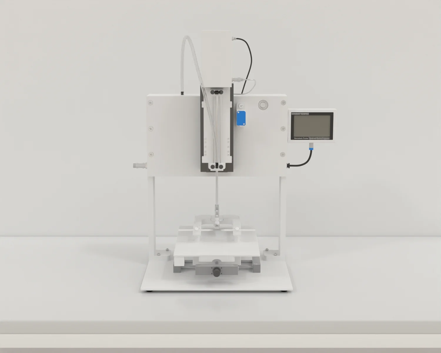
Conduct Science offers a wide range of high-quality, durable stereotaxic apparatus and neuroscience equipment that are efficient and ultraprecise. Manifold stereotaxic instruments, drills, and impactors are available. The researchers can choose suitable apparatus depending on their experiment requirements.
A general surgery procedure involving implanting a cannula or probe into the rodent brain employs stereotaxic apparatus and follows underlying steps (Geiger et al., 2021).
Optogenetics involves merging optical and genetic methods to obtain the loss or gain of functions of specific cells of biological tissues in well-defined events. Generally, optogenetics incorporates core technology that utilizes certain tools that control and target specific cells that respond to light and allows this technology to:
The principle of optogenetics is based on the ability of microbial opsin genes to impart light detection ability in a single’ readily targetable module’ to the neurons. Some microorganisms can produce visible light-gated proteins that control the ionic flow across the cell membrane. The researchers suggested that the addition of microbial opsin genes makes the neurons precisely responsive to light (Deisseroth, 2010).
Optogenetic tools have entirely changed the way neuroscience influences biological systems. Optogenetic lasers monitored by laser power meters are used to induce diseases such as ophthalmic disorders in rodent models to study the mechanisms of the respective disorders. The use of safety goggles while working with optogenetic systems is a must as these lasers can cause serious eye diseases and even loss of sight.
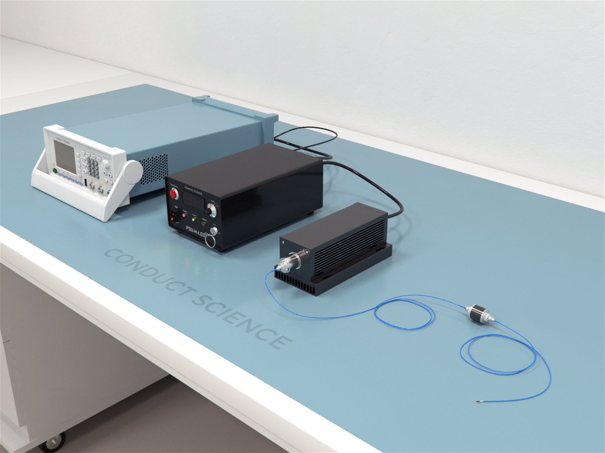
Conduct Science offers optogenetic lasers to be used in conjunction with a laser power meter. Accessories to join and establish a complete optogenetic system and personal protective equipment are also available.
Restraining refers to the procedure of immobilizing animals during experimental procedures. Proper restraint techniques are vital for reducing stress on lab animals. Restraining is required in various psychological experiments for drug administration, anesthesia injection, and studying the effect of stress on animals. As a potential source of stress, animal restrainers should be designed to allow minimum restraint stress and be safe for both the animal and the handler.
Restraining procedures depend on the animal’s size, age, species, and temper. The animals can be immobilized chemically, manually, or mechanically. Manual restraining of rodents involves grasping the animal firmly at the tail’s base. However, the animal can easily escape this type of restraint. Therefore, for a steady immobilization, grasp the whole body between the index and middle fingers along the sides of the head and thumb and the remaining fingers under the axilla. These methods allow easier access to the facial area. However, manual restraining techniques like scruffing and gripping over or under the shoulders are not safe for the researchers. Therefore, the experimenter should ensure minimal arousal of the animals. Otherwise, a little mishandling stimulates aggression in animals, and they can bite the handler readily (Buerge and Weiss, 2004).
Mechanical restrainers are used in stereotaxic surgeries. These restrainers, made of high-quality acrylic, polycarbonate, or Perspex, vary in design and size depending on the animal’s size, species, strain, and age. The restrainer’s size should be appropriate, neither too small to suffocate the animal nor too large to let the animal escape. Different types of animal lab restrainers offered by Conduct Science include ferret restrainers, flat bottom restrainers, guinea pig restrainers, Broome style restrainers, etc.
For restraining the animals in a physical restrainer, lift the animal by grasping from the tail, place it into the entry groove and push the animal gently from behind into the restrainer. Close the restrainer’s tailgate to immobilize the animal.
Apart from this, rodents also need to be immobilized for the successful administration of intravenous tail injections. In rodents, intravenous injections are administered via lateral tail veins. Vein visibility is critical in this process, especially when working with black mice strains due to the lack of contrast with their skin. Conduct Science provides light arc, nose cone with light, and Broome handlers to overcome this problem.
Anesthesia administration during stereotaxic surgeries in rodents can cause hypothermia. Anesthetics suppress the body’s heat conservation mechanisms, leading to impaired thermoregulation before, during, or after surgery. Furthermore, vivarium and housing cages have ambient temperatures of 23.5oC, whereas the normal body temperature of rodents ranges between 36.2-37.5oC. This temperature difference can also lead to hypothermia in rodents. Animal labs are equipped with heat and temperature controllers, also known as homeothermic monitoring systems, to prevent this pre-operative and post-operative heat loss.
Homeothermic monitoring systems are closed-loop systems comprising a rectal thermometer inserted into the rectum for monitoring temperature connected to a temperature controller that monitors the temperature. The temperature controller is also connected to a heating pad kept under the cages or the subject to prevent hypothermia. The temperature controller keeps a balance between the heating pad temperature and the temperature measured by a rectal thermometer to ensure homeothermic conditions in rodents (Schuster and Pang, 2017).
Mechanical ventilation is used to support spontaneous breathing in animals with respiratory system impairments. Long-term anesthesia processes also necessitate the use of ventilators. Anesthetics and neuromuscular blocking (NMB) agents cause depression in the respiratory system. Different types of mechanical ventilators based on animal size and weight are available.
Despite the life-saving respiratory assistance provided by mechanical ventilators, the risk of ventilator-induced lung injury is undeniable. In addition to adults, neonates, and infants born with respiratory disorders also require assisted ventilation. However, the risk of pulmonary hypertension, chronic lung disease, and nutritional and developmental issues associated with mechanical ventilation in neonates is higher (Hopper and Powell, 2013).
Mechanical ventilation involves the placement of an endotracheal tube or tracheal cannula. Once the tube or cannula is implanted, it can be connected to a ventilator. The subject should be properly anesthetized before and during the surgery. Conduct Science offers ventilators for both small and large size animals.
The use of mazes in studying behavioral and learning responses in mammals is quite old. Willard S. Small performed the first maze experiment in the 20th century. Many animal models, including rodents, are subjected to maze experiments in psychology laboratories. Mazes are also used to assess cognitive impairments in animal models. The two aspects of maze learning include the discovery process in which the subject explores the maze and the fixation process defined by the animal’s ability to perform the maze task better than before. Maze experiments are influenced by various factors, including subject conditions and maze conditions.
Maze tasks employ a valuable feature of the subject’s memory system: their ability to navigate their environment. This navigation can be allocentric, egocentric, or spatial. The allocentric navigation relies upon distal cues, whereas egocentric navigation depends on proximal and internal cues. Egocentric navigation can be route-based or path integration. Researchers performed experiments by injuring a part of the brain and studying its impact on maze learning abilities. These experiments concluded that spatial navigation depends on the hippocampus and entorhinal cortex, whereas head direction cells of the brain’s separated parts are responsible for egocentric navigation.
Conduct Science offers a wide range of dry and wet mazes depending on the experimental needs to observe anxiety behaviors, social interactions, motor functions, and spatial learning in animals like rats, mice, ferrets, and zebrafish.
Dry Mazes
Dry mazes include the Simple Maze, T maze, Elevated Plus Maze, Radial Arm Maze, Barnes mazes, and much more.
Simple Maze
The simple maze was introduced in 1917 by Lashley and Franz to study the behavior of rodents and other vertebrates. It consists of two-choice arms; one is a cul-de-sac, and the other leads to the reward. The experiments with this maze are simple and less time-consuming.
Elevated Plus Maze
The Elevated Plus Maze (EPM) is used in psychology experiments to observe anxiety behavior in rodents. Montgomery, in 1955 described that rats show more retreats in a maze with an elevated open alley than in mazes with an enclosed alley. Later, in 1984, Hanley and Mithani modified this maze task into an elevated maze with four arms, two open and two closed to form a plus shape. The anxiety behavior of rodents is analyzed by determining the ratio of time spent in open arms to the time spent in closed arms.
T Maze
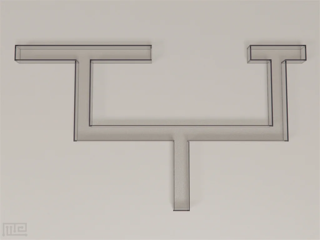
The T maze contains a longitudinal start arm that branches into two perpendicular choice arms forming a T-shaped apparatus. The subject is placed in the start arm and is allowed to enter either choice arm. It is a spatial maze that reflects individual differences in learning ability. Animals can be tested using different cues including, visual and olfactory cues. In addition, it can be also applied to other behavioral tests. Deacon and Rawlins (2006) used it to assess the cognitive ability of rodents by using spontaneous alternation.
Radial Arm Maze
The Radial Arm Maze is a traditional eight-arm maze, first designed by Olton and Samuelson in 1976 to assess the spatial working ability of rodents. The place learning ability of animals is tested in the presence of extra-maze and intra-maze cues. The purpose of assessing spatial learning is incomplete due to surrounding odors and proximal intra-maze cues. This problem led to the development of the Barnes maze.
Barnes Maze
The Barnes Maze consists of a circular platform (1.22m in diameter) and eighteen or twenty evenly distributed holes around the platform’s edge. A small, dark chamber is present under the platform of the designated target hole. The platform is rotatable from its center point, allowing the location of the target hole to be varied. Under the center of the circular platform, a cylindrical dark start chamber is present. The rodent is acclimated to the start chamber for a few minutes before trials and released in the center of the platform. Rodents must utilize both intra-maze and extra-maze cues to find the target hole. The Barnes Maze is useful for hippocampal-dependent learning.
Water mazes employ “visuospatial” environmental cues to conduct experiments driven by water escape and aversion escape rewards. One such maze is Morris Water Maze.
Morris Water Maze
Richard Morris developed the Morris Water Maze in 1981. The apparatus consists of a circular water pool and an escape platform. The pool is filled with water so that the platform can be seen 1cm above the water level. The water can either be clear or turned opaque by adding milk to conceal the platform completely. A rodent is released into the pool during testing and is tasked to locate the hidden platform. The aversiveness of water for rodents motivates them to find the platform quickly. The Morris Water Maze Task is widely used to assess the spatial learning and memory abilities of rodents. It relies on the animal’s spatial localization system to guide its navigation.
The fruit fly, Drosophila melanogaster, has long been used in genetics research as a model organism. Short life cycle, simple reproduction, and inexpensive maintenance are the traits that make Drosophila an ideal organism to be used for research. The use of fruit flies in laboratories started in the early 1900s by Thomas Hunt Morgan, an American embryologist, who mainly studied fertilization and embryo formation. Currently, Drosophila is cultured in labs to study gene regulation and signaling, mutational analysis, and developmental and neurological processes. Culturing Drosophila does not require standard lab equipment. They are grown in bottles and vials. Bottles can maintain large fruit fly populations, whereas vials are destined to keep smaller populations and make genetically significant crosses. Both plastic and glass bottles available in different sizes can be used. Bottles are covered by cotton or foam plugs.
For growing fruit flies in a vial, cooked or readymade, dehydrated media is added to the bottles to supplement the growth. The media is moistened by using water, brought to room temperature, and a few grains of yeast are added to the surface of the hydrated media. The flies are then transferred to the vial for growth and reproduction, and the vials are closed using cotton plugs. Their food is changed every two weeks (Parveen, 2018).
Conduct Science offers a wide range of equipment to grow Drosophila optimally in labs. This equipment ranges from small narrow vials to large bottles and foam plugs to cover the mouth of these vials. Yeast, cornmeal, and molasses to supplement the growing flies with food and nutrition are also available.
Let's work together!
Have questions? Ask anything!