
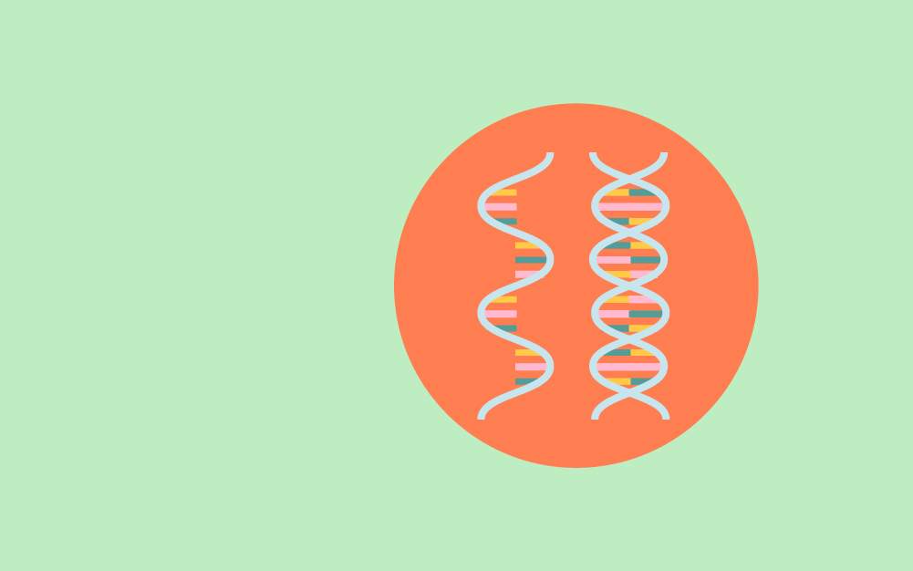
Nucleic acids such as deoxyribonucleic acid (DNA) and ribonucleic acid (RNA) are macromolecules that carry genetic information in organisms.
DNA consists of double nucleotide strands that are complementary to one another. The sequence of nucleotides signifies genetic elements, including genes, transcription factors, and regulators.
RNA only has one nucleotide strand transcribed from one DNA strand. RNA sequences indicate the functional portions of the genome such as genes in messenger RNAs (mRNAs), amino acid decoders in transfer RNA (tRNA), and coded protein assemblers in ribosomal RNA (rRNA).
To learn more about the structures, properties, and functions of nucleic acids, check out our article on nucleic acids!
Extracts of nucleic acids allow scientists and practitioners to assess the genetics and function of cells and organisms. Isolated DNA and RNA are the input of several techniques in molecular biology like polymerase chain reactions (PCR), Sanger, and next-generation sequencing.
In general, the extraction of nucleic acids consists of the following steps:[1]
The first step in nucleic acid extraction is to disrupt the cells and expose the DNA or RNA to the extraction buffer. Nucleic acids are then purified from proteins and other cellular components.
Cell lysis is performed using a lysis buffer containing one form of a non-ionic detergent such as sodium dodecyl sulfate (SDS), Triton X-100, and Tween-20. The detergent lyses the cells by disrupting the cell membrane and denaturing the proteins.
This step can be approached in several manners, depending on the organism of interest and the material used as nucleic acid sources.
Microorganisms such as bacteria are cultured in a liquid medium where the cells can be harvested by centrifugation. The cell pellets are lysed using a surfactant-containing lysis buffer. Some microorganisms with thick cell walls may need to be digested by cell-wall digesting enzyme mix or crushed by glass beads before adding the extraction buffer.[2]
Fungal nucleic acids can be acquired from hyphae, mycelia, and conidia grown on agar plates, are collected by overlaying detergent-containing extraction buffers, and filtered or centrifuged to remove cell debris. The supernatant contains nucleic acids, which are purified in subsequent steps.
Fungi with thick cell walls are usually sonicated or frozen using liquid nitrogen before using a mortar and pestle to grind the tissue into powders. After pulverization, the detergent-containing extraction buffer is added to lyse the cell membrane and release the nucleic acids into the buffer.[3]
Plant tissue is typically crushed before the detergent-containing nucleic acid extraction buffer is added. This is often achieved by grinding the material in liquid nitrogen using mortar and pestle, homogenizer, or high-frequency shaking with stainless steel or glass beads. The liquid nitrogen flash-freezes the sample and inhibits cellular enzyme activities, preserving the integrity of the nucleic acids.
Most plant nucleic acid extraction buffers are based on sodium chloride solution that uses cetyltrimethylammonium bromide (CTAB) or SDS as the nonionic detergent. Plant materials with high polysaccharide content and secondary metabolites are often pre-treated with a different buffer after pulverization but before the nucleic acid extraction buffer is added. This step is to ensure that carbohydrates and other metabolites are removed and cannot interfere with the subsequent steps.
Mammalian nucleic acids are extracted from whole tissue or cultured cells using similar methods.
In cases of whole tissue, the tissue is first excised and immediately flash-frozen in liquid nitrogen. The excised tissue is crushed using a homogenizer or a mortar and pestle. The powdered tissue is resuspended in an extraction buffer with an appropriate detergent.
Cultured mammalian cells suspended in a liquid medium can be collected by centrifugation. Oftentimes, the cells are trypsinized to loosen any cell that adheres to the surface before the liquid medium is collected for centrifugation. The pellets are resuspended and lysed using a detergent-containing extraction buffer.
The cell lysis step is essential in obtaining nucleic acids with a sufficient amount and good quality with minimal shearing. Treating samples with liquid nitrogen before cell lysis can inhibit any cellular nuclease activity that degrades nucleic acids.
Along the same line, some nucleic acid extraction buffers contain additional components such as proteinase K and beta-mercaptoethanol, which inactivate nucleases that digest nucleic acids.
After cell lysis, the nucleic acids are released into the extraction buffer along with proteins and other cellular components. The extracts can be centrifuged at high-speed to remove high molecular-weight components.
The resulting aqueous supernatant contains nucleic acids and some low molecular-weight components, which can be removed using the following approaches:[1,4]
Liquid-liquid separation is the conventional nucleic acid extraction approach. It is a separation technique that relies on the ability of the molecules to be solubilized in a particular solvent.
In this approach, a mixture of organic solvents with differing properties is mixed with the cell lysate and centrifuged. The cellular components are removed from the aqueous phase and partitioned into the organic phase below.
The aqueous phase contains nucleic acids, which are removed by precipitation with alcohol solvents such as ethanol or isopropanol. The precipitated nucleic acid is packed into a pellet by centrifugation, washed, and air-dried before the pellet is dissolved.
Solid-phase separation, also known as a column-based approach, is a technique that uses spin columns containing membranes from silica or resin. It is widely used in commercial nucleic acid extraction kits.
Centrifugation removes the cell lysate from cell debris, and the supernatant containing the nucleic acids is mixed with a binding buffer before the mixture is added to the column. The membrane absorbs the mixture, and the nucleic acid is bound to the membrane when the column is centrifuged.
The nucleic acid-bound membranes are washed with various solutions and centrifuged several times to remove contaminants. Nucleic acids are finally removed by adding an elution buffer to the membrane and centrifuging.
Magnetic separation uses paramagnetic beads and a magnetic bar to single out nucleic acids in the cell lysates.
This separation approach is based on the nucleic acids’ ability to reversibly bind to a charged surface under high salt concentration. The magnetic bar attracts the nucleic acid-bound paramagnetic beads while they are successively washed with buffers containing an alcohol solvent. Finally, nucleic acids are eluted from the beads with an elution buffer containing low salts or water.
Unlike the other two approaches, magnetic separation has no centrifugation step and does not need to remove supernatants or exchange containers, allowing full automation of the extraction process.
After nucleic acid extraction, the quantity and quality of the extracts are frequently inspected by spectrophotometry. It relays the concentration of the extracts, proteins, and solvent contamination in the final extract based on 260/280 and 260/230 absorbance ratios.
Spectrophotometer-based evaluation is generally sufficient for typical polymerase chain reactions (PCR). For applications such as direct sequencing and quantitative PCR (qPCR or qRT-PCR), which require a high quantity of intact nucleic acids, the extracts should be inspected by agarose gel electrophoresis to check the integrity of the obtained nucleic acids.
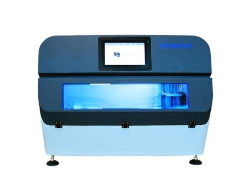
Figure: An automated nucleic acid extraction system
Extraction of nucleic acids can be lengthy and laborious, especially in screening or diagnostic laboratories that receive tens and hundreds of samples per day. The laborious and time-consuming task can be reduced by semi- or full automatization of the nucleic acid extraction methods.
In most semi-automated nucleic acid extraction systems, cell lysis is automatized through a user-controlled sample disruptor or homogenizer. It replaces manual grinding and pulverization of the samples for cell lysis, reducing the labor necessary to prepare and process samples for nucleic acid extraction.
Fully automated nucleic acid extraction systems are benchtop instruments that automatize all nucleic acid extraction steps. Automated devices significantly reduce the time and labor for manually extracting nucleic acids from the same number of samples. They also circumvent human error introduction, which can occur during manual extraction.
Nonetheless, all automated nucleic acid extraction instruments commercially available to date are designed specifically for a column-based nucleic acid extraction kit or magnetic separation, which restricts users’ flexibility in choosing the extraction approach.[4]
Nucleic acids are keys to the genetic information and function of organisms. They can be extracted by lysing cells, isolating, and purifying the nucleic acids from proteins and other cellular components using liquid-liquid, solid phase, and magnetic separation.
The solid-phase and magnetic-based nucleic acid extraction workflow have been fully automated into nucleic acid extraction devices, which can perform high-throughput extraction with minimal supervision, saving time and labor compared to manual extraction.
If you are looking for automated magnetic-based nucleic acid extraction systems, check out our programmable nucleic acid extraction devices, which can handle up to 32, 48, or 96 samples in one round of extraction.

Monday – Friday
9 AM – 5 PM EST
DISCLAIMER: ConductScience and affiliate products are NOT designed for human consumption, testing, or clinical utilization. They are designed for pre-clinical utilization only. Customers purchasing apparatus for the purposes of scientific research or veterinary care affirm adherence to applicable regulatory bodies for the country in which their research or care is conducted.