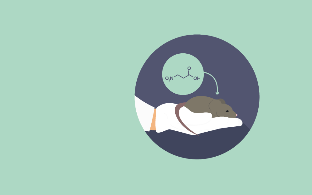

3-Nitropropionic acid (3-NP) is a selective striatal neurotoxin used in rodents to mimic pathological features of Huntington’s disease. 3-Nitropropionic acid interrupts the mitochondrial electron transport chain. This mycotoxin is an inhibitor of succinate dehydrogenase, an enzyme responsible for the oxidation of succinate to fumarate. This process reduces ATP production and causes oxidative stress. Generally, the brain lesions caused by systemic 3-nitropropionic acid are specific to the striatum. However, the hippocampus and thalamus could also be affected. Oxidative stress and excitotoxicity are two hallmarks of several neurodegenerative diseases and are relevant to the striatal cell loss observed in Huntington’s disease. 3-NP is a natural toxin produced by fungi and plants; it crosses the blood-brain barrier and could be administered systemically. Its administration by using a subcutaneous osmotic pump or direct subcutaneous or intraperitoneal injection is effective for systemic infusion, although the doses should be adjusted according to the animal’s weight.
The 3-NP model could reproduce the hyperkinetic and hypokinetic symptoms of Huntington’s disease, depending on the time and dose administered; therefore, allowing the assessment of initial and late phases of the disease. The effects of acute treatment (20 mg/kg) are observed after the first two injections, although its histopathology is different from that of the initial stages of HD, accompanied by extra-striatal lesions. Chronic administration of 3-NP at low doses (10 mg/kg/day, 3–6 weeks) leads to a sustained state of metabolic alterations and display some other features shown by the HD patients. 3-NP-triggered neurodegeneration efficiently mimics the chain of processes leading to cell death in HD; therefore, it has been proposed as a powerful phenotypic model to investigate different aspects of HD and its potential treatments (Borlongan., Koutouzis., & Sanberg., 1997).
In Huntington’s disease, glucose metabolism is altered, and ATP production is reduced. Several enzymes involved in the electron transport chain and tricarboxylic acid cycle (TCA) are also triggered in the course of HD. It has also been studied that aconitase and complex II, III, and IV activities are reduced in the striatal nucleus of HD patients. Therefore, damage to mitochondrial complexes together with a redistribution of cytochrome c occurs following the loss of membrane potential. 3-NP induces caspase-9 activation, which in turn triggers the presence of Apaf-1, cytochrome c, and ATP. 3-NP is also known to induce the expression of different mitochondrial factors associated with apoptosis, such as cytochrome c and Smac/DIABLO protein. For oxidative damage, 3-NP alters oxidative metabolism by excessive ROS/RNS production or antioxidants depletion. Therefore, the oxidative damage is largely linked with the neuronal loss in the 3-NP model (Túnez., Tasset., & Santamaría., 2010).
It has been known that glutamatergic innervation due to excitotoxicity triggers 3-NP-induced striatal degeneration. Glutamate is found to be increased in rat brains treated with 3-NP. Lactate levels are also elevated, indicating mitochondrial energy disruption in this model. Taking this together, the 3-NP induces excitotoxicity by sensitizing the neurons to basal glutamate levels. Glutamate levels are controlled by sodium-dependent transporters (Na+) in neurons and glia. These transporters rely on the transmembrane sodium concentration gradient generated by Na+/K+ ATPase. Glutamate is metabolized to glutamine after being internalized in the glia and is released into the extracellular space, where it is then reconverted into glutamate. Therefore, the glutamatergic homeostasis depends on the balance and regulation of all the components involved; its alteration could lead to excessive NMDA-R stimulation, prompting excitotoxicity-induced neuronal death. It has also been reported that during ischemia, nitric oxide (NO) production starts after the stimulation of NMDA-R, a process involving neuronal-NOS (nNOS) activation. 3-NP-induced increase in intracellular Ca2+ levels is more significant in astrocytes than in neurons, thus potentiating primary astrocyte degradation. In astroglia, these changes are catalyzed by the sodium-calcium exchange system. Also, the administration of 3-NP to astrocyte cultures causes substantial increases in intracellular calcium levels. In vivo administration prompts a decrease in astrocytes, as well as a loss of axons and white matter, together with the damage of oligodendrocytes. This data shows that in the 3-NP rat model, excitotoxicity is induced by both necrosis and apoptosis (Túnez., Tasset., & Santamaría., 2010).
Note: For 20 rats × 0.350 kg × 20 mg/kg × 5 days = 700 mg of 3-NP powder.
Steady subcutaneous delivery of 3-NP using osmotic pumps
Dose calculations
For example: Prepare 20 rats × 2.5 ml of original 3-NP solution, i.e., 50 ml at the dose of 89.14 mg/ml. This makes 50 ml × 89.14 mg/ml = 4.457 g of 3-NP powder. Total weight 4.460 g.
Solution preparation
Osmotic pump filling
Subcutaneous pump implantation
Follow-up
Removing osmotic pump
Mimicking Huntington’s disease neuropathology (Vis et al., 1999)
The mitochondrial toxin 3-nitropropionic acid (3-NP) creates selective striatal lesions and serves as a powerful experimental model of Huntington’s disease (HD). In the study, hematoxylin-eosin (HE) and Nissl stains along with immunohistochemical labeling of striatal neurons and astrocytes were performed to examine the neurotoxic effects of 3-NP. Generally, chronic systemic administration of 3-NP creates bilateral striatal lesions ranging from mild to severe, together with subtle, but noticeable behavioral lesions. Severe lesions showed marked neuronal loss and an increase in glial fibrillary acidic protein (GFAP) in astrocytes surrounding the lesioned area, whereas, in the core, it was absent. The mild type lesion was characterized by a substantial loss of striatal neurons and an increased expression of GFAP-positive astrocytes throughout the lesion. In the striatum of the tested animals, compromised rk’ neurons were observed, suggesting subtle and early 3-NP-induced excitotoxicity. Similar dark neurons were also observed in mild and severe lesions and were categorized as gamma-aminobutyric acid (GABA) and substance P containing spiny neurons. These results suggest that systemic administration of 3-NP in rats could result in a spectrum of striatal pathology closely resembling the characteristic HD neuropathology.
Exploring the neuroprotective effects of Praeruptorin C on HD (Wang et al., 2017)
Huntington’s disease (HD) is an autosomal dominant genetic disease characterized by psychiatric, movement, and cognitive disorders. Praeruptorin C (Pra-C) is known to be an effective component in the root of Peucedanum praeruptorum dunn, which could have neuroprotective properties. The study was aimed to evaluate the effectiveness of Pra-C to treat HD-like symptoms induced by 3-nitropropionic acid (3-NP) and to explore the possible mechanisms of the drug’s activity in mice. For this, the test animals were injected with 3-NP with two different doses of Pra- C (1.5 and 3.0 mg/kg) for 3 days. Motor activities were tested using the rotarod test and open field test (OFT), while the psychiatric symptoms were tested using the tail suspension test (TST) and the forced swimming test (FST). It was found that Pra-C alleviated the depression-like behavior and motor symptoms in the 3-NP-treated mice, and protected the neurons from excitotoxicity. Western blot analysis revealed the upregulation of BDNF, DARPP32, and huntingtin protein in the striatum of 3-NP-treated mice. The study suggested that Pra-C has therapeutic potential to treat movement, psychiatric, and cognitive symptoms of Huntington’s disease.
Investigation of the effects of Probucol (Colle., et al., 2013)
Huntington’s disease (HD) is a genetic neurodegenerative disease characterized by the loss of striatal and cortical neurons. The molecular mechanisms mediating this neuronal death involve mitochondrial dysfunction and oxidative stress. Administration of 3-NP, an irreversible inhibitor of the mitochondrial enzymes, in rodents is a useful experimental model of HD. This study investigated the effects of Probucol, a lipid-lowering agent with antioxidant and anti-inflammatory properties, on oxidative stress and the behavioral parameters related to motor function in a 3-NP-HD model in rats. The animals were treated with 3.5 mg/kg of Probucol in drinking water for 2 months and received 3-NP (25 mg/kg, i.p.) once a day for 6 days. Bodyweight loss corresponded with the motor ability impairment, mitochondrial complex II activity inhibition, and oxidative stress in the striatum. Probucol protected against behavioral and striatal biochemical changes induced by 3-NP and attenuated motor impairments and striatal oxidative stress without affecting 3-NP-induced mitochondrial complex II inhibition. Probucol also increased the activity of glutathione peroxidase (GPx), an enzyme mediating the detoxification of peroxides in the brain. This data could be efficiently extrapolated to human neurodegenerative processes involving mitochondrial dysfunction and validates the neuroprotective effects of Probucol.
Assessment of the impact of growth hormone on slowing down the HD progression (Park., Lee., & Kim., 2013)
The growth hormone (GH) has been used to control the aging process in healthy individuals because of its slowing effect on senescence-associated degeneration. Aging is also related to mitochondrial dysfunction, and one of the chemical models of HD could be induced by a mitochondrial toxin. To determine the potential application of GH to modify the progression of Huntington’s disease (HD), the effects of GH on the striatal damage induced by 3-nitropropionic acid (3NP) were assessed. 3-Nitropropionic acid (63 mg/kg/day) was delivered to test animals using osmotic pumps for five consecutive days, and the rats received i.p. administration of GH or saline throughout the experiment. Behavioral deficits and body weight were monitored. 3NP-treated rats presented progressive neurologic deficits with striatal damage. Growth hormone accelerated behavioral deterioration, particularly between day 3 and day 5, causing reduced survival outcomes. These results suggested that the growth hormone deteriorates the progression of functional deficits in a 3NP-induced experimental model of HD, possibly by disturbing mitochondrial activities.
