x
[quotes_form]

Cranial Nerve Exam

As an Amazon Associate Conductscience Inc earns revenue from qualifying purchases

Introduction
  1. The neurological exam allows for the detection of symptoms that are indicative of underlying neurological pathologies and abnormalities. The exam is also necessary to determine the range of disability in neurological function.
  2. A properly conducted neurological examination allows for the localization of the pathology to particular areas in the central and peripheral nervous system.
  3. The examination serves as a necessary preliminary step to determine which diagnostic test should be prescribed.
  4. Sensory dysfunction as a consequence of systemic illness can also be screened through neurological examinations.

The key parts covered in the neurological exam include:

  1. Cranial Nerves
  2. Muscle strength, tone, and bulk
  3. Reflexes
  4. Coordination
  5. Sensory Function
  6. Gait
  7. Mental status testing

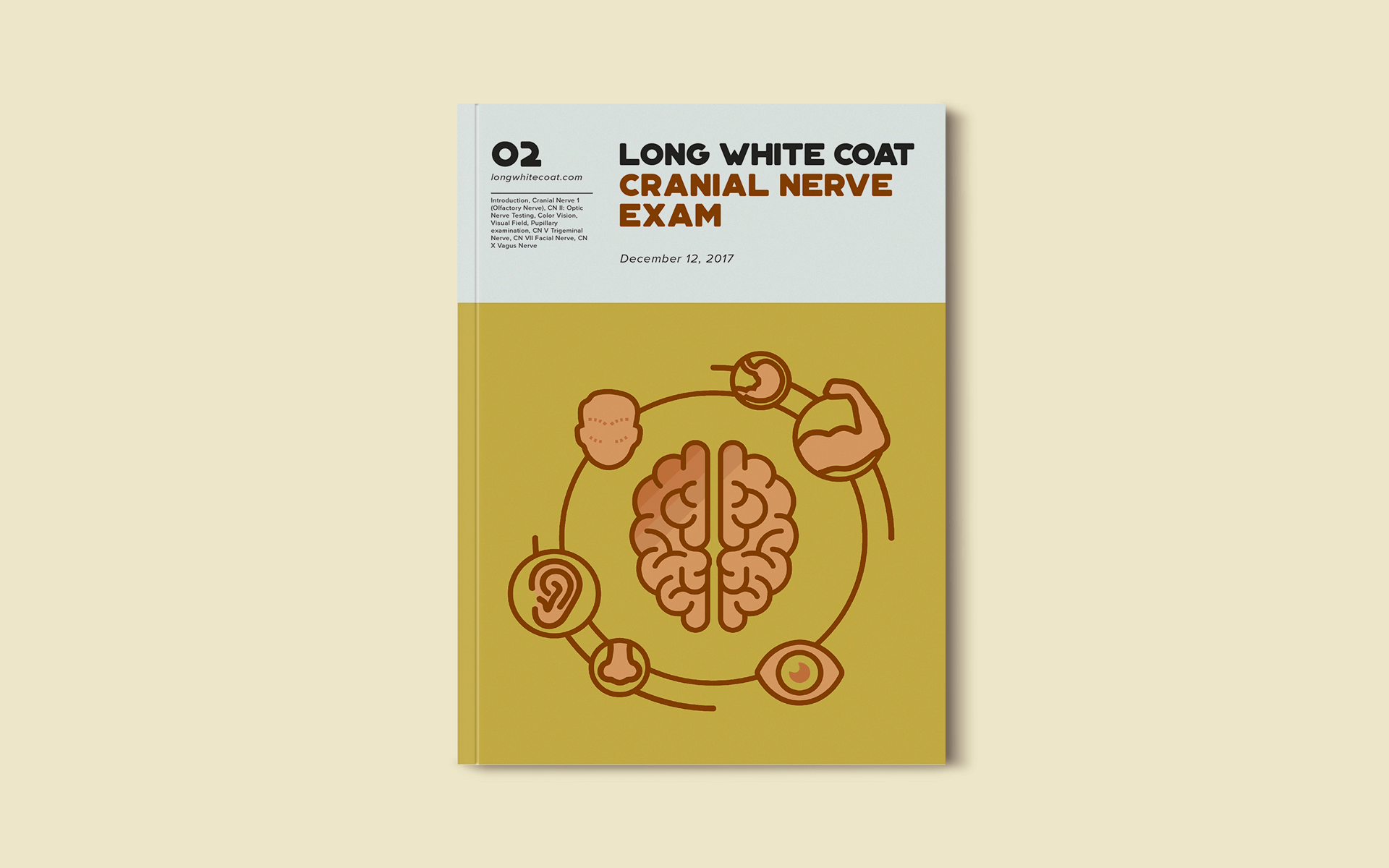
There are twelve cranial nerves that control different aspects of facial functions. Damage to different nuclei or different sections of some nerves can lead to the presentation of different signs and symptoms.

Cranial Nerve 1 (Olfactory Nerve)

There is little value in routinely testing olfaction as it is uncommon to detect an abnormality. Hence testing olfaction is usually omitted from a clinical exam unless there are specific clinical suspicions. A sub frontal tumor such as a meningioma may cause unilateral anosmia. Subfrontal meningioma can cause bilateral anosmia. Olfactory nerve fibers pass through the cribriform plate; hence head injury resulting in a fracture of the cribriform plate can lead to permanent bilateral anosmia. Anosmia is commonly neurodegenerative, occurring particularly in Lewy body disease. Olfaction is also tested when Kallman’s syndrome is suspected.

  1. Before starting the examination, the patency of the nostrils and the ability of the patient to smell should be checked.
  2. The patient is first told to close their eyes.
  3. Test each nostril separately.
  4. One nostril should be closed while the other is being tested.
  5. Irritating smells like ammonia should not be used.
  6. Present common smells like peppermint, soap, clove oil, etc.
CN II: Optic Nerve Testing

Testing the optic nerve consists of several components: visual acuity, color vision, visual field, pupillary examination, and fundus examination.
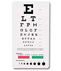
Visual Acuity

  1. To assess visual acuity, a Snellen’s chart is used, and each eye is tested separately.
  2. Ask the patient to read the lowest possible line or successively lower lines to identify efficiently which line can be read with 100% accuracy.
  3. If the patient cannot read even the topmost line, show him your hand and ask him to count fingers.
  4. If the patient fails, wave your hand and ask if the patient can detect the hand movement.
  5. If the patient fails, direct light into his/her eyes to check if the patient can perceive the light.
Color Vision

Acquired unilateral loss of color vision is a characteristic feature of optic neuropathy, and loss of color vision can occur even if visual acuity is intact. Hence, testing color vision using Ishihara test chart books may be a sensitive bedside test for mild optic neuropathy.

Visual Field

Visual field defects in one eye indicate a retinal or optic nerve defect. Lesions at the optic chiasm or lesions behind the optic chiasm in the optic tracts, visual radiations or occipital cortex will result in visual field defects in both eyes.

  1. Sit about a foot away from the patient and face them nose to nose. Ask the patient to close their eye, and you should close the corresponding eye on the ipsilateral side so that you are both looking into each other’s eyes.
  2. Move your hand to the periphery of the visual field.
  3. Bring your fingers into the visual field slowly. Both you and the patient should be able to see the fingers at the same time.
  4. Repeat the above step and check the visual field in all four corners.
  5. Repeat the above procedure for the other eye.

Visual fields are divided into nasal and temporal halves. On each side, the nasal visual field is projected to the temporal half of the retina, and the temporal visual field is projected to the nasal half of the retina. Similarly, the upper half of the visual field is projected to the lower part of the retina and vice versa. Visual field defects can occur due to lesions of various parts of the visual pathway:

  • Optic nerve: complete loss of vision on the affected side
  • Optic chiasm: lesions involving crossing fibers lead to bitemporal hemianopia, and lesions involving uncrossed fibers lead to bi-nasal hemianopia.
  • Optic tract: right optic tract lesion leads to loss of nasal half visual field of the right side and temporal half of visual field of the left side – left homonymous hemianopia (as there is a loss of left halves of visual fields of both sides). Similarly, left optic tract lesion leads to right homonymous hemianopia.
  • Optic radiations: temporal optic radiations represent the upper quadrants of the visual field and damage here leads to superior quadrantanopia of the opposite side. The parietal optic radiations represent lower quadrants of the visual field and lesions lead to inferior quadrantanopia of the opposite side.
  • Visual cortex: there is homonymous hemianopia of the opposite side with macular sparing.
Pupillary examination

Examination of the pupils and their responses to light and accommodation provides information not only about specific neurological syndromes but also information about the integrity of the anterior visual pathways, the brainstem, and the efferent parasympathetic and sympathetic pathways to the pupillary sphincter and dilator muscles, respectively. The third cranial nerve contributes the efferent portion of the visual pathway; hence the pupillary examination takes into account the pathologies of both the optic and oculomotor nerves.

  1. The test is best conducted in a dim-lit room in which the eyes will be relatively dilated, and the constriction can hence be properly noted.
  2. Test each eye separately.
  3. Notice pupil shape and size at rest.
  4. Ask the patient to place one hand on their nose to block light from entering into the other eye (to accurately determine consensual light reflex).
  5. Shine a penlight into one eye and note the constriction in this eye as well as in the other eye from which the light is blocked.
  • Repeat the procedure on the other eye.
  • In order to check for pupillary constriction during accommodation, place your finger close to the patient’s face. Ask the patient to first look at a distant point and then focus on your finger. Notice if the pupils construct upon focusing on your finger.

Asymmetry of the pupils is referred to as anisocoria. Some people with anisocoria have no underlying neuropathology. In this setting, the asymmetry will have been present for a long time without change, and the patient will have no other neurological signs or symptoms. The direct and consensual responses should be preserved.

Some conditions can also affect the size of the pupils. Medications/ intoxications which cause generalized sympathetic activation will result in dilatation of both pupils. Other drugs (e.g., narcotics) cause symmetric constriction of the pupils. These findings can provide important clues when dealing with an agitated or comatose patient suffering from a medication overdose. Eye drops known as mydriatic agents are employed to paralyze the muscles, resulting in marked dilatation of the pupils on the affected side. They are used during a detailed eye examination, allowing a clear view of the retina. Additionally, any process which causes increased intracranial pressure can result in a dilated pupil that does not respond to light. Other conditions like anxiety, pontine lesion, and even simply old age can cause bilateral pupillary dilation.

If the afferent nerve is not working, neither pupil will respond when the light is shined in the affected eye. Light shined in the normal eye, however, will cause the affected pupil to constrict. That’s because the efferent (signal to constrict) response, in this case, is generated by the afferent impulse received by the normally functioning eye. This is referred to as an afferent pupil defect.

If the efferent nerve is not working, the pupil will appear dilated at baseline and will have neither direct nor consensual pupillary responses

CN III, IV, and VI Oculomotor, Trochlear and Abducent Nerves

These three cranial nerves control the movement of the extraocular muscles and hence are tested together. The oculomotor nerve controls the movement of the inferior oblique, inferior rectus, superior rectus, and medial rectus, along with controlling the movement of the upper lid and mediating the constriction of the pupil. The trochlear nerve controls the movement of the superior oblique. The abducent nerve controls the movement of the lateral rectus. Movements are described as elevation (pupil directed upwards), depression (pupil directed downwards), abduction (pupil directed laterally), adduction (pupil directed medially), extorsion (top of eye rotating away from the nose), and intorsion (top of eye rotating towards the nose).

Movements of the extraocular muscles:

Lateral rectus: abduction

Medial rectus: adduction

Inferior rectus: depression, extorsion, adduction

Superior rectus: elevation, intorsion, adduction

Inferior oblique: elevation, extorsion, abduction

Superior oblique: depression, intorsion, abduction

Examination

The examination of the cranial nerves should be done in such a manner that it is easy for you to observe the eye movements of the patient.

  1. Stabilize the patient’s head with one hand. Ask the patient to look straight at your finger, which is held at a distance of two feet, and follow its movements.
  2. Trace an imaginary “H” using your finger and make sure your finger travels far enough for you to be able to detect significant eye movement.
  3. To check for accommodation bring your finger closer to the patient, causing the patient to look cross-eyed.
Pathology

Third nerve paralysis

There is ptosis due to paralysis of levator palpebrae superioris. Ptosis can be caused by third cranial nerve palsy, Myasthenia gravis, or Horner’s syndrome.

Superior, inferior, and medial recti and inferior oblique are paralyzed. The eyeball becomes deviated laterally and slightly downwards due to the unopposed action of the superior oblique and lateral rectus. The pupil is dilated and fixed. Light and accommodation reflexes will be absent

Third nerve palsy can be caused by diabetes, aneurysm of the posterior communicating artery, midbrain lesion, or cavernous sinus thrombosis.

Fourth nerve paralysis

The superior oblique is paralyzed, leading to double vision

Sixth nerve paralysis

The lateral rectus is paralyzed, and the eyeball deviates medially. Because of its long intracranial route, the sixth cranial nerve is often involved in raised intracranial pressure of any etiology. When the patient is asked to move the eye laterally, there is nystagmus in internuclear ophthalmoplegia, and the opposite eye cannot move medially. This occurs due to the lesion in the medial longitudinal bundle on the side of the weakness of adduction. Bilateral internuclear ophthalmoplegia is characteristic of multiple sclerosis.

CN V Trigeminal Nerve

This is a mixed motor and sensory nerve. The neve is divided into three portions: ophthalmic division (sensory), maxillary division (sensory), and mandibular division (mixed). The motor portion supplies the muscles of mastication: masseter, temporalis, and the pterygoids. The sensory portion carries sensations of touch, pain, and temperature from the face, the anterior part of the head, and inside the mouth.

Motor examination

  1. Place your hands on the sides of the patient’s cheek, fingers being on the temple. Ask the patient to clench their teeth. You should be able to feel the contraction of the masseter and temporalis muscles. In unilateral paralysis, muscles of the affected side will not contract. In bilateral paralysis, the jaw hangs loosely.
  2. Ask the patient to open the jaw against resistance to test the pterygoids on both sides. The jaw will deviate towards the weaker side.
  3. Jaw jerk: ask the patient to open their mouth and hang the jaw loosely. Place your thumb over the chin and strike it with a clinical hammer. There is a closure of the jaw if the reflex is present. Normally this is not eligible but is brisk in upper motor neuron paralysis of the trigeminal nerve.

Sensory examination

  1. Use a sharp implement (e.g., broken wooden handle of a cotton-tipped applicator).
  2. Ask the patient to close their eyes so that they receive no visual cues.
  3. Touch the sharp tip of the stick to the right and the left side of the forehead, assessing the ophthalmic branch.
  4. Touch the tip to the right and the left side of the cheek area, assessing the Maxillary branch.
  5. Touch the tip to the right and the left side of the jaw area, assessing the Mandibular branch.
  6. Temperature can be assessed by touching the different areas of the face described above with test tubes filled with cold and hot water and asking the patient to describe what sensation they feel.

The patient should be able to identify the sharp end touching their face. Due to the sensitivity of the face, the object should not be pushed too hard. The ophthalmic branch of CN V also receives sensory input from the surface of the eye. To assess this component the corneal and conjunctival reflexes need to be tested:

  • Pull out a wisp of cotton and twist into a thin strand.
  • While the patient is looking straight ahead, gently brush the wisp against the lateral aspect of the sclera.
  • This should cause the patient to blink. Blinking also requires that the facial nerve is functioning normally, as it controls eyelid closure.
  • The corneal reflex can also be elicited by blowing into the eye of the patient.

Pathology

  1. Trigeminal neuralgia is the most common disease of the fifth nerve. It can be idiopathic or caused by multiple sclerosis, tumors, or herpes zoster.
  2. Cerebellopontine angle tumors can involve the trigeminal nerve
  3. The ophthalmic division can be involved in lesions of the cavernous sinus.
  4. Bilateral motor paralysis may occur in bulbar palsy (causing wasting of muscles of mastication) and pseudobulbar palsy (causing jaw jerk to be brisk).
 
CN VII Facial Nerve

The facial nerve innervates most of the muscles of facial expression.

Motor Function Examination

  1. If the facial nerve is paralyzed, the patient may complain of the inability to close the eyelid, the collection of food in the mouth, and the dribbling of saliva on the affected side so be sure to take those complaints into account.
  2. Before beginning the examination, inspect the face of the patient and compare the symmetry of both sides of the face. The number of wrinkles should be equal on both sides, the palpebral fissures should be the same on both sides (the palpebral fissure may be wide on the paralyzed side), the nasolabial folds should be equal (the nasolabial fold may be flattened on the paralyzed side) and the angles of the mouth should be of the same height.
  3. Ask the patient to frown or wrinkle the forehead. There will be no wrinkling on the affected side.
  4. Ask the patient to close the eyes; the affected side will remain open, and there will be brisk upwards rolling of the eyeball. To test the power of the orbicularis oculi, ask the patient to close their eyes as strongly as possible while you try to open the upper eyelids. The affected side will be weak.
  5. Ask the patient to inflate the cheeks and tap on both sides with the finger. The weak side will be deflated easily.
  6. Ask the patient to show the teeth. The angle of the mouth would deviate towards the healthy side. In an unconscious patient, deviation of the angle of the mouth can be demonstrated by applying pressure on the supraorbital notch.
  7. Ask the patient to whistle. The patient cannot whistle as air escapes from the paralyzed side.
  8. The patient will complain of unusually loud sounds on the paralyzed side if the nerve to stapedius muscle is involved.
  9. Pouting of the lips and transverse smile are the manifestations of bilateral facial weakness.
  10. Also, perform the corneal/conjunctival reflex as the sensory component of the reflex is carried by the facial nerve

Taste Examination

  1. The taste test should be of the anterior two-thirds of the tongue.
  2. Get solutions of four common tastes – sweet, salt, sour and bitter.
  3. Instruct the patient to identify the taste, either by writing or raising fingers, e.g., one finger if the taste is sweet, two fingers if salty, and so on.
  4. Ask the patient to protrude the tongue. Hold it with gauze, dry it and test each side separately.
  5. Put a drop of each solution one by one and ask for the response.
  6. Test bitter at the end.

The facial nerve is the most commonly affected cranial nerve by lesion of both upper motor neuron and lower motor neuron. This clinical distinction is very important, as central (UMN lesion) vs. peripheral dysfunction (LMN lesion) carry different prognostic and treatment implications. Bell’s palsy (peripheral facial nerve dysfunction) tends to happen in patients over 50 and often responds to treatment with Acyclovir (an anti-viral agent) and Prednisone (a corticosteroid). Over the course of weeks or months, there is usually improvement and often complete resolution of symptoms. Assessment of acute central facial nerve dysfunction would require quite a different approach (e.g., neuroimaging to determine etiology).

Upper Motor Neuron Lesion

Manifestations are on the opposite side. The upper half of the face is less severely affected because the part of the facial nerve nucleus which supplies muscles of the upper half of the face is connected with both cerebral hemispheres. The part of the facial nerve nucleus which supplies muscles of the lower half of the face is connected only with the contralateral cerebral hemisphere. Smiling and other emotional movements are usually preserved in UMN lesions because there is a separate path for these movements. An upper motor neuron lesion may occur with a central nervous system event, such as a stroke. The patient will present with preserved ability to wrinkle the forehead, but the corner of the mouth will deviate on the contralateral side.

Lower Motor Neuron Lesion

The whole ipsilateral half of the face is affected. Bell’s palsy is the most common cause of isolated lower motor neuron facial palsy. The lesion is in the facial canal. The facial nerve has a long route and gives off branches at various sites, the site of the lesion can be localized with considerable precision.

  1. If the lesion is after the nerve exits from the skull, there is the only weakness of facial muscles. This presents with an inability to wrinkle the forehead, inability to close eyelids completely, decreased nasolabial fold, and deviated angle of the corner of the mouth.
  2. If the lesion is in the facial canal, between the chords tympani and branch to the stapedius, in addition to motor weakness, there is a loss of taste as well.
  3. If the lesion is between the branch of the stapedius muscle and internal auditory meatus, there is hyperacusis on the affected side, in addition to motor weakness and loss of taste. The geniculate ganglion can be infected by herpes zoster (Ramsay Hunt Syndrome). In addition to other features of facial palsy, there are vesicles in that part of the external auditory meatus which get sensory supply from the facial nerve.
  4. If the lesion is in the internal auditory meatus, in addition to the features of facial nerve palsy, the eighth nerve is also paralyzed.
CN VIII Vestibulocochlear Nerve

The vestibulocochlear nerve has two components, the cochlear component which deals with hearing, and the vestibular component which deals with a sense of balance. The examination of all these components is hence separate as well.

For the cochlear division the following tests are performed:

Whisper-Test

  1. Ask the patient to close their eyes.
  2. Whisper the patient’s name or some other words while standing just behind one ear. The patient should be able to repeat those words back.
  3. Repeat the same on the other ear.
  4. Alternatively, you can place your fingers close to the ears (about a distance of 5 cm away from the ears) and rub them. The patient should be able to hear that.

The Rinne’s and Weber’s tests are carried out using tuning forks, which help to differentiate between conductive deafness and sensorineural deafness.

Rinne’s Test

  1. Strike the 512 Hz tuning fork on a hard surface.
  2. Place the base of the vibrating tuning fork on the mastoid process. If the patient cannot hear, they have sensorineural deafness.
  3. If the patient can hear, indicate to them when they stop hearing the vibration.
  4. Then bring the tuning fork close to the external auditory meatus. If the patient can still hear something that means that air conduction is better than bone conduction and the Rinne’s test is positive.
  5. If the patient cannot hear anything, perform the test in reverse order by first bringing the tuning fork to the auditory meatus and then placing it on the mastoid process. If the patient can hear the noise when the tuning fork is placed on the mastoid process; Rinne’s test is negative.
  6. Repeat the same process on the other ear.

A positive Rinne’s test is normal. In sensory neural deafness, air conduction is better than bone conduction, but both are reduced – reduced positive Rinne’s test. In conductive deafness Rinne’s test is negative. In mixed deafness, bone conduction is better than air conduction, but both are reduced.

Weber’s Test

  1. Place a vibrating tuning fork in the middle of the forehead or the vertex.
  2. Ask the patient whether the hearing is better in the right or left ear.

If it is equal on both sides, the test is central, and it indicates either normal hearing or equal deafness on both sides. In sensorineural deafness, it is lateralized to the normal ear while in conductive deafness it is lateralized to the diseased ear.

For the vestibular division the following tests are done:

Revolving Chair Test

  1. Ask the patient to sit on a revolving chair with open eyes and head flexed at 30˚ forwards, chin almost touching the neck.
  2. Rotate the chair quickly to the right for 20 seconds or about 10 revolutions.
  3. Note the initial nystagmus and then ask the subject to close their eyes.
  4. Stop the chair abruptly and ask the patient to open their eyes.
  5. Now note the direction of nystagmus.

Caloric Test

  1. Ask the patient to lie down in a supine position.
  2. The head of the subject is elevated at 30˚.
  3. 5-10 ml of ice water is instilled into the external ear of the subject with a
    [amazon link=”B01LWX4FEC” title=”Syringe” /]

    or [amazon link=”B0055QNVDM” title=”Soft Rubber Catheter” /].

  4. Quick nystagmus occurs in the direction of the irrigated ears. The patient may experience vertigo, nausea, or vomiting.
CN IX Glossopharyngeal Nerve

The motor part of the nerve supplies the stylopharyngeus muscle and the sensory part carries sensations from the pharynx, tonsillar region, and the posterior one-third of the tongue. It also carries secretomotor fibers to the parotid gland. The motor function of the nerve cannot be tested separately from the vagus nerve hence only the sensory component examination is mentioned here.

Gag Reflex

  1. Ask the patient to open the mouth and depress the tongue with a
    [amazon link=”B005Z4SBZW” title=”Tongue Depressor” /]
  2. Touch the posterior pharyngeal wall with a [amazon link=”B000BI3M60″ title=”Cotton Tip Applicator” /]
  3. First, touch one side of the midline and then the other.
  4. There will be contraction and elevation of the pharyngeal wall on that side. The sensory component of this reflex arc is the glossopharyngeal nerve, and the motor component is the vagus nerve.

Palatal Reflex

  1. Similarly, touch the soft palate. The soft palate should move upwards.
  2. Test each side separately.
CN X Vagus Nerve

The vagus nerve mainly carries the parasympathetic fibers to the organs of the chest and abdomen, but there is also a motor component that supplies the muscles of the soft palate and pharynx and the intrinsic muscles of the larynx. In the examination, only the motor function of the nerve is tested.

Speech Examination

  1. If the larynx is paralyzed, there is hoarseness but if the soft palate is paralyzed the voice becomes nasal.
  2. Ask the patient to cough. If there is paralysis of the vagus nerve, the cough will become nasal or bovine.

Soft palate Examination

  1. Ask the patient to open their mouth.
  2. Depress their tongue with a tongue depressor to visualize the uvula. Ask the patient to say “ah.” In bilateral paralysis, the soft palate will not move. In unilateral paralysis, the affected side will remain immobile, and the uvula will deviate towards the normal side. A peritonsillar abscess, among other causes, can also cause the deviation, so these need to be ruled out as well.
  3. Ask the patient to puff out their cheek. Normally, the palate elevates and occludes the nasopharynx. However, in 10th nerve paralysis air will audibly escape from the nose.
  4. The patient will also give a history of dysphagia and nasal regurgitation of fluid. This can be confirmed by asking the patient to take a drink.

Posterior Pharyngeal Wall Examination

  1. Observe movements of the posterior pharyngeal wall when the patient says “ah.” If one side is paralyzed, the wall will move more laterally towards the normal side.
  2. Also, check the gag and palatal reflexes.

The recurrent laryngeal nerve (a branch of the vagus nerve) can be damaged during thyroid surgery or by malignant tumors. Bilateral vagus nerve palsy occurs in bulbar and pseudobulbar palsy.

CN XI Accessory Nerve

The spinal accessory nerve innervates the trapezius and sternocleidomastoid muscles. The trapezius muscle is responsible for shrugging shoulders, whereas the sternocleidomastoid helps in turning the head laterally.

Examination of Sternocleidomastoid Function

  1. Ask the patient to bend their head downwards as you try to oppose this action and offer resistance using your hand. This action is carried out by both sternocleidomastoid muscles
  2. Each sternocleidomastoid muscle pushes the head towards the opposite side. Ask the patient to turn their head towards the left while you resist this movement with your hand placed on the left side of the face. This will test the right sternocleidomastoid muscle
  3. Repeat the same for the other side.

Examination of Trapezius Function

  1. Before beginning the examination inspect the patient from behind. The upper portion of the scapula will deviate away from the spine, whereas the lower portion will deviate towards the spine. The whole arm will droop, and the fingers will appear to be closer to the ground on the affected side compared to the unaffected side.
  2. Examine the patient by asking the patient to shrug their shoulders against resistance you offer by pressing down on their shoulders.

The accessory nerve tends to get paralyzed along with other nerves in bulbar palsy.

CN XII Hypoglossal Nerve

The hypoglossal nerve innervates the muscles of the tongue. Each hypoglossal nerve innervates one-half of the tongue.

  1. Ask the patient to open the mouth and inspect the tongue as it lies on the floor of the mouth for size, shape, wasting, and fasciculation.
  2. Ask the patient to protrude the tongue; it will deviate towards the paralyzed side as normal genioglossus will push it towards the opposite side.
  3. Ask the patient to press the tongue against the cheek while you resist with finger pressure on the outside of the cheek. In unilateral paralysis movements towards the normal side will be weak.

In bilateral UMN lesion, the tongue looks small and conical and is immobile. In unilateral UMN lesion, the tongue may sometimes deviate towards the paralyzed side when protruded. No wasting is seen in UMN lesions. In bilateral LMN lesion, there is generalized wasting with fasciculation. In unilateral LMN lesions, wasting and fasciculation are present on the affected side only.

Learn More about our Services and how can we help you with your research!

Introduction

In behavioral neuroscience, the Open Field Test (OFT) remains one of the most widely used assays to evaluate rodent models of affect, cognition, and motivation. It provides a non-invasive framework for examining how animals respond to novelty, stress, and pharmacological or environmental manipulations. Among the test’s core metrics, the percentage of time spent in the center zone offers a uniquely normalized and sensitive measure of an animal’s emotional reactivity and willingness to engage with a potentially risky environment.

This metric is calculated as the proportion of time spent in the central area of the arena—typically the inner 25%—relative to the entire session duration. By normalizing this value, researchers gain a behaviorally informative variable that is resilient to fluctuations in session length or overall movement levels. This makes it especially valuable in comparative analyses, longitudinal monitoring, and cross-model validation.

Unlike raw center duration, which can be affected by trial design inconsistencies, the percentage-based measure enables clearer comparisons across animals, treatments, and conditions. It plays a key role in identifying trait anxiety, avoidance behavior, risk-taking tendencies, and environmental adaptation, making it indispensable in both basic and translational research contexts.

Whereas simple center duration provides absolute time, the percentage-based metric introduces greater interpretability and reproducibility, especially when comparing different animal models, treatment conditions, or experimental setups. It is particularly effective for quantifying avoidance behaviors, risk assessment strategies, and trait anxiety profiles in both acute and longitudinal designs.

What Does Percentage of Time in the Centre Measure?

This metric reflects the relative amount of time an animal chooses to spend in the open, exposed portion of the arena—typically defined as the inner 25% of a square or circular enclosure. Because rodents innately prefer the periphery (thigmotaxis), time in the center is inversely associated with anxiety-like behavior. As such, this percentage is considered a sensitive, normalized index of:

  • Exploratory drive vs. risk aversion: High center time reflects an animal’s willingness to engage with uncertain or exposed environments, often indicative of lower anxiety and a stronger intrinsic drive to explore. These animals are more likely to exhibit flexible, information-gathering behaviors. On the other hand, animals that spend little time in the center display a strong bias toward the safety of the perimeter, indicative of a defensive behavioral state or trait-level risk aversion. This dichotomy helps distinguish adaptive exploration from fear-driven avoidance.

  • Emotional reactivity: Fluctuations in center time percentage serve as a sensitive behavioral proxy for changes in emotional state. In stress-prone or trauma-exposed animals, decreased center engagement may reflect hypervigilance or fear generalization, while a sudden increase might indicate emotional blunting or impaired threat appraisal. The metric is also responsive to acute stressors, environmental perturbations, or pharmacological interventions that impact affective regulation.

  • Behavioral confidence and adaptation: Repeated exposure to the same environment typically leads to reduced novelty-induced anxiety and increased behavioral flexibility. A rising trend in center time percentage across trials suggests successful habituation, reduced threat perception, and greater confidence in navigating open spaces. Conversely, a stable or declining trend may indicate behavioral rigidity or chronic stress effects.

  • Pharmacological or genetic modulation: The percentage of time in the center is widely used to evaluate the effects of pharmacological treatments and genetic modifications that influence anxiety-related circuits. Anxiolytic agents—including benzodiazepines, SSRIs, and cannabinoid agonists—reliably increase center occupancy, providing a robust behavioral endpoint in preclinical drug trials. Similarly, genetic models targeting serotonin receptors, GABAergic tone, or HPA axis function often show distinct patterns of center preference, offering translational insights into psychiatric vulnerability and resilience.

Critically, because this metric is normalized by session duration, it accommodates variability in activity levels or testing conditions. This makes it especially suitable for comparing across individuals, treatment groups, or timepoints in longitudinal studies.

A high percentage of center time indicates reduced anxiety, increased novelty-seeking, or pharmacological modulation (e.g., anxiolysis). Conversely, a low percentage suggests emotional inhibition, behavioral avoidance, or contextual hypervigilance. reduced anxiety, increased novelty-seeking, or pharmacological modulation (e.g., anxiolysis). Conversely, a low percentage suggests emotional inhibition, behavioral avoidance, or contextual hypervigilance.

Behavioral Significance and Neuroscientific Context

1. Emotional State and Trait Anxiety

The percentage of center time is one of the most direct, unconditioned readouts of anxiety-like behavior in rodents. It is frequently reduced in models of PTSD, chronic stress, or early-life adversity, where animals exhibit persistent avoidance of the center due to heightened emotional reactivity. This metric can also distinguish between acute anxiety responses and enduring trait anxiety, especially in longitudinal or developmental studies. Its normalized nature makes it ideal for comparing across cohorts with variable locomotor profiles, helping researchers detect true affective changes rather than activity-based confounds.

2. Exploration Strategies and Cognitive Engagement

Rodents that spend more time in the center zone typically exhibit broader and more flexible exploration strategies. This behavior reflects not only reduced anxiety but also cognitive engagement and environmental curiosity. High center percentage is associated with robust spatial learning, attentional scanning, and memory encoding functions, supported by coordinated activation in the prefrontal cortex, hippocampus, and basal forebrain. In contrast, reduced center engagement may signal spatial rigidity, attentional narrowing, or cognitive withdrawal, particularly in models of neurodegeneration or aging.

3. Pharmacological Responsiveness

The open field test remains one of the most widely accepted platforms for testing anxiolytic and psychotropic drugs. The percentage of center time reliably increases following administration of anxiolytic agents such as benzodiazepines, SSRIs, and GABA-A receptor agonists. This metric serves as a sensitive and reproducible endpoint in preclinical dose-finding studies, mechanistic pharmacology, and compound screening pipelines. It also aids in differentiating true anxiolytic effects from sedation or motor suppression by integrating with other behavioral parameters like distance traveled and entry count (Prut & Belzung, 2003).

4. Sex Differences and Hormonal Modulation

Sex-based differences in emotional regulation often manifest in open field behavior, with female rodents generally exhibiting higher variability in center zone metrics due to hormonal cycling. For example, estrogen has been shown to facilitate exploratory behavior and increase center occupancy, while progesterone and stress-induced corticosterone often reduce it. Studies involving gonadectomy, hormone replacement, or sex-specific genetic knockouts use this metric to quantify the impact of endocrine factors on anxiety and exploratory behavior. As such, it remains a vital tool for dissecting sex-dependent neurobehavioral dynamics.
The percentage of center time is one of the most direct, unconditioned readouts of anxiety-like behavior in rodents. It is frequently reduced in models of PTSD, chronic stress, or early-life adversity. Because it is normalized, this metric is especially helpful for distinguishing between genuine avoidance and low general activity.

Methodological Considerations

  • Zone Definition: Accurately defining the center zone is critical for reliable and reproducible data. In most open field arenas, the center zone constitutes approximately 25% of the total area, centrally located and evenly distanced from the walls. Software-based segmentation tools enhance precision and ensure consistency across trials and experiments. Deviations in zone parameters—whether due to arena geometry or tracking inconsistencies—can result in skewed data, especially when calculating percentages.


  • Trial Duration: Trials typically last between 5 to 10 minutes. The percentage of time in the center must be normalized to total trial duration to maintain comparability across animals and experimental groups. Longer trials may lead to fatigue, boredom, or habituation effects that artificially reduce exploratory behavior, while overly short trials may not capture full behavioral repertoires or response to novel stimuli.


  • Handling and Habituation: Variability in pre-test handling can introduce confounds, particularly through stress-induced hypoactivity or hyperactivity. Standardized handling routines—including gentle, consistent human interaction in the days leading up to testing—reduce variability. Habituation to the testing room and apparatus prior to data collection helps animals engage in more representative exploratory behavior, minimizing novelty-induced freezing or erratic movement.


  • Tracking Accuracy: High-resolution tracking systems should be validated for accurate, real-time detection of full-body center entries and sustained occupancy. The system should distinguish between full zone occupancy and transient overlaps or partial body entries that do not reflect true exploratory behavior. Poor tracking fidelity or lag can produce significant measurement error in percentage calculations.


  • Environmental Control: Uniformity in environmental conditions is essential. Lighting should be evenly diffused to avoid shadow bias, and noise should be minimized to prevent stress-induced variability. The arena must be cleaned between trials using odor-neutral solutions to eliminate scent trails or pheromone cues that may affect zone preference. Any variation in these conditions can introduce systematic bias in center zone behavior. Use consistent definitions of the center zone (commonly 25% of total area) to allow valid comparisons. Software-based segmentation enhances spatial precision.

Interpretation with Complementary Metrics

Temporal Dynamics of Center Occupancy

Evaluating how center time evolves across the duration of a session—divided into early, middle, and late thirds—provides insight into behavioral transitions and adaptive responses. Animals may begin by avoiding the center, only to gradually increase center time as they habituate to the environment. Conversely, persistently low center time across the session can signal prolonged anxiety, fear generalization, or a trait-like avoidance phenotype.

Cross-Paradigm Correlation

To validate the significance of center time percentage, it should be examined alongside results from other anxiety-related tests such as the Elevated Plus Maze, Light-Dark Box, or Novelty Suppressed Feeding. Concordance across paradigms supports the reliability of center time as a trait marker, while discordance may indicate task-specific reactivity or behavioral dissociation.

Behavioral Microstructure Analysis

When paired with high-resolution scoring of behavioral events such as rearing, grooming, defecation, or immobility, center time offers a richer view of the animal’s internal state. For example, an animal that spends substantial time in the center while grooming may be coping with mild stress, while another that remains immobile in the periphery may be experiencing more severe anxiety. Microstructure analysis aids in decoding the complexity behind spatial behavior.

Inter-individual Variability and Subgroup Classification

Animals naturally vary in their exploratory style. By analyzing percentage of center time across subjects, researchers can identify behavioral subgroups—such as consistently bold individuals who frequently explore the center versus cautious animals that remain along the periphery. These classifications can be used to examine predictors of drug response, resilience to stress, or vulnerability to neuropsychiatric disorders.

Machine Learning-Based Behavioral Clustering

In studies with large cohorts or multiple behavioral variables, machine learning techniques such as hierarchical clustering or principal component analysis can incorporate center time percentage to discover novel phenotypic groupings. These data-driven approaches help uncover latent dimensions of behavior that may not be visible through univariate analyses alone.

Total Distance Traveled

Total locomotion helps contextualize center time. Low percentage values in animals with minimal movement may reflect sedation or fatigue, while similar values in high-mobility subjects suggest deliberate avoidance. This metric helps distinguish emotional versus motor causes of low center engagement.

Number of Center Entries

This measure indicates how often the animal initiates exploration of the center zone. When combined with percentage of time, it differentiates between frequent but brief visits (indicative of anxiety or impulsivity) versus fewer but sustained center engagements (suggesting comfort and behavioral confidence).

Latency to First Center Entry

The delay before the first center entry reflects initial threat appraisal. Longer latencies may be associated with heightened fear or low motivation, while shorter latencies are typically linked to exploratory drive or low anxiety.

Thigmotaxis Time

Time spent hugging the walls offers a spatial counterbalance to center metrics. High thigmotaxis and low center time jointly support an interpretation of strong avoidance behavior. This inverse relationship helps triangulate affective and motivational states.

Applications in Translational Research

  • Drug Discovery: The percentage of center time is a key behavioral endpoint in the development and screening of anxiolytic, antidepressant, and antipsychotic medications. Its sensitivity to pharmacological modulation makes it particularly valuable in dose-response assessments and in distinguishing therapeutic effects from sedative or locomotor confounds. Repeated trials can also help assess drug tolerance and chronic efficacy over time.
  • Genetic and Neurodevelopmental Modeling: In transgenic and knockout models, altered center percentage provides a behavioral signature of neurodevelopmental abnormalities. This is particularly relevant in the study of autism spectrum disorders, ADHD, fragile X syndrome, and schizophrenia, where subjects often exhibit heightened anxiety, reduced flexibility, or altered environmental engagement.
  • Hormonal and Sex-Based Research: The metric is highly responsive to hormonal fluctuations, including estrous cycle phases, gonadectomy, and hormone replacement therapies. It supports investigations into sex differences in stress reactivity and the behavioral consequences of endocrine disorders or interventions.
  • Environmental Enrichment and Deprivation: Housing conditions significantly influence anxiety-like behavior and exploratory motivation. Animals raised in enriched environments typically show increased center time, indicative of reduced stress and greater behavioral plasticity. Conversely, socially isolated or stimulus-deprived animals often show strong center avoidance.
  • Behavioral Biomarker Development: As a robust and reproducible readout, center time percentage can serve as a behavioral biomarker in longitudinal and interventional studies. It is increasingly used to identify early signs of affective dysregulation or to track the efficacy of neuromodulatory treatments such as optogenetics, chemogenetics, or deep brain stimulation.
  • Personalized Preclinical Models: This measure supports behavioral stratification, allowing researchers to identify high-anxiety or low-anxiety phenotypes before treatment. This enables within-group comparisons and enhances statistical power by accounting for pre-existing behavioral variation. Used to screen anxiolytic agents and distinguish between compounds with sedative vs. anxiolytic profiles.

Enhancing Research Outcomes with Percentage-Based Analysis

By expressing center zone activity as a proportion of total trial time, researchers gain a metric that is resistant to session variability and more readily comparable across time, treatment, and model conditions. This normalized measure enhances reproducibility and statistical power, particularly in multi-cohort or cross-laboratory designs.

For experimental designs aimed at assessing anxiety, exploratory strategy, or affective state, the percentage of time spent in the center offers one of the most robust and interpretable measures available in the Open Field Test.

Explore high-resolution tracking solutions and open field platforms at

References

  • Prut, L., & Belzung, C. (2003). The open field as a paradigm to measure the effects of drugs on anxiety-like behaviors: a review. European Journal of Pharmacology, 463(1–3), 3–33.
  • Seibenhener, M. L., & Wooten, M. C. (2015). Use of the open field maze to measure locomotor and anxiety-like behavior in mice. Journal of Visualized Experiments, (96), e52434.
  • Crawley, J. N. (2007). What’s Wrong With My Mouse? Behavioral Phenotyping of Transgenic and Knockout Mice. Wiley-Liss.
  • Carola, V., D’Olimpio, F., Brunamonti, E., Mangia, F., & Renzi, P. (2002). Evaluation of the elevated plus-maze and open-field tests for the assessment of anxiety-related behavior in inbred mice. Behavioral Brain Research, 134(1–2), 49–57.

Written by researchers, for researchers — powered by Conduct Science.