
As an Amazon Associate Conductscience Inc earns revenue from qualifying purchases
Otoscopy, an integral part of every doctor’s routine physical examination, is a medical procedure that involves taking a look into our ears and down our ear canals using an instrument called an otoscope. The purpose of otoscopy is to examine whether the different parts of the ear, which include the pinna, the external and internal auditory canal, and the eardrum, are all in good condition. Looking into the state of these structures can spell the difference between ignorance and prevention of some serious medical illnesses associated with hearing loss. Aside from general check-up procedures and routine screening, otoscopy is also done during extensive hearing assessments, before taking an impression of the ear for hearing aid fitting, and in the assessment of hearing aids. Usually sold in sets with ophthalmoscopes, otoscopes are standard tools in every doctor’s arsenal.
The otoscope is a tool began in the mid-1300s in France, used to study the aural and nasal passages. During the 19th century, many other otoscope varieties arose, all serving the same, original purpose. The funnel-shaped speculum, which is how the modern otoscope looks today, was invented in 1838, and the pneumatic otoscope was invented in 1864, used to observe how the tympanic membrane reacts to air pressure. The binocular otoscope, meanwhile introduced for surgical use, was invented in 1872 and revolutionized ear surgery for its time. Today, because of the tool’s versatility, the modern otoscope can also be used to observe the nasal passages of patients, as well as the upper throat and the oral cavity. What used to be a simple tool addressing the cause of ear pain in patients has now evolved into a sophisticated instrument that serves a variety of uses in the medical setting today.
Though seemingly a complex instrument, the key concept upon which the otoscope operates is simple–it is a tool that provides a concentrated source of illumination with magnification incorporated in the easy-to-hold structural design. Otoscopes usually consist of three primary parts–the handle, the head, and the cone. The handle is where the power source is located, either in the form of an electrical component to be plugged into a wall socket, or an enclosure for batteries. The head of the otoscope includes the most important component of the instrument, the manual focus halogen or incandescent bulb, with easily adjustable intensity of light. The head of the otoscope is also where the eyepiece is located. Meanwhile, the cone part of the otoscope’s head is appropriately shaped to fit inside a patient’s ear, as well as the nose and throat. This particular part of the instrument is designed to be fitted with specular or ear tips, which are separate, detachable components of the otoscope usually made of plastic and disposable material. Shaped like a cone with holes at both ends, the speculum goes on top of the head of the otoscope and is used to focus the light onto the ear canal. Specula are produced in various diameters and lengths to fit different patients’ ear sizes, permitting safe and sanitary visualization of the inner ear. It is recommended that the largest possible size of the speculum be used to allow for the most amount of light to enter the patient’s ear canal. Common sizes for disposable speculums are 2 mm for infants, and, 3 mm, 4 mm, and 5 mm for adults.
The head of the otoscope is also equipped with a hole for an insufflator, another separate piece that provides a small air vent connection that lets the doctor to puff air into the ear canal. This technique, called insufflation or pneumatic otoscopy, allows the doctor to assess the mobility of the eardrum by observing how it responds to varying levels of air pressure.
A complete assessment of the ear requires a systematic examination of its external and internal structures. The first step to the otoscopic examination is the preparation of tools and procedures using hygienic and sanitary methods. Next, since otoscopy as a procedure is one that may cause discomfort or pain to the patient due to a very close proximity with the doctor as well as the naturally uncomfortable process of having a foreign object stuck into one’s ear, it is important to establish proper informed consent before the examination can begin, as with any medical procedure. It can be of large assistance to inform the patient beforehand as to what shall happen during the examination, and what they must keep in mind. Such reminders include sitting still and speaking up if it gets too uncomfortable for the patient. It must also be noted that this preliminary step is especially important when dealing with children and babies because the risk for complications in the procedure could be high. When dealing with younger patients, it is better to proceed with the examination because any preliminary information might scare them or make them refuse to cooperate. It is better to work with a parent or primary caregiver in keeping them steady for the procedure.
Ask the client to tilt their head slightly while seated. The first thing to be examined is the outer ear, with careful observation of the auricle, pinna, and surrounding areas for any abnormalities such as skin lesions, tenderness, foul odor, and tilt of the ears. For instance, the ear position of a child is normally at a 10-degree tilt, and positions outside of this range could be a clinical marker for serious medical conditions associated with hearing loss. Any tenderness or pain that the patient could experience during this preliminary inspection could be a sign not to go through with the otoscopy and should, therefore, be noted accordingly.
After the external visual inspection is done, the internal structures are examined using the otoscope. First, equip the otoscope with the properly-sized speculum, preferably the largest. For adults, it is generally recommended to use speculum of 4-6 mm in size. Turn the device on and hold it delicately, as if you are holding a pen. When examining the right ear of the patient, use your right hand. Balance the otoscope lightly between thumb and index finger, and extend your pinky and rest it against the patient’s face to track movement. This is so that if the patient suddenly moves, you may follow with your hand and otoscope to avoid any accidental harm or injury. During the procedure, the pinna must be pulled gently upwards, outwards, and backward to straighten the auditory canal. Finally, insert the otoscope into the external auditory meatus, look through the view piece, and inspect the tympanic membrane or the eardrum. In general inspection procedures, it is useful to take note of the 4Ds, namely, discharge, displacement, discoloration, and deformity. After examining the first ear, repeat the whole process with the other ear and proceed to do some other hearing tests to complete the ear examination.
Abnormal findings from observations of the outer ear and external auditory canal may include flaky linings and symptoms of itchiness suggestive of eczema, or a swollen auditory canal with discharge often accompanied by an unpleasant smell, suggestive of infection. Take note of the normal structures that must be seen in the tympanic membrane during the otoscopic procedure. In addition to identifying these structures, other contents of the inner ear such as foreign bodies or wax must also be noted.
Primary care physicians require the use of otoscopes for screening illnesses and looking closely at symptoms of the ear, and it is, therefore, an indispensable instrument in the medical field. Specialty instruments such as these are often sold for high prices in the market, and for a good reason–as healthcare practitioners, the service one provides should not be compromised by tools of low quality. Thus, here are some important selection criteria one should keep in mind when choosing what otoscope to purchase.
In selecting the best otoscope in the market, the first thing to decide is one’s budget. Otoscopes are no cheap tools, and it should, therefore, be one’s goal to look for a compromise between quality and cost. The performance of otoscopes meanwhile must be judged regarding light source technology and strength of magnification, both very important features of an effective instrument for patient examination. Next, as these tools must ideally be designed for repeated use, the comfort of both patient and user should be factored in its structural design. How easy is it to use? How comfortable is it for the patient? These are questions that must be answered sufficiently to make a confident otoscope purchase. Finally, another important feature of otoscopes is how long they will last. In buying an instrument as expensive as these specialty tools, one must look into the product’s lifespan and available warranty from the brand.
Now that we’ve listed all there is to think about, let’s look at the best otoscopes in the market today. Though there are many different types of otoscopes such as pocket, full-size, and video otoscopes, this list shall cover all types and identify which ones are the best-recommended purchases.
The Heine Mini 3000 Otoscope is the most popular choice of an otoscope for students in the field because of its friendly price. Equipped with Xenon Halogen bulbs for bright illumination, the Mini 3000 Otoscope is a reliable tool for use in hospitals and diagnostic clinics. The unique features of this product include reflect-free illumination, allowing for a clearer visualization of the tympanic membrane and auditory canal. It is also optimally designed for easy use and comfort, with the structurally incorporated swiveling viewing window. All in all, this flexible and high-performing instrument is the true compromise between quality and cost.
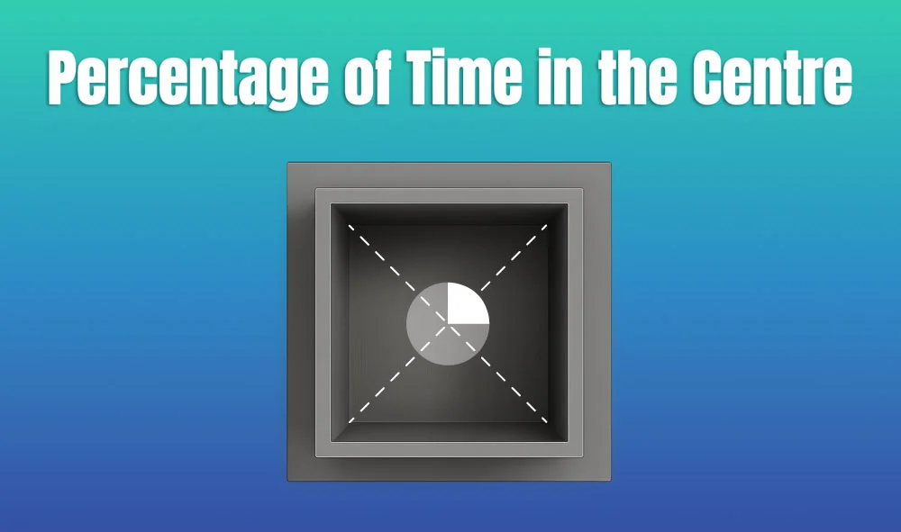
In behavioral neuroscience, the Open Field Test (OFT) remains one of the most widely used assays to evaluate rodent models of affect, cognition, and motivation. It provides a non-invasive framework for examining how animals respond to novelty, stress, and pharmacological or environmental manipulations. Among the test’s core metrics, the percentage of time spent in the center zone offers a uniquely normalized and sensitive measure of an animal’s emotional reactivity and willingness to engage with a potentially risky environment.
This metric is calculated as the proportion of time spent in the central area of the arena—typically the inner 25%—relative to the entire session duration. By normalizing this value, researchers gain a behaviorally informative variable that is resilient to fluctuations in session length or overall movement levels. This makes it especially valuable in comparative analyses, longitudinal monitoring, and cross-model validation.
Unlike raw center duration, which can be affected by trial design inconsistencies, the percentage-based measure enables clearer comparisons across animals, treatments, and conditions. It plays a key role in identifying trait anxiety, avoidance behavior, risk-taking tendencies, and environmental adaptation, making it indispensable in both basic and translational research contexts.
Whereas simple center duration provides absolute time, the percentage-based metric introduces greater interpretability and reproducibility, especially when comparing different animal models, treatment conditions, or experimental setups. It is particularly effective for quantifying avoidance behaviors, risk assessment strategies, and trait anxiety profiles in both acute and longitudinal designs.
This metric reflects the relative amount of time an animal chooses to spend in the open, exposed portion of the arena—typically defined as the inner 25% of a square or circular enclosure. Because rodents innately prefer the periphery (thigmotaxis), time in the center is inversely associated with anxiety-like behavior. As such, this percentage is considered a sensitive, normalized index of:
Critically, because this metric is normalized by session duration, it accommodates variability in activity levels or testing conditions. This makes it especially suitable for comparing across individuals, treatment groups, or timepoints in longitudinal studies.
A high percentage of center time indicates reduced anxiety, increased novelty-seeking, or pharmacological modulation (e.g., anxiolysis). Conversely, a low percentage suggests emotional inhibition, behavioral avoidance, or contextual hypervigilance. reduced anxiety, increased novelty-seeking, or pharmacological modulation (e.g., anxiolysis). Conversely, a low percentage suggests emotional inhibition, behavioral avoidance, or contextual hypervigilance.
The percentage of center time is one of the most direct, unconditioned readouts of anxiety-like behavior in rodents. It is frequently reduced in models of PTSD, chronic stress, or early-life adversity, where animals exhibit persistent avoidance of the center due to heightened emotional reactivity. This metric can also distinguish between acute anxiety responses and enduring trait anxiety, especially in longitudinal or developmental studies. Its normalized nature makes it ideal for comparing across cohorts with variable locomotor profiles, helping researchers detect true affective changes rather than activity-based confounds.
Rodents that spend more time in the center zone typically exhibit broader and more flexible exploration strategies. This behavior reflects not only reduced anxiety but also cognitive engagement and environmental curiosity. High center percentage is associated with robust spatial learning, attentional scanning, and memory encoding functions, supported by coordinated activation in the prefrontal cortex, hippocampus, and basal forebrain. In contrast, reduced center engagement may signal spatial rigidity, attentional narrowing, or cognitive withdrawal, particularly in models of neurodegeneration or aging.
The open field test remains one of the most widely accepted platforms for testing anxiolytic and psychotropic drugs. The percentage of center time reliably increases following administration of anxiolytic agents such as benzodiazepines, SSRIs, and GABA-A receptor agonists. This metric serves as a sensitive and reproducible endpoint in preclinical dose-finding studies, mechanistic pharmacology, and compound screening pipelines. It also aids in differentiating true anxiolytic effects from sedation or motor suppression by integrating with other behavioral parameters like distance traveled and entry count (Prut & Belzung, 2003).
Sex-based differences in emotional regulation often manifest in open field behavior, with female rodents generally exhibiting higher variability in center zone metrics due to hormonal cycling. For example, estrogen has been shown to facilitate exploratory behavior and increase center occupancy, while progesterone and stress-induced corticosterone often reduce it. Studies involving gonadectomy, hormone replacement, or sex-specific genetic knockouts use this metric to quantify the impact of endocrine factors on anxiety and exploratory behavior. As such, it remains a vital tool for dissecting sex-dependent neurobehavioral dynamics.
The percentage of center time is one of the most direct, unconditioned readouts of anxiety-like behavior in rodents. It is frequently reduced in models of PTSD, chronic stress, or early-life adversity. Because it is normalized, this metric is especially helpful for distinguishing between genuine avoidance and low general activity.
Environmental Control: Uniformity in environmental conditions is essential. Lighting should be evenly diffused to avoid shadow bias, and noise should be minimized to prevent stress-induced variability. The arena must be cleaned between trials using odor-neutral solutions to eliminate scent trails or pheromone cues that may affect zone preference. Any variation in these conditions can introduce systematic bias in center zone behavior. Use consistent definitions of the center zone (commonly 25% of total area) to allow valid comparisons. Software-based segmentation enhances spatial precision.
Evaluating how center time evolves across the duration of a session—divided into early, middle, and late thirds—provides insight into behavioral transitions and adaptive responses. Animals may begin by avoiding the center, only to gradually increase center time as they habituate to the environment. Conversely, persistently low center time across the session can signal prolonged anxiety, fear generalization, or a trait-like avoidance phenotype.
To validate the significance of center time percentage, it should be examined alongside results from other anxiety-related tests such as the Elevated Plus Maze, Light-Dark Box, or Novelty Suppressed Feeding. Concordance across paradigms supports the reliability of center time as a trait marker, while discordance may indicate task-specific reactivity or behavioral dissociation.
When paired with high-resolution scoring of behavioral events such as rearing, grooming, defecation, or immobility, center time offers a richer view of the animal’s internal state. For example, an animal that spends substantial time in the center while grooming may be coping with mild stress, while another that remains immobile in the periphery may be experiencing more severe anxiety. Microstructure analysis aids in decoding the complexity behind spatial behavior.
Animals naturally vary in their exploratory style. By analyzing percentage of center time across subjects, researchers can identify behavioral subgroups—such as consistently bold individuals who frequently explore the center versus cautious animals that remain along the periphery. These classifications can be used to examine predictors of drug response, resilience to stress, or vulnerability to neuropsychiatric disorders.
In studies with large cohorts or multiple behavioral variables, machine learning techniques such as hierarchical clustering or principal component analysis can incorporate center time percentage to discover novel phenotypic groupings. These data-driven approaches help uncover latent dimensions of behavior that may not be visible through univariate analyses alone.
Total locomotion helps contextualize center time. Low percentage values in animals with minimal movement may reflect sedation or fatigue, while similar values in high-mobility subjects suggest deliberate avoidance. This metric helps distinguish emotional versus motor causes of low center engagement.
This measure indicates how often the animal initiates exploration of the center zone. When combined with percentage of time, it differentiates between frequent but brief visits (indicative of anxiety or impulsivity) versus fewer but sustained center engagements (suggesting comfort and behavioral confidence).
The delay before the first center entry reflects initial threat appraisal. Longer latencies may be associated with heightened fear or low motivation, while shorter latencies are typically linked to exploratory drive or low anxiety.
Time spent hugging the walls offers a spatial counterbalance to center metrics. High thigmotaxis and low center time jointly support an interpretation of strong avoidance behavior. This inverse relationship helps triangulate affective and motivational states.
By expressing center zone activity as a proportion of total trial time, researchers gain a metric that is resistant to session variability and more readily comparable across time, treatment, and model conditions. This normalized measure enhances reproducibility and statistical power, particularly in multi-cohort or cross-laboratory designs.
For experimental designs aimed at assessing anxiety, exploratory strategy, or affective state, the percentage of time spent in the center offers one of the most robust and interpretable measures available in the Open Field Test.
Written by researchers, for researchers — powered by Conduct Science.


Monday – Friday
9 AM – 5 PM EST
DISCLAIMER: ConductScience and affiliate products are NOT designed for human consumption, testing, or clinical utilization. They are designed for pre-clinical utilization only. Customers purchasing apparatus for the purposes of scientific research or veterinary care affirm adherence to applicable regulatory bodies for the country in which their research or care is conducted.