x
[quotes_form]
Biomolecules

Classes and Benefits of Peptides

What Are Peptides?

The biological significance of proteins was recognized more than two centuries ago.[1] They were considered the primary material required for living organisms. But from the beginning of the 20th century, the significance of protein-like molecules, peptides, became apparent in several life processes.[1]

Name and year of peptides discovered in the 20th century

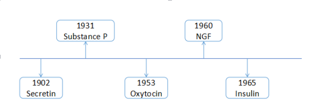
Figure: The illustration of name and year of peptides discovered in the 20th century.

Source: Creative Peptides[2]

Like proteins, peptides also consist of chains of amino acids and are held together by peptide bonds. Peptides have the same properties as protein molecules.[2] However, unlike proteins, their classes of molecules are small, simpler, and of lower molecular weight. They consist of 2-50 amino acids, unlike proteins which consist of more than 50 amino acids.[2]

Emil Fischer is considered the father of the chemistry of peptides. He coined the term “peptide,” which originated from the word “pepsis,” meaning digestion products of proteins.[1]

This article poses a classification of peptides, their classes, actions, and essential peptide functions in living organisms.

Structure and Classification of Peptides

Peptides are formed by linking two or more amino acids through an amide linkage, called a peptide bond.[3] The formation of peptide bonds occurs by the removal of a hydroxyl group (-OH) from one alpha-amino group and hydrogen (-H) atom from another alpha-amino group, forming a water molecule (H2O). This reaction is called a dehydration reaction.[3]

Peptides are sub-categorized into two groups based on the number of amino acids present in their structures: oligopeptides and polypeptides.[3]

Oligopeptides

When two or more (but less than 20) amino acids are linked together with the loss of a water molecule, they are called oligopeptides. It also includes dipeptides, tripeptides, tetrapeptides, and pentapeptides. Some examples of naturally occurring oligopeptides are microviridin, cyanopeptolin, microcystins, etc.[4]

An illustrative structure of dipeptide and tripeptide

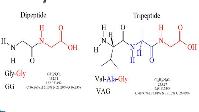
Figure: An illustrative structure of dipeptide and tripeptide.

Source: Online Biology notes.[3]

The oligopeptides are synthesized through non-ribosomal pathways, except for cyclamates and microviridin synthesized through ribosomal pathways.[4] There are several ways to identify peptides, and these ways include gel chromatography, HPLC, HPLC-mass spectroscopy, and ion-exchange chromatography.[4]

Polypeptides

When 20 or more amino acids are linked together through covalent peptide bonds, they are called polypeptides.[4] One or more polypeptides are involved in the formation of proteins. They have two terminals present in their structure: N-terminal containing an amino group and C-terminal containing a carboxyl group. Some examples of polypeptides include insulin and growth hormones.[4]

Polypeptides are arranged in different structural forms to create different functional proteins. So, according to the number and arrangement of polypeptides, the structure of proteins are categorized into four groups:[4]

  1. Primary Structure: Only a single polypeptide chain is involved in building this structure with peptide bond formation.
  2. Secondary Structure: It is formed due to the folding of the polypeptide chain by forming hydrogen bonds between amide hydrogens and carbonyl oxygens of the peptide backbone. Two major secondary structures are alpha-helix and beta-sheet structures.
  3. Tertiary Structure: It’s a 3-D structure of proteins in which the side chains of amino acids are linked together and folded in several ways. This is via hydrophobic bonds, hydrogen bonds, ionic bonds, disulfide bonds, and Van der Waals interactions.[4]
  4. Quaternary Structure: It is formed by joining two or more polypeptides together. The chains are held together by hydrogen bonds and Van der Waals forces between nonpolar side chains.

Classes of Peptides and Their Biological Significance

Peptides, based on their functional properties, are categorized into many small groups. Here’s a list of the most commonly studied classes of peptides in organisms, along with their functions and some examples.[5]

1. Antimicrobial Peptides

Antimicrobial peptides, also known as host defense peptides, are a class of peptides that play a role in the innate immune response of all organisms.[6] They are classified into two groups: ribosomally synthesized peptides and non-ribosomally synthesized peptides.[6]

  • Non-ribosomally synthesized peptides are elaborated in different organisms (bacteria, fungi, and streptomycetes containing two or more moieties derived from amino acids) and composed of multienzyme complexes. Examples include penicillin, cephalosporin C, vancomycin, and teicoplanin.[6]
  • Ribosomally synthesized peptides are produced by nearly all organisms including mammals, amphibians, insects, plants, bacteria, and viruses.[6] They are typically synthesized on ribosomes. Examples include gramicidin S, bacitracin, polymyxin B, human beta-defensin 1, and cattle indolicidin.[6]

2. Bacterial Peptides

As the name suggests, bacterial peptides are fragments of proteins produced by bacteria. They include flagellar peptides, lipoproteins, enterotoxins, and several enzymes.[7]

The peptides secreted from both gram-positive and gram-negative bacteria are both cationic and neutral.[6] These types of peptides are included within bacteriocin that kills specific competitor bacteria, protecting the host bacterium.[6]

Examples include Escherichia coli 7-amino-acid peptide microcin C7 (inhibits protein synthesis), Lactococcus peptide mersacidin (inhibits peptidoglycan biosynthesis), nisin, and epidermin (permeabilizes target cell membrane).[6]

3. Neuropeptides

These are small proteins synthesized by neurons to act on receptors and modulate synaptic transmission.[8] The neuropeptides are synthesized by large, inactive precursor proteins, called pre-propeptides.

These proteins are cleaved into several active peptides and produce multiple copies of different neuropeptides.[8] Most neuropeptides act on G protein-coupled receptors (GPCRs) and fall into two families: the rhodopsin-like and the secretin families.

Examples include acetylcholine, epinephrine, norepinephrine, dopamine, serotonin, and Gamma-aminobutyric acid (GABA).[8]

4. Anticancer Peptides

Anticancer peptides (ACPs) are small peptides with a short amino acid sequence that are selective and toxic to cancer cells.[9] The predominant amino acids in anticancer peptides include glycine, lysine, and leucine.[9]

The anticancer peptides are a highly preferred choice among all the other available anticancer therapeutics due to their high selectivity, high penetration, and easy modifications.[9]

The peptides destroy cancer cells via apoptosis and necrosis by lysing or forming pores in the membranes of cancerous cells. These types of peptides, depending on their structure, mode of action, selectivity, and efficacy to specific cancer cells, are divided into three categories:[9]

  • Molecularly targeted peptides: They directly act on cancer cells via cytotoxic, anti-proliferative, and apoptotic activities. Examples include Mastoparan I, anticancer peptide SVS-1, and tubulysin analog KEMTUB10.[9]
  • ‘Guiding missile’ peptides or binding peptides: They are drug binding peptides that deliver drugs to the targeted cancer cells. Examples are CP-EPS8-NLS (a synthetic peptide derived from nuclear localization signal NLS and epidermal growth factor receptor pathway substrate 8 EPS8) and cell-penetrating peptide TAT-conjugated gambogic acid (GA-TAT).[9]
  • Cell-stimulating peptides: They indirectly kill cancer cells by stimulating other cells via immunomodulatory activities and hormone receptors. It includes the E75 peptide breast cancer vaccine (Her2 p369-p377), a melittin-RADA32 hybrid peptide hydrogel-linked doxorubicin, and Tyrosinase-related protein 2 melanoma antigen peptide nanovaccine.[9]

5. Cardiovascular Peptides

Cardiovascular peptides have a role in physiological and pathological conditions in the cardiovascular system.[10] They are implicated in controlling vascular tone, blood pressure, congestive heart failure, atherosclerosis, coronary artery diseases, and pulmonary and systemic hypertension.[10] Below are some of the examples of cardiovascular peptides with their brief functional roles:[11]
  • Adrenomedullin peptide: It’s a 52 amino acid peptide playing multiple roles in cardiovascular actions, including reducing blood pressure, anti-inflammation, vasodilation, stimulation of nitric oxide production, and inhibition of myocardial hypertrophy and fibrosis.[11]
  • Angiotensin II peptide: It’s the central product of the renin-angiotensin system (RAS).[11] It plays a significant role in causing hypertension, myocyte hypertrophy, myocyte gene reprogramming, fibroblast proliferation, extracellular matrix (ECM) protein accumulation, and other pathophysiology of cardiovascular diseases in humans.[11]
  • CGRP: It’s a 37 amino acid neuropeptide belonging to the family of structurally related peptides like adrenomedullin (AM) and amylin (AMY).[11] It binds with GPCRs, known as calcitonin receptor-like receptors (CLR), to activate its functional signaling pathway. It acts as a potent vasodilator and is involved in cardiovascular homeostasis. Any mutation in CGRP may lead to its functional disturbances that can cause diseases like a migraine.[11]
  • Natriuretic peptides: In mammals, it’s a peptide family consisting of atrial (A-type) natriuretic peptide (ANP), brain (B-type) natriuretic peptide (BNP), and C-type natriuretic peptide (CNP).[11] ANP and BNP peptides are abundantly produced in cardiomyocytes, while CNP is synthesized in endothelial cells and cardiac fibroblasts. They are involved in dilating blood vessels and inducing diuresis/natriuresis by increasing the intracellular cGMP concentration.[11]
  • Urocortins: It’s a paralogue (a particular class of homologous gene) of corticotropin-releasing hormone.[11] They regulate pressure and volume in different organs, including the heart, kidneys, adrenals, and vasculature.[11] It’s an ongoing research interest for scientists, and it promises a better understanding of the pathophysiology of ischemia-reperfusion injury, hypertension, and heart failure.[11]
  • Urotensin peptides: It’s a peptide hormone consisting of urotensin I (UI) and urotensin II (UII).[11] It was initially discovered in fish urophysis. UI plays several roles in different organisms, including stimulation of cell proliferation and hypertrophy, positive inotropic action, and CNS actions on cardiovascular control. Whereas UII has a vital role in congestive heart failure, hypertension, end-stage renal disease, and diabetes mellitus.[11]

6. Endocrine Peptides

They are short amino acid-chained peptide hormones synthesized and secreted by specialized cells in the endocrine.[11] They are stored in membrane-bound secretory vesicles, which enable their rapid secretion whenever required.

They are water-soluble, and this makes it difficult for them to cross the hydrophobic cell membranes.[11] Thus, they need specific receptors on the cell surface to exert their actions.

Some examples of endocrine peptides include:

  • Adiponectin (APN): It’s an anorexigenic peptide involved in multiple functions, including stimulating fatty acid oxidation and glucose uptake in skeletal muscle and adipose tissue. It also improves whole-body insulin sensitivity and increases energy expenditure; however, it suppresses hepatic glucose output by activating AMP-activated protein kinase (AMPK) signaling.[11]
  • Leptin: It is majorly produced by adipocytes. It acts as a signaling molecule between peripheral organs and the central nervous system.[11] It has several functions that include regulating numerous endocrine functions relevant for the maintenance of energy expenditure, appetite regulation, body weight control, and the functioning of endocrine organs.[11]
  • Atrial natriuretic peptide (ANP): It was initially purified from the rat’s heart and was the first natriuretic peptide (NP).[11] It is involved in several activities, including antiproliferation, anti-fibrosis, anti-inflammation, insulin-like functions, cardiovascular homeostasis, and regulation of bone growth.[11]
  • Orexins: It has two subtypes: Orexins A and B. These are involved in regulating feeding, wakefulness, sleep, and energy homeostasis. They are synthesized in multiple organs, including the intestines, pancreas, adrenal gland, reproductive tract, and adipose tissue.[11]
  • Pituitary adenylate cyclase-activating polypeptide (PACAP): It belongs to the superfamily of the vasoactive intestinal polypeptide (VIP)-glucagon peptides. PACAP exerts neuroendocrine, paracrine, and autocrine control of the pituitary gland activity, thyroid glands, testis, ovary, adrenal medulla, adrenal cortex, and endocrine pancreas.[11]

7. Antifungal Peptides

Antifungal peptides are peptides produced against fungi and isolated from other organisms. Fungi cause several infections and diseases in plants, humans, and other animals.[11] So, the proteinaceous or peptidic molecules produced by other organisms against any specific fungi are isolated for antifungal strategies.[11] A diversity of antifungal peptides are available with different molecular masses, N-terminal or complete amino acid sequences, specificity, and mechanism of antifungal actions.[11] Given below is a list of different types of fungal peptides and their functions in organisms:[11]
  • Peptaibols: It consists of four categories of peptides having both antifungal and antibacterial properties. There are nine membrane-active peptaibols, two nonadecapeptide peptaibols, four nonadecapeptide peptaibols with antibacterial and antifungal activities, and two linear 19-amino-acids.[11]
  • Cyclic antifungal peptides: These are antifungal peptides having cyclic structures.[11] Their examples include Isarfelin (having inhibitory activity against the fungi, Rhizoctonia solani, and Sclerotinia sclerotiorum), eujavanicin A (suppresses growth in the human pathogenic filamentous fungus Aspergillus fumigatus), and echinocandin type l antifungal lipopeptide (used in the therapy of deep-seated mycoses).[11]
  • Fungal peptides with ribosome-inactivating activity: Fungi, like molds and mushrooms, synthesize variant peptides with ribonuclease- and ribosome-inactivating activities. They include RNases and ubiquitin-like peptides from different fungi.[11]

8. Opiate Peptides

The endogenous and exogenous opioids exert several physiological and pharmacological effects through receptors of four different subtypes.[12] It include μ (μ1, μ2), 𝜹 (𝜹1, 𝜹2), 𝜿 (𝜿1, 𝜿2), and ε (ε1, ε2).[12] 

They regulate other endocrine systems like the hypothalamic-pituitary-adrenocortical axis and the phenomenon of stress-induced analgesia.[12]

Endogenous opiate peptides are better studied than exogenous peptides. They consist of three families of peptides based on their origin:[12]

  • The proopiomelanocortin (POMC) family of peptides
  • The proenkephalin A (PA) family of peptides
  • The prodynorphin (PD) family of peptides

Some peptides like cholecystokinin (CCK), neuropeptide FF (NPFF), and melanocyte inhibiting factor (MIF)-related peptides possess anti-opioid properties.[12] But these peptide families also include some peptides harboring opioid-like properties, and that’s why they are also known as “opioid modulating” peptides.[12]

9. Plant Peptides

Plant peptides are peptides that originated in plants and they possess significant health benefits in humans. They lower blood pressure and cholesterol levels and inhibit enzymes within the renin-angiotensin-aldosterone system (RAAS).

Other benefits include their anti-inflammatory activity, anticancer and immunomodulatory activities, prevention of and protection against oxidative damage through free radical scavenging activities, and antimicrobial activity.[13]

Plant peptides are categorized into three groups based on their functional response:[13]

  • Plant-derived peptides for cardiovascular health: Examples include ACE-I- and renin-inhibiting bioactive peptides generated from plants, including potato, yams, rapeseeds, lentils, red seaweed, and many kinds of cereal.[13]
  • Plant-derived antioxidant peptides: Examples include glutathione peptide, β -conglycinin, and trypsin hydrolysate. Most of the antioxidants are rich in amino acids like histidine (His), tryptophan (Trp), tyrosine (Tyr), and lysine (Lys).[13]
  • Anticancer and antimicrobial plant-derived peptides: Antimicrobial peptides derived from plants are a novel alternative for cancer treatment. It includes peptides like thionins, plant defensins, cyclotides, and small cationic peptides.[13]
  • Plant-derived peptides in controlling type II diabetes: These peptides work against diabetes by inhibiting the enzyme dipeptidyl peptidase-IV.[14] It includes peptides that are inhibitors of dipeptidyl peptidase-IV and are commonly extracted from plants like Opuntia streptacantha, Trigonella foenum-graecum, Momordica charantia, Ficus benghalensis, Polygala senega, and Gymnema sylvestre.[13]

10. Venom Peptides

Toxins developed in animals are strategies to protect themselves from different predators and/or capture their prey.[14]

Several of these venoms studied are found in animals with envenomation apparatus like cone snails, spiders, scorpions, snakes, the Gila monster lizard, and sea anemone.[14]

Venom peptides act as natural ligands of ions channels and different receptors they bind to initiate physiological responses.[14] Based on these characteristics, they are categorized into eight groups:[14]

  • Calcium channel peptides
  • Sodium-channel toxins
  • Potassium-channel toxins
  • Chloride-channel toxins
  • Toxins inhibiting nicotinic acetylcholine receptors
  • Noradrenaline transporter inhibitors
  • NMDA receptor antagonists
  • Neurotensin receptor agonists

The application of these peptides in healthcare sectors demands many issues associated with their safety, pharmacokinetics, and delivery to be addressed.[13]

Conclusion

Peptides are a class of biological molecules that have essential roles in fundamental physiological processes and are required for many biochemical processes. They are small molecules made by sequential arrangements of 2-50 amino acids. The difference between peptide and protein is that proteins are large chains of amino acid sequence (50 or more) with different specialized structures.

The benefits of peptides have established a special place in the pharmaceutical landscape.[15] And the innovations in peptide therapeutics are believed to rise in the future by expanding into new indications and molecular targets, exploiting novel chemistry strategies to broaden molecular diversity, and engineering enhanced pharmaceutical properties.[15]

Further, the study of peptide benefits and chemistry opens a door for scientists to unravel its hidden potentials. It can be identifying novel targets and receptors of peptides, the discovery of more peptides in organisms and their functional properties, or the discovery of sustainable peptide-based drugs.[15]

References:

  1. The world of peptides. Retrieved from https://www.springer.com/gp/book/9783642758522.
  2. The research history of peptides (2017). Retrieved from https://www.creative-peptides.com/blog/the-research-history-of-peptide/.
  3. Karki Gaurab (2018). Peptides: types and functions. Retrieved from https://www.onlinebiologynotes.com/peptide-types-functions/.
  4. Madhu (2020). Difference Between Oligopeptide and Polypeptide. Retrieved from https://www.differencebetween.com/difference-between-oligopeptide-and-polypeptide/.
  5. Helemenstine M. Anne (2018). What Is a Peptide? Definition and Examples. Retrieved from https://www.thoughtco.com/what-is-a-peptide-definition-examples-4177787#.
  6. Hancock, R. E., & Chapple, D. S. (1999). Peptide antibiotics. Antimicrobial agents and chemotherapy, 43(6), 1317–1323. https://doi.org/10.1128/AAC.43.6.1317.
  7. Bacterial peptides. Retrieved from https://www.biosyn.com/catalog-peptides/bacterial-peptides.aspx.
  8. Neuropeptide. Retrieved from https://en.wikipedia.org/wiki/Neuropeptide.
  9. Chiangjong, W., Chutipongtanate, S., & Hongeng, S. (2020). Anticancer peptide: Physicochemical property, functional aspect, and trend in the clinical application (Review). International journal of oncology, 57(3), 678–696. https://doi.org/10.3892/ijo.2020.5099.
  10. Grieco, P., & Gomez-Monterrey, I. (2018). Natural and synthetic peptides in cardiovascular diseases: An update on diagnostic and therapeutic potentials. Archives of Biochemistry and Biophysics. doi:10.1016/j.abb.2018.11.021.
  11. Abba J Kastin – Handbook of biologically active peptides-Elsevier Academic Press (2013).
  12. Cesselin, F. (1995). Opioid and anti-opioid peptides. Fundamental & Clinical Pharmacology, 9(5), 409–433. doi:10.1111/j.1472-8206.1995.tb00517.x.
  13. Hayes, M., & Bleakley, S. (2018). Peptides from plants and their applications. Peptide Applications in Biomedicine, Biotechnology, and Bioengineering, 603–622. doi:10.1016/b978-0-08-100736-5.00025-9.
  14. Lewis, R., Garcia, M. Therapeutic potential of venom peptides. Nat Rev Drug Discov 2, 790–802 (2003). https://doi.org/10.1038/nrd1197.
  15. Lau, J. L., & Dunn, M. K. (2018). Therapeutic peptides: Historical perspectives, current development trends, and future directions. Bioorganic & Medicinal Chemistry, 26(10), 2700–2707. doi:10.1016/j.bmc.2017.06.052.

Learn More about our Services and how can we help you with your research!

Introduction

In behavioral neuroscience, the Open Field Test (OFT) remains one of the most widely used assays to evaluate rodent models of affect, cognition, and motivation. It provides a non-invasive framework for examining how animals respond to novelty, stress, and pharmacological or environmental manipulations. Among the test’s core metrics, the percentage of time spent in the center zone offers a uniquely normalized and sensitive measure of an animal’s emotional reactivity and willingness to engage with a potentially risky environment.

This metric is calculated as the proportion of time spent in the central area of the arena—typically the inner 25%—relative to the entire session duration. By normalizing this value, researchers gain a behaviorally informative variable that is resilient to fluctuations in session length or overall movement levels. This makes it especially valuable in comparative analyses, longitudinal monitoring, and cross-model validation.

Unlike raw center duration, which can be affected by trial design inconsistencies, the percentage-based measure enables clearer comparisons across animals, treatments, and conditions. It plays a key role in identifying trait anxiety, avoidance behavior, risk-taking tendencies, and environmental adaptation, making it indispensable in both basic and translational research contexts.

Whereas simple center duration provides absolute time, the percentage-based metric introduces greater interpretability and reproducibility, especially when comparing different animal models, treatment conditions, or experimental setups. It is particularly effective for quantifying avoidance behaviors, risk assessment strategies, and trait anxiety profiles in both acute and longitudinal designs.

What Does Percentage of Time in the Centre Measure?

This metric reflects the relative amount of time an animal chooses to spend in the open, exposed portion of the arena—typically defined as the inner 25% of a square or circular enclosure. Because rodents innately prefer the periphery (thigmotaxis), time in the center is inversely associated with anxiety-like behavior. As such, this percentage is considered a sensitive, normalized index of:

  • Exploratory drive vs. risk aversion: High center time reflects an animal’s willingness to engage with uncertain or exposed environments, often indicative of lower anxiety and a stronger intrinsic drive to explore. These animals are more likely to exhibit flexible, information-gathering behaviors. On the other hand, animals that spend little time in the center display a strong bias toward the safety of the perimeter, indicative of a defensive behavioral state or trait-level risk aversion. This dichotomy helps distinguish adaptive exploration from fear-driven avoidance.

  • Emotional reactivity: Fluctuations in center time percentage serve as a sensitive behavioral proxy for changes in emotional state. In stress-prone or trauma-exposed animals, decreased center engagement may reflect hypervigilance or fear generalization, while a sudden increase might indicate emotional blunting or impaired threat appraisal. The metric is also responsive to acute stressors, environmental perturbations, or pharmacological interventions that impact affective regulation.

  • Behavioral confidence and adaptation: Repeated exposure to the same environment typically leads to reduced novelty-induced anxiety and increased behavioral flexibility. A rising trend in center time percentage across trials suggests successful habituation, reduced threat perception, and greater confidence in navigating open spaces. Conversely, a stable or declining trend may indicate behavioral rigidity or chronic stress effects.

  • Pharmacological or genetic modulation: The percentage of time in the center is widely used to evaluate the effects of pharmacological treatments and genetic modifications that influence anxiety-related circuits. Anxiolytic agents—including benzodiazepines, SSRIs, and cannabinoid agonists—reliably increase center occupancy, providing a robust behavioral endpoint in preclinical drug trials. Similarly, genetic models targeting serotonin receptors, GABAergic tone, or HPA axis function often show distinct patterns of center preference, offering translational insights into psychiatric vulnerability and resilience.

Critically, because this metric is normalized by session duration, it accommodates variability in activity levels or testing conditions. This makes it especially suitable for comparing across individuals, treatment groups, or timepoints in longitudinal studies.

A high percentage of center time indicates reduced anxiety, increased novelty-seeking, or pharmacological modulation (e.g., anxiolysis). Conversely, a low percentage suggests emotional inhibition, behavioral avoidance, or contextual hypervigilance. reduced anxiety, increased novelty-seeking, or pharmacological modulation (e.g., anxiolysis). Conversely, a low percentage suggests emotional inhibition, behavioral avoidance, or contextual hypervigilance.

Behavioral Significance and Neuroscientific Context

1. Emotional State and Trait Anxiety

The percentage of center time is one of the most direct, unconditioned readouts of anxiety-like behavior in rodents. It is frequently reduced in models of PTSD, chronic stress, or early-life adversity, where animals exhibit persistent avoidance of the center due to heightened emotional reactivity. This metric can also distinguish between acute anxiety responses and enduring trait anxiety, especially in longitudinal or developmental studies. Its normalized nature makes it ideal for comparing across cohorts with variable locomotor profiles, helping researchers detect true affective changes rather than activity-based confounds.

2. Exploration Strategies and Cognitive Engagement

Rodents that spend more time in the center zone typically exhibit broader and more flexible exploration strategies. This behavior reflects not only reduced anxiety but also cognitive engagement and environmental curiosity. High center percentage is associated with robust spatial learning, attentional scanning, and memory encoding functions, supported by coordinated activation in the prefrontal cortex, hippocampus, and basal forebrain. In contrast, reduced center engagement may signal spatial rigidity, attentional narrowing, or cognitive withdrawal, particularly in models of neurodegeneration or aging.

3. Pharmacological Responsiveness

The open field test remains one of the most widely accepted platforms for testing anxiolytic and psychotropic drugs. The percentage of center time reliably increases following administration of anxiolytic agents such as benzodiazepines, SSRIs, and GABA-A receptor agonists. This metric serves as a sensitive and reproducible endpoint in preclinical dose-finding studies, mechanistic pharmacology, and compound screening pipelines. It also aids in differentiating true anxiolytic effects from sedation or motor suppression by integrating with other behavioral parameters like distance traveled and entry count (Prut & Belzung, 2003).

4. Sex Differences and Hormonal Modulation

Sex-based differences in emotional regulation often manifest in open field behavior, with female rodents generally exhibiting higher variability in center zone metrics due to hormonal cycling. For example, estrogen has been shown to facilitate exploratory behavior and increase center occupancy, while progesterone and stress-induced corticosterone often reduce it. Studies involving gonadectomy, hormone replacement, or sex-specific genetic knockouts use this metric to quantify the impact of endocrine factors on anxiety and exploratory behavior. As such, it remains a vital tool for dissecting sex-dependent neurobehavioral dynamics.
The percentage of center time is one of the most direct, unconditioned readouts of anxiety-like behavior in rodents. It is frequently reduced in models of PTSD, chronic stress, or early-life adversity. Because it is normalized, this metric is especially helpful for distinguishing between genuine avoidance and low general activity.

Methodological Considerations

  • Zone Definition: Accurately defining the center zone is critical for reliable and reproducible data. In most open field arenas, the center zone constitutes approximately 25% of the total area, centrally located and evenly distanced from the walls. Software-based segmentation tools enhance precision and ensure consistency across trials and experiments. Deviations in zone parameters—whether due to arena geometry or tracking inconsistencies—can result in skewed data, especially when calculating percentages.


  • Trial Duration: Trials typically last between 5 to 10 minutes. The percentage of time in the center must be normalized to total trial duration to maintain comparability across animals and experimental groups. Longer trials may lead to fatigue, boredom, or habituation effects that artificially reduce exploratory behavior, while overly short trials may not capture full behavioral repertoires or response to novel stimuli.


  • Handling and Habituation: Variability in pre-test handling can introduce confounds, particularly through stress-induced hypoactivity or hyperactivity. Standardized handling routines—including gentle, consistent human interaction in the days leading up to testing—reduce variability. Habituation to the testing room and apparatus prior to data collection helps animals engage in more representative exploratory behavior, minimizing novelty-induced freezing or erratic movement.


  • Tracking Accuracy: High-resolution tracking systems should be validated for accurate, real-time detection of full-body center entries and sustained occupancy. The system should distinguish between full zone occupancy and transient overlaps or partial body entries that do not reflect true exploratory behavior. Poor tracking fidelity or lag can produce significant measurement error in percentage calculations.


  • Environmental Control: Uniformity in environmental conditions is essential. Lighting should be evenly diffused to avoid shadow bias, and noise should be minimized to prevent stress-induced variability. The arena must be cleaned between trials using odor-neutral solutions to eliminate scent trails or pheromone cues that may affect zone preference. Any variation in these conditions can introduce systematic bias in center zone behavior. Use consistent definitions of the center zone (commonly 25% of total area) to allow valid comparisons. Software-based segmentation enhances spatial precision.

Interpretation with Complementary Metrics

Temporal Dynamics of Center Occupancy

Evaluating how center time evolves across the duration of a session—divided into early, middle, and late thirds—provides insight into behavioral transitions and adaptive responses. Animals may begin by avoiding the center, only to gradually increase center time as they habituate to the environment. Conversely, persistently low center time across the session can signal prolonged anxiety, fear generalization, or a trait-like avoidance phenotype.

Cross-Paradigm Correlation

To validate the significance of center time percentage, it should be examined alongside results from other anxiety-related tests such as the Elevated Plus Maze, Light-Dark Box, or Novelty Suppressed Feeding. Concordance across paradigms supports the reliability of center time as a trait marker, while discordance may indicate task-specific reactivity or behavioral dissociation.

Behavioral Microstructure Analysis

When paired with high-resolution scoring of behavioral events such as rearing, grooming, defecation, or immobility, center time offers a richer view of the animal’s internal state. For example, an animal that spends substantial time in the center while grooming may be coping with mild stress, while another that remains immobile in the periphery may be experiencing more severe anxiety. Microstructure analysis aids in decoding the complexity behind spatial behavior.

Inter-individual Variability and Subgroup Classification

Animals naturally vary in their exploratory style. By analyzing percentage of center time across subjects, researchers can identify behavioral subgroups—such as consistently bold individuals who frequently explore the center versus cautious animals that remain along the periphery. These classifications can be used to examine predictors of drug response, resilience to stress, or vulnerability to neuropsychiatric disorders.

Machine Learning-Based Behavioral Clustering

In studies with large cohorts or multiple behavioral variables, machine learning techniques such as hierarchical clustering or principal component analysis can incorporate center time percentage to discover novel phenotypic groupings. These data-driven approaches help uncover latent dimensions of behavior that may not be visible through univariate analyses alone.

Total Distance Traveled

Total locomotion helps contextualize center time. Low percentage values in animals with minimal movement may reflect sedation or fatigue, while similar values in high-mobility subjects suggest deliberate avoidance. This metric helps distinguish emotional versus motor causes of low center engagement.

Number of Center Entries

This measure indicates how often the animal initiates exploration of the center zone. When combined with percentage of time, it differentiates between frequent but brief visits (indicative of anxiety or impulsivity) versus fewer but sustained center engagements (suggesting comfort and behavioral confidence).

Latency to First Center Entry

The delay before the first center entry reflects initial threat appraisal. Longer latencies may be associated with heightened fear or low motivation, while shorter latencies are typically linked to exploratory drive or low anxiety.

Thigmotaxis Time

Time spent hugging the walls offers a spatial counterbalance to center metrics. High thigmotaxis and low center time jointly support an interpretation of strong avoidance behavior. This inverse relationship helps triangulate affective and motivational states.

Applications in Translational Research

  • Drug Discovery: The percentage of center time is a key behavioral endpoint in the development and screening of anxiolytic, antidepressant, and antipsychotic medications. Its sensitivity to pharmacological modulation makes it particularly valuable in dose-response assessments and in distinguishing therapeutic effects from sedative or locomotor confounds. Repeated trials can also help assess drug tolerance and chronic efficacy over time.
  • Genetic and Neurodevelopmental Modeling: In transgenic and knockout models, altered center percentage provides a behavioral signature of neurodevelopmental abnormalities. This is particularly relevant in the study of autism spectrum disorders, ADHD, fragile X syndrome, and schizophrenia, where subjects often exhibit heightened anxiety, reduced flexibility, or altered environmental engagement.
  • Hormonal and Sex-Based Research: The metric is highly responsive to hormonal fluctuations, including estrous cycle phases, gonadectomy, and hormone replacement therapies. It supports investigations into sex differences in stress reactivity and the behavioral consequences of endocrine disorders or interventions.
  • Environmental Enrichment and Deprivation: Housing conditions significantly influence anxiety-like behavior and exploratory motivation. Animals raised in enriched environments typically show increased center time, indicative of reduced stress and greater behavioral plasticity. Conversely, socially isolated or stimulus-deprived animals often show strong center avoidance.
  • Behavioral Biomarker Development: As a robust and reproducible readout, center time percentage can serve as a behavioral biomarker in longitudinal and interventional studies. It is increasingly used to identify early signs of affective dysregulation or to track the efficacy of neuromodulatory treatments such as optogenetics, chemogenetics, or deep brain stimulation.
  • Personalized Preclinical Models: This measure supports behavioral stratification, allowing researchers to identify high-anxiety or low-anxiety phenotypes before treatment. This enables within-group comparisons and enhances statistical power by accounting for pre-existing behavioral variation. Used to screen anxiolytic agents and distinguish between compounds with sedative vs. anxiolytic profiles.

Enhancing Research Outcomes with Percentage-Based Analysis

By expressing center zone activity as a proportion of total trial time, researchers gain a metric that is resistant to session variability and more readily comparable across time, treatment, and model conditions. This normalized measure enhances reproducibility and statistical power, particularly in multi-cohort or cross-laboratory designs.

For experimental designs aimed at assessing anxiety, exploratory strategy, or affective state, the percentage of time spent in the center offers one of the most robust and interpretable measures available in the Open Field Test.

Explore high-resolution tracking solutions and open field platforms at

References

  • Prut, L., & Belzung, C. (2003). The open field as a paradigm to measure the effects of drugs on anxiety-like behaviors: a review. European Journal of Pharmacology, 463(1–3), 3–33.
  • Seibenhener, M. L., & Wooten, M. C. (2015). Use of the open field maze to measure locomotor and anxiety-like behavior in mice. Journal of Visualized Experiments, (96), e52434.
  • Crawley, J. N. (2007). What’s Wrong With My Mouse? Behavioral Phenotyping of Transgenic and Knockout Mice. Wiley-Liss.
  • Carola, V., D’Olimpio, F., Brunamonti, E., Mangia, F., & Renzi, P. (2002). Evaluation of the elevated plus-maze and open-field tests for the assessment of anxiety-related behavior in inbred mice. Behavioral Brain Research, 134(1–2), 49–57.

Written by researchers, for researchers — powered by Conduct Science.