Automated Cell Counting: Methods, Principles & How to Choose
How automated cell counters work, the trade-offs between hardware counters and image-analysis software, and what to weigh before you commit to a workflow.
What Automated Cell Counting Is
Cell counting is routine work in life-science research and clinical diagnostics. Cells can be counted by hand with a hemocytometer or a colony-forming unit (CFU) plating method, but manual counting is slow, subjective, and prone to distributional bias when sample volumes are large.[1]
Automated cell counting removes most of that manual burden. Broadly, it splits into two families: hardware counters that draw a sample through an aperture or a laser and report a number, and image-analysis software that detects and counts cells in a micrograph. This guide covers the principles behind both, the main counting methods, and how to decide which fits your lab.
Counting from images?
If your cells are already on a slide, in a well plate, or under a microscope, image-analysis software counts them from the image you have — no dedicated benchtop instrument required.
Explore ConductVision Cell Count (Fluorescence) →How Automated Cell Counters Work
Hardware cell counters are built on one of two physical principles: electrical impedance or light scattering.[2][3]
Electrical Impedance (the Coulter Principle)
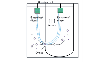
Each time a cell passes through a narrow aperture between two electrodes, it displaces conductive fluid and briefly raises the electrical resistance. The size of that impedance spike is proportional to the cell's volume, so the counter can both tally cells and estimate their size in a given sample volume.[3]
Light Scattering
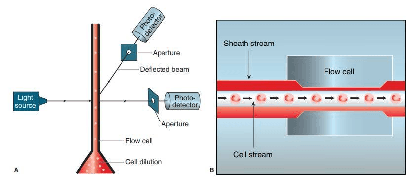
When a light beam crosses a stream of diluted cells, each cell deflects the light at a small angle. A photodetector registers those deflections and converts them into a count for the sample volume.[4]
Types of Automated Cell Counting Methods
Building on those principles, four methods cover most lab applications.[1]
1. Coulter Counter
Uses the impedance principle to report both cell count and cell volume in an electrolyte. It is cheaper than a flow cytometer and is used in particle characterization, hematology, and counting fat cells, plant-cell aggregates, bacteria, and stem-cell embryoid bodies.[5]
2. Image Analysis
A microscope captures the field and a classification algorithm detects and counts cells directly in the image.[1] This is the method that modern software such as ConductVision Cell Count (Fluorescence) automates — you supply the micrograph and the software returns the count, rather than passing the sample through a dedicated instrument.
3. Flow Cytometry
Cells flow single-file past a laser; the scattered signal is read on a detector that keeps the count. Flow cytometry also reports cell shape, internal and external structure, and protein or biochemical content — and is the most expensive of the automated methods.[1]
4. Stereological Cell Counting
Counts cells in histological sections using systematic, random sampling to estimate object frequency.[1] It is only semi-automated, since sampling and inclusion decisions still involve manual judgment.
Hardware Counters vs. Image-Analysis Software
The choice is not just brand or budget — it is a choice of workflow.
- Hardware counters (Coulter, flow cytometers, benchtop counters) draw a prepared suspension through the instrument. They are fast for suspension samples and report size distributions, but they add an instrument, consumables such as disposable slides, and running costs.
- Image-analysis software counts cells from images you already capture on a microscope or plate reader. It suits adherent cells, stained or fluorescent samples, and any workflow where you want to keep the image as a record. ConductVision Image is image-analysis software, not a benchtop instrument — it analyzes the micrograph rather than aspirating the sample.
If your cells live on slides or in well plates and you want a reproducible, auditable count without buying dedicated hardware, image analysis is usually the lower-friction path.
Benefits and Limitations
Benefits
- Faster, more objective, and statistically stronger than manual counting, without the distributional bias of hand counts.
- Reliable, reproducible data.
- Efficient and cost-effective across large sample numbers.
- Multiple assays can run on a single platform.
- Substantially reduces hands-on labor.[6]
Limitations
- Some counters miscount — for example, failing to distinguish nucleated red blood cells from small platelet clumps.
- Hardware counters carry high running costs.
- Those costs can make the overall workflow expensive.[6]
Preparing Samples and Running a Count
Instruments differ, so always follow the manual for your specific counter. A typical benchtop hardware workflow looks like this, with or without trypan blue staining:[7]
- Without trypan blue: pipette about 10 µl of cell suspension into the outer opening of the counting-slide chamber.
- With trypan blue: mix 10 µl of cell suspension with 10 µl of trypan blue, then pipette about 10 µl of the mixture into the chamber.
- Insert the slide into the instrument's slot.
- The counter reports total cells per ml for the sample volume.
If the screen reads "value out of range," inspect the captured image and dilute the suspension if needed.[7] Dispose of used counting slides as biohazardous waste. With image-analysis software, the parallel step is simply loading the micrograph and letting the algorithm segment and count the cells.
How to Choose
Whether you are comparing instruments or software, weigh these factors before committing.
- Counting accuracy. A larger counted area and more sophisticated algorithms reduce data variability and correct for hardware inequality.[8]
- Simple interface. Pick a system your team can operate reliably and that returns consistent, accurate results quickly.[9]
- Reproducibility. A defined measuring area keeps captured fields the same size and cuts field-of-view variation.[8]
- Accurate focusing. Autofocus and high camera resolution help separate live cells (dark edges, bright centers) from debris and dead cells.[8]
- Declustering. Look for software that recognizes clustered cells and counts each one rather than the clump.[8]
- Result storage and analysis. For high-throughput routines, favor systems with generous storage and easy export.[8]
- Reliable supplier. A known supplier means up-to-date software, support, and repair — and help matching the tool to your budget.[9]
- Lab space. Hardware counters range from handheld to bulky benchtop units; software needs none of that footprint.
- Cost. Account for consumables (such as disposable slides), maintenance, and service contracts, not just the sticker price.
References
- Cell counting. Wikipedia.
- Types of Automated Cell Counters. Bio-Rad.
- Electrical Impedance Methodology. Hematology Academy.
- Principles of automated blood cell counters. Clinical Science.
- Coulter counter. Wikipedia.
- Manual vs. Automated Cell Counting. ChemoMetec.
- What to look for in an automated cell counter. Westburg.
- How to choose a hematology cell counter. China Care Medical.
Count Cells from the Images You Already Have
ConductVision Cell Count (Fluorescence) detects and counts cells directly in your micrographs — reproducible, auditable, and no dedicated benchtop counter required.