Introduction
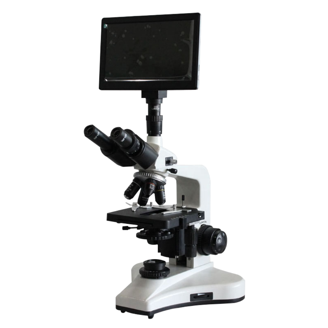
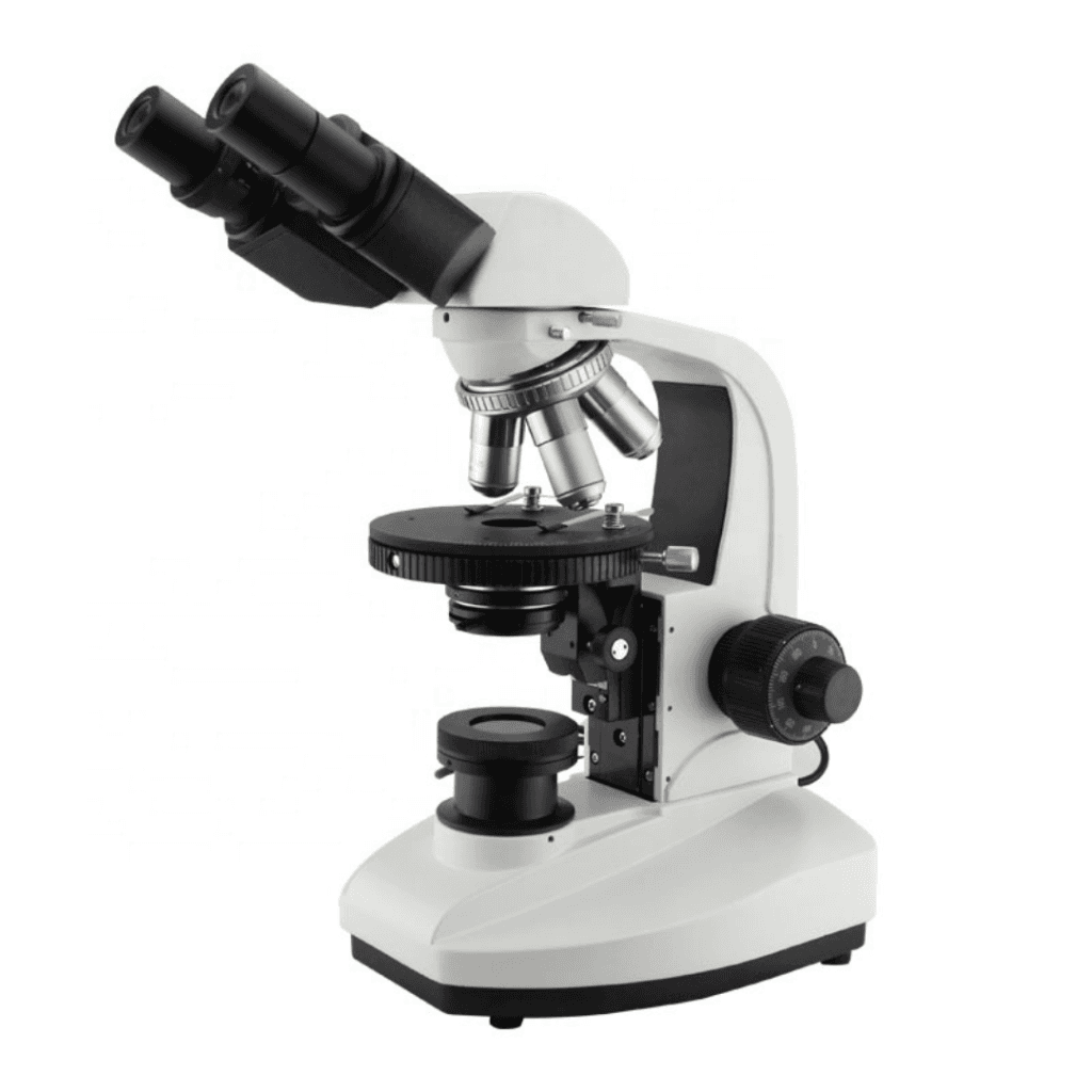
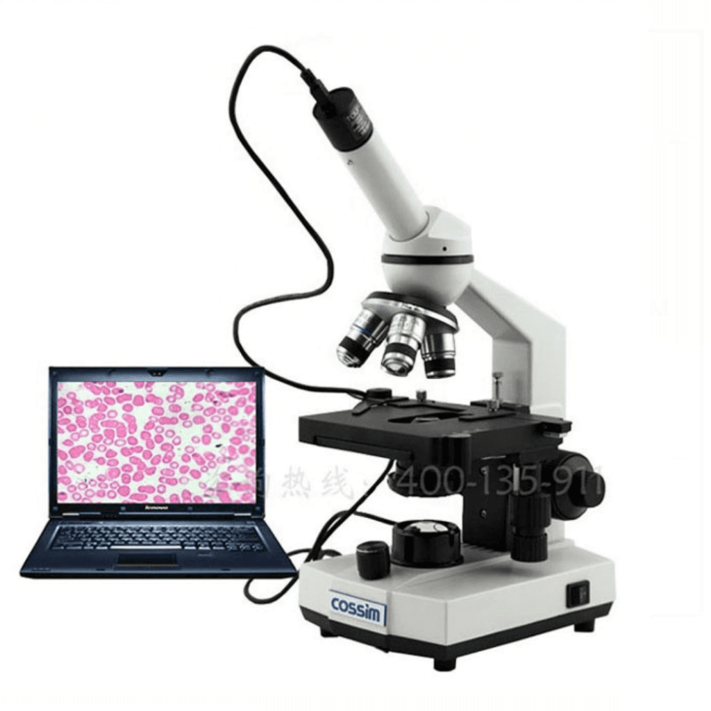
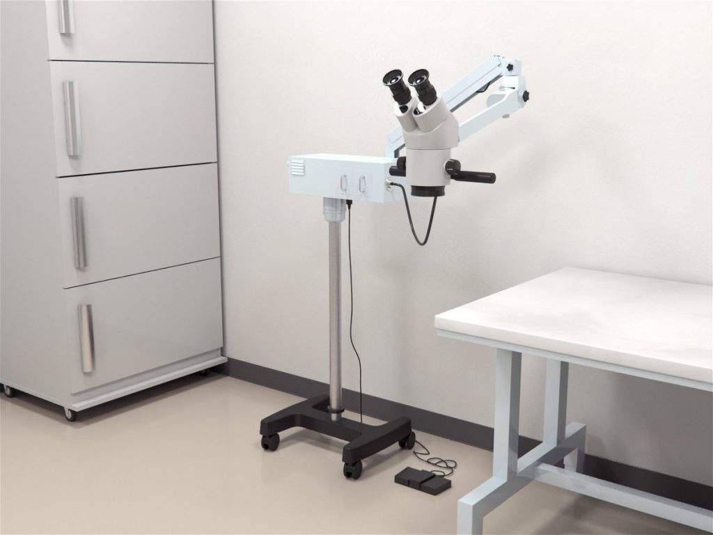
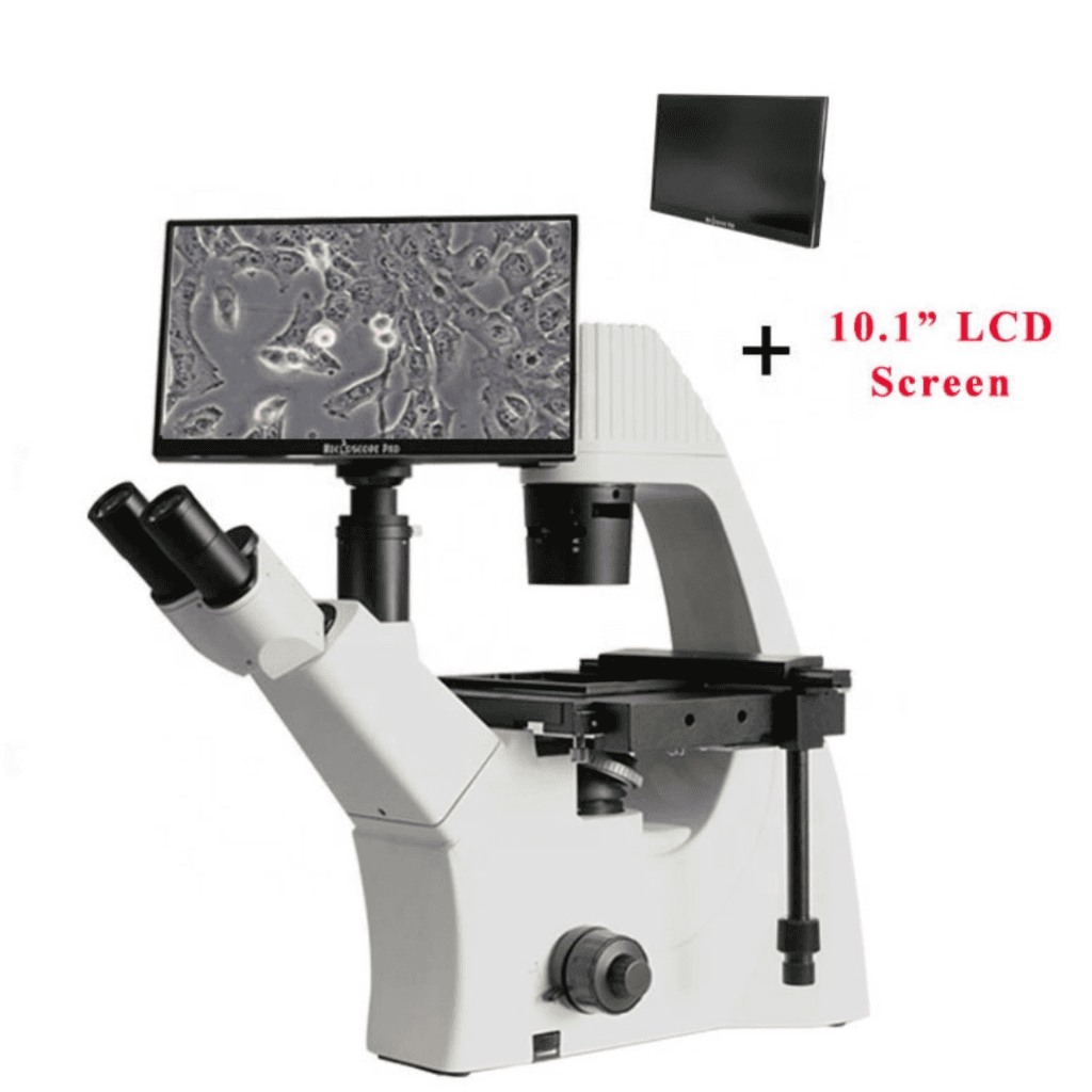
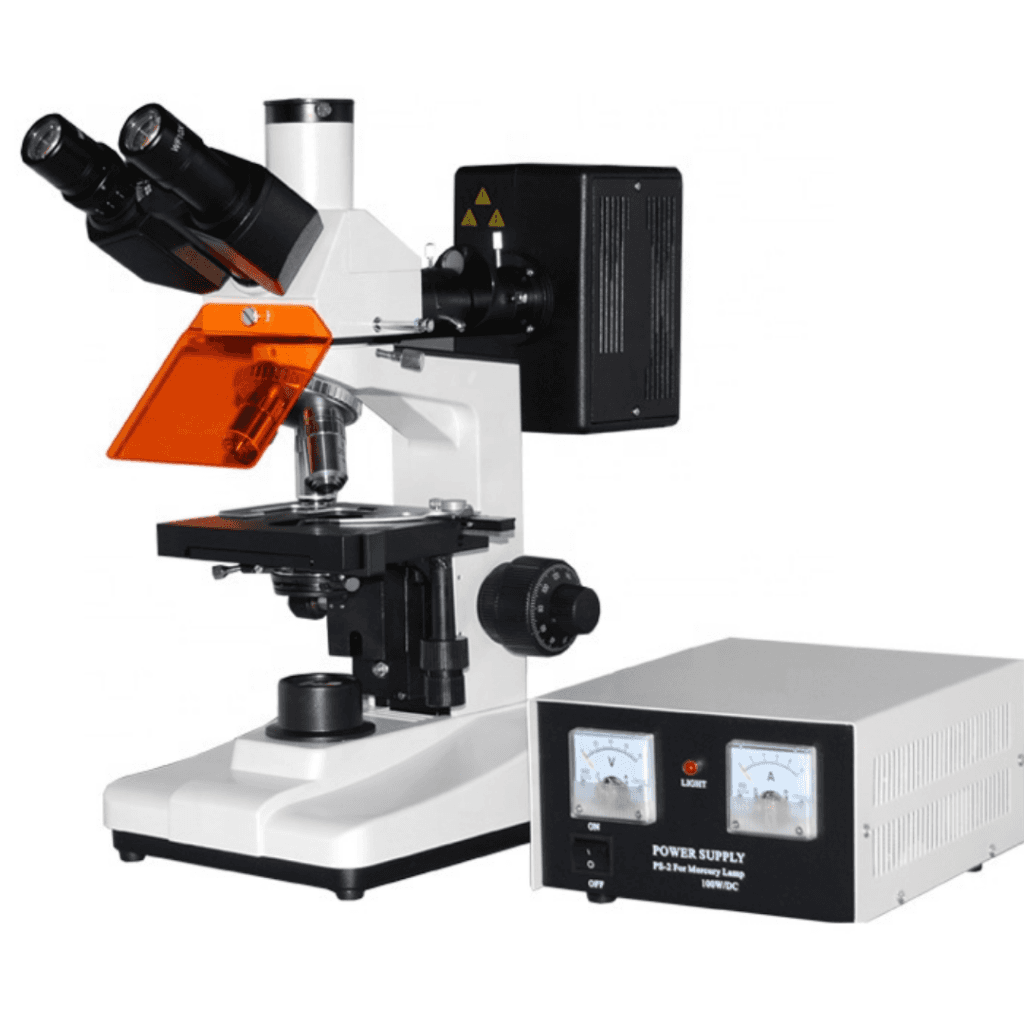
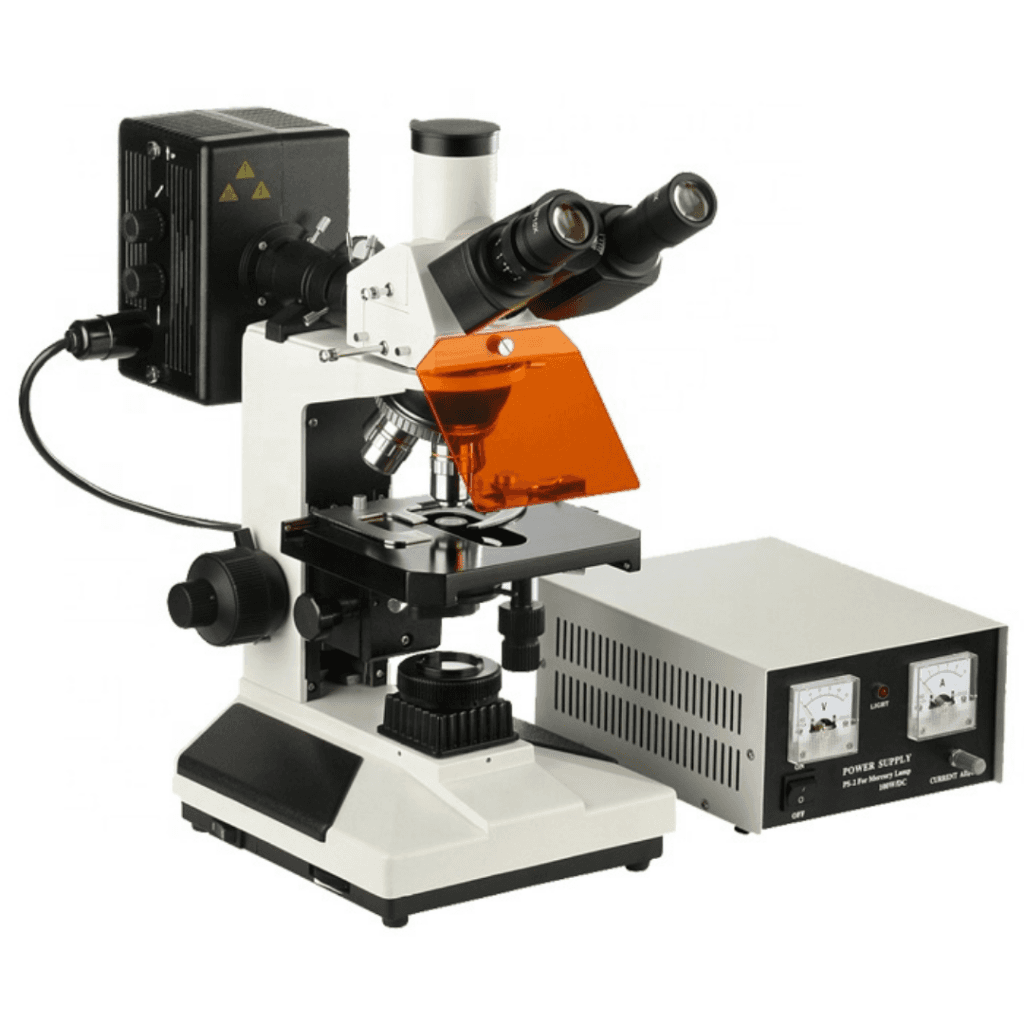
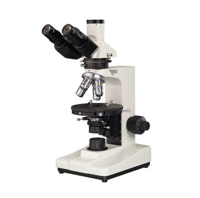
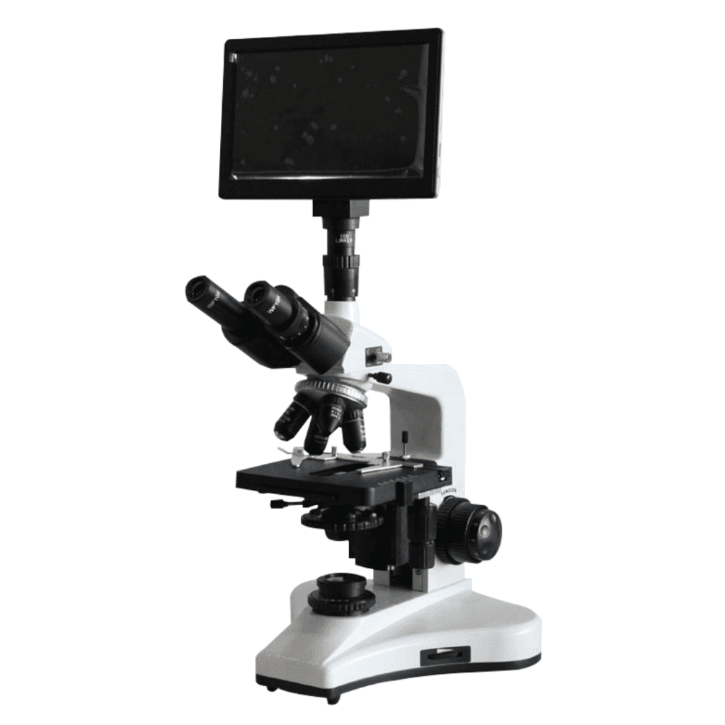
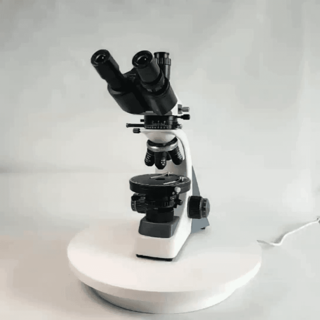
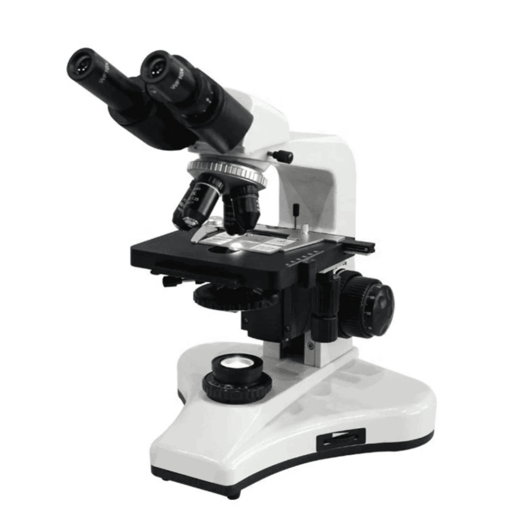
The LCD Display Screen Digital Video Microscope is essentially a Trinocular microscope with an LCD Display Screen and camera integrated. The LCD Display Screen allows live display and easy analysis of specimens and allows the experimenter to view the specimen without looking into the eyepieces, which can reduce eye fatigue. Moreover, the LCD Display Screen allows several experimenters to view the specimen simultaneously. Since the screen and camera are integrated into the microscope, the need for attaching a microscope camera to the laptop is also eliminated, which helps save bench space and time from setting up several technological devices.
The LCD Display Screen Digital Video Microscope also has several other benefits. The LCD screen has a high definition of 1920 × 1080 at 30 fps. It includes an SD card slot, allowing photos and videos to be saved directly on an SD card instantly. The camera includes several functions, such as digital amplification (Max. 10x), picture freezing, and horizontal/vertical mirroring. It also includes a cross-line feature in which the color, line thickness, and bloom are adjustable. Additionally, a mesh line feature comprising four groups of grid lines is also present. The microscope also has a built-in charging system, allowing it to be used for 50 hours without electricity.
Apparatus
The LCD Display Screen Digital Video Microscope measures 282.20×56.20×180.60mm and weighs 0.7 kg. It includes 2 eyepieces and an additional eyepiece tube where the LCD Display Screen is placed. The specimen can be viewed under 4 objective lenses with a total magnification of 40X-1600X. Coarse and fine focusing adjustment knobs, a quadruple nosepiece, a double layer mechanical stage, an ABBE condenser, and blue, green, and yellow filters are present. A built-in charging system and LED-Light source with adjustable iris diaphragm are also present. The LCD screen produces high-definition images and videos of 1920 × 1080 at 30 fps. Its brightness, contrast, saturation, white balance, black and white switch, and HDR-wide dynamic can be adjusted. The user interface includes full mouse operation. It allows picture previews and photos and videos to be instantly saved with dynamic contrast on an SD card. The camera also has other functions, including digital amplification (Max. 10x), horizontal/vertical mirroring, and picture freezing. Cross line and mesh line features are also included.
Protocol
- Fix the slide onto the microscope’s stage
- Adjust the focusing and illumination
- Observe the specimen under the desired objective lens
- View the image of the specimen on the LCD display screen
- Capture images or videos and save them on the SD card.
Applications
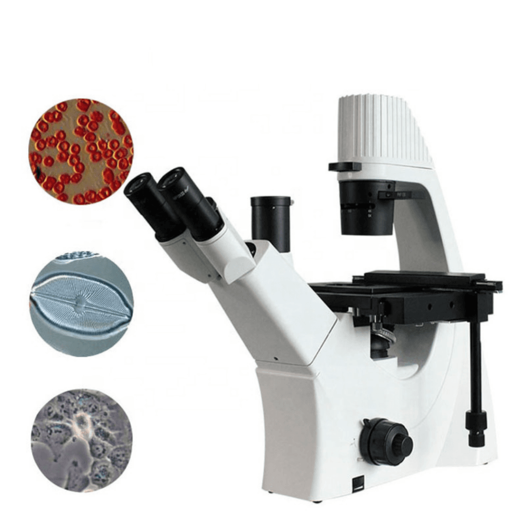
The LCD Display Screen Digital Video Microscope can be utilized for viewing and analyzing the various specimens. The LCD Display Screen allows several people to view the sample under observation for diagnosis and teaching purposes. It can be used in research, medicine, education, forensics, and industrial manufacturing settings. The following studies have utilized the LCD Display Digital Microscope:
Evaluation of sperm viability of West African Dwarf goat bucks during cryopreservation
Daramola et al. (2016) investigated the effects of cucumber, pineapple, and orange juices on sperm viability of buck spermatozoa during cryopreservation. An LCD Display Screen Digital Video Microscope was used to observe sperm motility, acrosome integrity, sperm membrane integrity, and sperm morphology at 400X magnification. The results indicated that the sperm extenders supplemented with pineapple and orange juice at 10% consistently improved the parameters tested and reduced sperm abnormality compared to controls.
Investigation of the effects of the interchange of electromagnetic forces (EMF) within an organism in the development of diseases
Embi (2016) investigated the effect of EMF emissions on cellular respiration oxidation-reduction reactions as a causative agent of diseases. In the experiment, human abdominal hairs were exposed to powder catalase and continuous (Redox) reactions triggered in a processed meat sample through a glass barrier. A solution of Prussian Blue Stain and iron nanoparticles (PBS Fe 2K) was used to stain the hair follicles. The LCD Digital Microscope was used to view and capture still pictures and record videos at 4X magnification during and after evaporation of the PBS Fe2K solution.
Investigation of the two-phase system involved in the conversion of refined palm oil to biodiesel with alkaline catalyst
Thoai, Chanakaewsomboon, Prasertsit, Photaworn, and Tongurai (2019) utilized the LCD Digital microscope to examine the two-phase system in alkaline catalyzed methanol transesterification. The microscope was used periodically to view images of refined palm oil that had alkoxide-methanol solution added to it until the alcohol phase was in overflow. The microscope was also used to view and photograph methanolysis.
Strengths
The LCD Display Screen Digital Video Microscope can be used in various disciplines, including biology, microbiology, and chemistry, for diagnosis and teaching purposes. It allows live viewing of the sample on the LCD screen, making it convenient for individual viewing or group work. Moreover, viewing the sample on the screen reduces eye strain achieved using traditional eyepieces. Since the LCD and camera are integrated into the microscope, an external computer or laptop and camera are not needed, which helps save bench space. The microscope’s LED adjustable illumination and LCD’s save feature allow high-quality pictures and videos to be taken that can instantly be saved onto an SD card.
Precautions
- Keep the microscope away from water and other liquids. Do not use the microscope with wet hands.
- Do not attempt to modify the microscope in any way.
- Turn off the power immediately if you feel like the microscope is overheating or you notice smoke or any strange odor coming from the microscope.
Summary
- The LCD Display Screen Digital Video Microscope is essentially a Trinocular microscope with an LCD Display Screen and camera integrated.
- The LCD Display Screen allows direct live display of specimen where several experimenters can view the specimen simultaneously.
- The microscope can be used in research, medicine, education, forensics, and industrial manufacturing settings. It is suitable for both diagnosis and teaching purposes.
- It helps save bench space since the LCD screen and camera are integrated into the microscope.
- The specimen’s pictures and videos can instantly be saved on an SD card.
References
- Daramola, J. O., Adekunle, E. O., Onagbesan, O. M., Oke, O. E., Ladokun, A. O., Abiona, J. A., … & Adeleke, M. A. (2018). Protective effects of fruit-juices on sperm viability of West African Dwarf goat bucks during cryopreservation. Animal Reproduction (AR), 13(1), 7-13. DOI: 10.4322/1984-3143-AR726
- Embi, A. A. (2016). Cellular respiration oxidation-reduction reactions electromagnetic fields emissions as the possible causative agent in diseases: a chronic bombardment theory. Phys J, 2(3), 226-30.
- Maude, R. J., Koh, G. C., & Silamut, K. (2008). Taking photographs with a microscope. The American journal of tropical medicine and hygiene, 79(3), 471–472. https://doi.org/10.4269/ajtmh.2009.08-0256
- Thoai, D. N., Chanakaewsomboon, I., Prasertsit, K., Photaworn, S., & Tongurai, C. (2019). A novel inspection of mechanisms in the conversion of refined palm oil to biodiesel with alkaline catalyst. Fuel, 256, 115831. https://doi.org/10.1016/j.fuel.2019.115831


Reviews
There are no reviews yet.