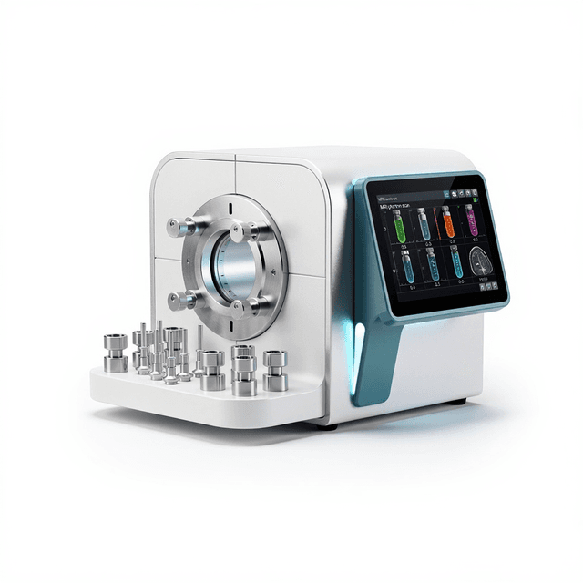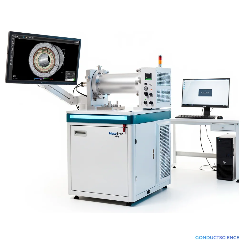
MesoScan MRI System
0.5 Tesla benchtop MRI system providing up to 1 micron resolution imaging for non-destructive analysis of materials, pharmaceuticals, and geological specimens up to 30 mm diameter.

Louise Corscadden, PhD
Director of Science · ConductScience
Ask Louise about MesoScan MRI System fit, setup, configuration, or quote prep.
Already working with us? Sign in to connect this with My Scientist.
Key Specifications
Full details →- Model fit
- Configured during quote
- SKU family
- NMS-MR12-060H-I
- Sizing
- 4.72 x 1.18 x 100.0 cm
- Ordering
- Online checkout and quote request available
- Category
- MRI Systems
- Build notes
- Confirm accessories, station layout, and support needs before purchase
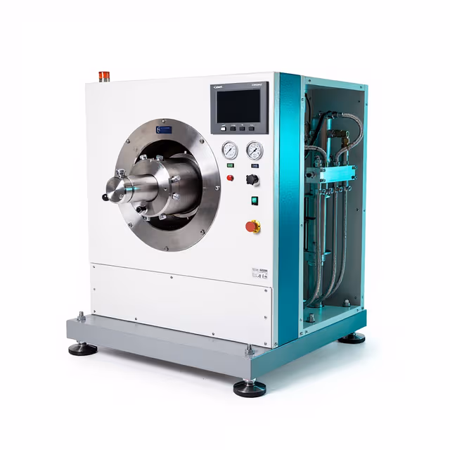
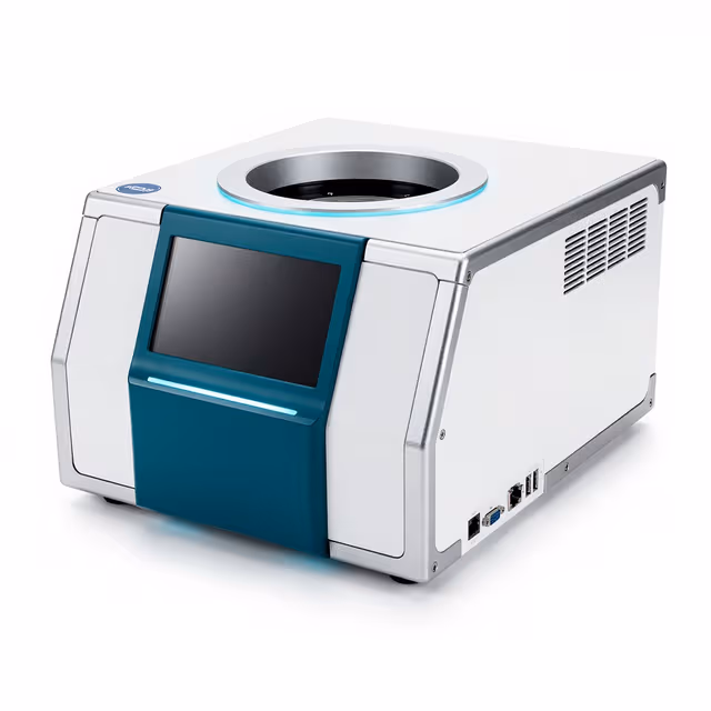
The MesoScan MRI System is a 0.5 Tesla benchtop magnetic resonance imaging analyzer designed for non-destructive structural analysis of materials, pharmaceutical formulations, and geological specimens. With spatial resolution capabilities down to 1 micron and accommodating samples up to 30 mm diameter by 120 mm length, the system provides detailed internal visualization without sample preparation or destruction.
Operating across a temperature range of -10°C to 60°C with maximum gradient strength of 20 G/cm, the system supports both ambient and controlled-temperature studies of material properties, phase transitions, and diffusion processes. The customizable pulse sequence engine and integrated spectroscopy mode enable researchers to develop application-specific protocols for their particular materials and analytical requirements.
How It Works
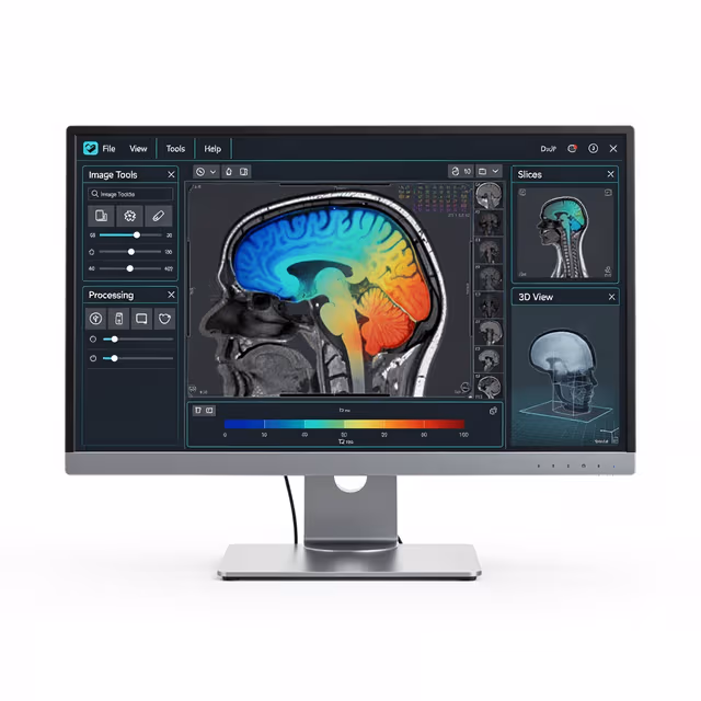
Magnetic resonance imaging operates on the principle of nuclear magnetic resonance, where hydrogen nuclei in the sample align with the 0.5 Tesla magnetic field and respond to radiofrequency pulses. When the RF pulse is removed, nuclei relax back to equilibrium, emitting signals that vary based on their local chemical environment and molecular mobility.
Spatial encoding is achieved through the application of gradient magnetic fields up to 20 G/cm in three orthogonal directions, allowing precise localization of signal origins within the 30 mm diameter sample volume. The customizable pulse sequences control the timing and intensity of RF pulses and gradients to generate contrast based on different tissue properties including T1 relaxation, T2 relaxation, proton density, and diffusion coefficients.
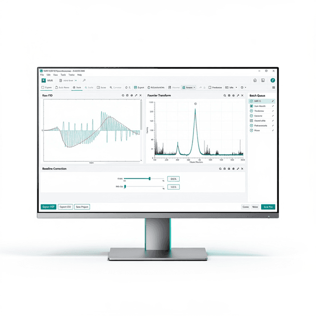
The spectroscopy mode enables chemical shift analysis, providing both spatial and chemical information simultaneously. Temperature control from -10°C to 60°C allows investigation of phase transitions, crystallization processes, and temperature-dependent molecular dynamics within the sample matrix.
Features & Benefits
Field of study
- Engineering
- Oil and Gas
- Pharmaceuticals
- Pharmacology
Product Application
- Analytical Instruments
- Environmental Studies
- Magnetic Resonance Imaging (MRI)
- Production and Quality Control
- Soil Study
magnetic_field_strength
- 0.5 Tesla
sample_diameter
- up to 30 mm
sample_length
- up to 120 mm
maximum_gradient_strength
- 20 G/cm
frequency
- 50Hz
software_interface
- user-friendly and customizable
pulse_sequence
- customizable
spectroscopy_mode
- available
Automation Level
- semi-automated
Brand
- Greenwaves Scientific
Power/Voltage
- 220V
- 7.5 kW
Temperature Range
- -10°C to 60°C
Accuracy
- up to 1 micron
Weight
- approximately 600 kg
Dimensions
- 1600 mm x 750 mm x 1250 mm
Research Domain
- Analytical Chemistry
- Cancer Research
- Environmental Monitoring
- Food Science
- Materials Science
- Pharmaceutical QC
Weight
- 175.0 kg
Dimensions
- L: 4.72 mm
- W: 1.18 mm
- H: 100.0 mm
| Feature | This Product | Typical Alternative | Advantage |
|---|---|---|---|
| Magnetic Field Strength | 0.5 Tesla permanent magnet | Entry-level systems often use 0.1-0.3 Tesla fields or require superconducting magnets | Provides improved signal-to-noise ratio and spectral resolution without cryogenic infrastructure requirements. |
| Spatial Resolution | Up to 1 micron resolution | Basic systems typically offer 10-50 micron resolution | Enables detailed microscale analysis of internal structures and interfaces not visible with lower resolution systems. |
| Sample Size Accommodation | 30 mm diameter, 120 mm length | Compact analyzers often limited to smaller sample volumes | Accommodates larger samples including intact cores, formulation prototypes, and extended geometries for comprehensive analysis. |
| Temperature Control Range | -10°C to 60°C operation | Many systems operate only at ambient temperature | Supports temperature-dependent studies of phase transitions, crystallization processes, and thermal stability. |
| Gradient Strength | 20 G/cm maximum | Lower-cost systems often provide weaker gradient performance | Delivers spatial encoding power necessary for high-resolution imaging and diffusion measurements in research applications. |
| Pulse Sequence Flexibility | Customizable pulse sequence engine | Fixed pulse sequences limit experimental flexibility in basic systems | Allows development of application-specific protocols optimized for particular materials and analytical objectives. |
The MesoScan combines 0.5 Tesla field strength with 1 micron resolution capability and customizable pulse sequences in a benchtop format that eliminates infrastructure requirements of superconducting systems. Temperature control capabilities and flexible sample accommodation support diverse research applications from pharmaceuticals to materials science.
Practical Tips
Perform gradient calibration using standard phantoms before high-resolution imaging sessions to ensure accurate spatial encoding.
Why: Gradient drift can compromise spatial accuracy and introduce geometric distortions in quantitative measurements.
Clean sample chambers and RF coils regularly with appropriate solvents to prevent contamination and signal artifacts.
Why: Residual sample material can create unwanted signals and reduce measurement accuracy in subsequent experiments.
Allow adequate thermal equilibration time when changing sample temperature to ensure uniform heating throughout the sample volume.
Why: Temperature gradients within samples can create artifacts and complicate interpretation of temperature-dependent measurements.
Optimize receiver gain settings for each sample type to maximize signal-to-noise ratio without introducing saturation artifacts.
Why: Proper gain adjustment is critical for quantitative analysis and ensures full dynamic range utilization.
If image quality degrades, check for ferromagnetic objects in the vicinity and verify sample positioning within the RF coil.
Why: Magnetic field distortions from nearby metal objects or improper sample placement can severely compromise image quality.
Ensure all personnel are trained on magnetic field safety protocols and maintain appropriate clearance zones around the system.
Why: The 0.5 Tesla field can affect pacemakers and attract ferromagnetic objects, presenting safety risks in laboratory environments.
Document pulse sequence parameters and acquisition settings for reproducible results across experimental sessions.
Why: Systematic documentation enables method validation and facilitates troubleshooting when comparing datasets over time.
Verify field homogeneity using water phantoms before beginning quantitative relaxometry or spectroscopy measurements.
Why: Field inhomogeneities can bias relaxation time measurements and affect spectral linewidths in quantitative applications.
Setup Guide
What’s in the Box
- MesoScan MRI System main unit
- Sample holder assembly
- Temperature control module
- RF coil set (typical)
- Power cables and connections
- User interface software
- Installation and operation manual
- Calibration phantoms (typical)
- Technical support documentation
Warranty
ConductScience provides standard 1-year manufacturer warranty covering parts and technical support, with extended service contracts available for ongoing maintenance and calibration services.
Compliance
What sample preparation is required for MRI analysis?
Minimal sample preparation is required - samples must fit within 30 mm diameter and 120 mm length constraints and be free of ferromagnetic materials. No sectioning, staining, or chemical treatment is necessary for most applications.
How does resolution compare to X-ray micro-CT systems?
The 1 micron resolution capability is comparable to high-end micro-CT systems, but MRI provides superior soft tissue contrast and chemical information through relaxometry and spectroscopy modes not available with X-ray techniques.
What is the typical imaging time for a complete dataset?
Acquisition times vary from minutes for basic structural imaging to hours for high-resolution 3D datasets, depending on desired resolution, signal averaging requirements, and pulse sequence complexity.
Can the system analyze liquid samples or only solids?
The system can analyze both liquid and solid samples within the dimensional constraints, making it suitable for emulsions, gels, porous materials saturated with fluids, and multi-phase systems.
What data formats are supported for export and analysis?
Consult product datasheet for specific file format compatibility and export options for integration with standard image analysis and visualization software packages.
How stable is the magnetic field over time?
The permanent magnet design provides excellent field stability without the drift associated with resistive electromagnets, though specific stability specifications should be confirmed from technical documentation.
What maintenance is required for optimal performance?
Regular cleaning of sample chambers, periodic gradient calibration, and annual service calibration are typically required, with specific maintenance schedules detailed in the operation manual.
Have a question about this product?
Have a question? Just ask.
Send it over and we'll email you a personalized answer — no call, no scheduling.
Prefer to talk it through?