Tail Flick Test
Automated thermal nociception testing system for measuring analgesic response latency in mice and rats with precise heat stimulus control and rapid detection capabilities.

Louise Corscadden, PhD
Director of Science · ConductScience
Ask Louise about Tail Flick Test fit, setup, configuration, or quote prep.
Already working with us? Sign in to connect this with My Scientist.
Key Specifications
Full details →- Model fit
- Mouse, Rat
- SKU family
- ME-5601
- Sizing
- 43.2 x 38.0 x 27.9 cm
- Ordering
- Online checkout and quote request available
- Category
- Behavioral Mazes
- Build notes
- Confirm accessories, station layout, and support needs before purchase
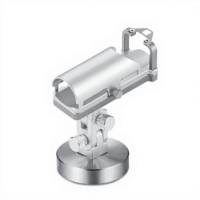
The Tail Flick Test (ME-5601) is a standardized thermal nociception assay system for measuring analgesic response in rodents. This apparatus employs a controlled radiant heat source with pulse width modulation light control to deliver precise thermal stimuli to the animal's tail, automatically measuring latency to withdrawal reflex. The system features a capacitive touch screen interface with sub-3ms response time and laser sensor detection capability of less than 0.01 seconds for accurate latency measurements.
The instrument includes dedicated restrainers for both mouse and rat subjects, integrated data logging with CSV export functionality, and thermal printer for immediate result documentation. Power adjustment occurs in 0.5W increments from 20W to 100W, enabling precise stimulus calibration. The high-grade filter lens maximizes heat radiation while minimizing visible light interference, ensuring consistent thermal stimulus delivery across experimental sessions.
How It Works
The tail flick test operates on the principle of thermal nociception, where radiant heat applied to the tail elicits a spinal reflex withdrawal response. The apparatus uses pulse width modulation to control light intensity from a halogen source, delivering consistent thermal stimuli through a high-grade filter lens system that maximizes infrared heat radiation while minimizing visible light artifacts.
A laser sensor system with sub-10ms detection capability monitors tail position and automatically records withdrawal latency when movement occurs. The capacitive touch screen interface allows real-time parameter adjustment and data visualization, while built-in algorithms account for baseline tail temperature variations that can influence response thresholds.
Thermal cutoff mechanisms prevent tissue damage by limiting maximum exposure duration, typically 10-15 seconds depending on protocol requirements. The system measures response latency from stimulus onset to tail movement initiation, providing quantitative assessment of nociceptive sensitivity with 99% measurement accuracy.
Features & Benefits
touch_screen_response_speed
- less than 3ms
laser_sensor_detection_time
- less than 0.01S
light_power_adjustment_step
- 0.5w
screen_type
- capacitive touch screen
included_accessories
- ['USB cable', 'Foot switch', 'Printer', 'Printer power adapter and cable', 'Mouse refrainer', 'Rat refrainer']
required_driver
- UBS to 232 Driver
data_export_format
- CSV
light_control_method
- pulse width modulation
filter_lens_feature
- high-grade filter lens to partially absorb visible light while maximizing heat radiation
Behavioral Construct
- Nociception
- Thermal sensitivity
- Analgesic response
- Withdrawal reflex
Automation Level
- semi-automated
Power/Voltage
- 20w-100w
Accuracy
- 99%
Species
- Mouse
- Rat
Display Type
- Touch Screen
Research Domain
- Addiction Research
- Behavioral Pharmacology
- Neurodegeneration
- Neuroscience
- Pain Research
- Toxicology
Compatible Tracking Software
- ConductVision
Weight
- 21.0 kg
Dimensions
- L: 43.2 mm
- W: 38.0 mm
- H: 27.9 mm
| Feature | This Product | Typical Alternative | Advantage |
|---|---|---|---|
| Response Detection Speed | Less than 0.01 seconds laser sensor detection | Manual observation methods often introduce 100-500ms observer reaction delays | Eliminates observer bias and improves measurement precision for detecting subtle analgesic effects. |
| Touch Screen Interface Response | Sub-3ms capacitive touch screen response time | Basic models often use slower resistive screens or button interfaces | Enables real-time parameter adjustments during experiments without interrupting data collection workflow. |
| Power Control Precision | 0.5W adjustment increments across 20W-100W range | Entry-level systems typically offer larger adjustment steps or limited power ranges | Allows fine stimulus intensity titration for establishing precise baseline responses and dose-response curves. |
| Light Control Method | Pulse width modulation for consistent stimulus delivery | Basic systems often use simple voltage control with intensity fluctuations | Eliminates stimulus variability that can mask treatment effects in pharmacological studies. |
| Data Export Capability | Integrated CSV export with thermal printer documentation | Lower-end models may lack automated data logging or require manual transcription | Streamlines data collection and reduces transcription errors in multi-animal studies. |
| Filter Lens System | High-grade filter lens maximizing heat radiation while minimizing visible light | Basic models may use unfiltered sources with visible light stress artifacts | Reduces confounding visual stress responses that can interfere with pure nociceptive measurements. |
This system combines rapid laser detection, precise power control, and automated data logging to provide standardized thermal nociception testing. The pulse width modulation control and filtered light source ensure consistent stimulus delivery, while the sub-millisecond detection capability captures accurate withdrawal latencies essential for pharmacological research.
Practical Tips
Verify laser sensor alignment and power output calibration weekly or after any system movement.
Why: Maintains measurement accuracy and ensures consistent stimulus delivery across experimental sessions.
Allow 10-15 minutes for thermal equilibration after power-on before beginning measurements.
Why: Eliminates stimulus intensity drift during initial warm-up period that can affect baseline measurements.
Clean filter lens weekly with appropriate optical cleaning materials to maintain consistent heat transmission.
Why: Prevents accumulation of debris that can create hot spots or reduce stimulus intensity over time.
Record ambient temperature for each session and maintain consistent laboratory environmental conditions.
Why: Ambient temperature variations can affect baseline tail temperature and alter nociceptive thresholds.
If detection sensitivity appears reduced, verify laser sensor positioning and check for obstructions in the beam path.
Why: Proper sensor alignment is critical for accurate latency measurements and preventing false readings.
Always use the programmed cutoff times and never exceed manufacturer-recommended maximum exposure durations.
Why: Prevents tissue damage and ensures animal welfare compliance while maintaining experimental validity.
Establish individual baseline responses through multiple pre-treatment trials before drug administration.
Why: Accounts for individual variation in thermal sensitivity and improves statistical power for detecting treatment effects.
Use the foot switch for hands-free operation to minimize investigator movement that could disturb animals during testing.
Why: Reduces stress-induced artifacts and maintains consistent experimental conditions throughout the session.
Setup Guide
What’s in the Box
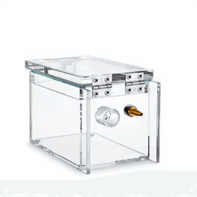
- Main tail flick apparatus
- USB cable
- Foot switch
- Thermal printer
- Printer power adapter and cable
- Mouse restrainer
- Rat restrainer
- User manual (typical)
- Calibration certificate (typical)
Warranty
ConductScience provides standard one-year manufacturer warranty covering defects in materials and workmanship, with technical support for setup, calibration, and operational guidance throughout the warranty period.
Compliance
References
Background reading relevant to this product:
What is the recommended baseline latency range for establishing individual animal responses?
Consult product datasheet for species-specific baseline parameters. Typical protocols establish individual baselines through multiple pre-treatment measurements, with cutoff times preventing tissue damage.
How does the laser detection system differentiate between tail movement and other body movements?
The laser sensor system monitors the specific tail exposure area with sub-0.01 second detection capability. Proper restrainer positioning minimizes whole-body movement artifacts while allowing natural tail positioning.
Can the system accommodate different tail diameters between species and individual animals?
Species-specific restrainers are included for mouse and rat subjects. The laser detection system and heat source positioning accommodate natural variation in tail dimensions within each species.
What data parameters are included in the CSV export files?
CSV export includes withdrawal latency measurements, stimulus parameters, trial timestamps, and session identifiers. Consult software manual for complete data field specifications and formatting details.
How frequently should the system be recalibrated for consistent results?
Calibration frequency depends on usage intensity and laboratory requirements. The system provides calibration verification through the touch screen interface for power output and sensor alignment confirmation.
What is the maximum number of animals that can be tested in a single session?
Testing throughput depends on protocol requirements including inter-trial intervals and recovery periods. The system supports continuous operation with individual animal data logging throughout extended sessions.
How does ambient temperature affect measurement consistency?
The high-grade filter lens system and pulse width modulation control minimize environmental temperature effects on stimulus delivery. Maintain stable laboratory temperature for optimal measurement reproducibility.
Have a question about this product?
Have a question? Just ask.
Send it over and we'll email you a personalized answer — no call, no scheduling.
Prefer to talk it through?